Right Iliac Fossa Mass Ayesha Nasrullah GI Radiology


- Slides: 12
Right Iliac Fossa Mass Ayesha Nasrullah GI Radiology Fellow
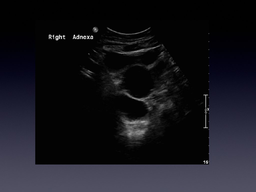
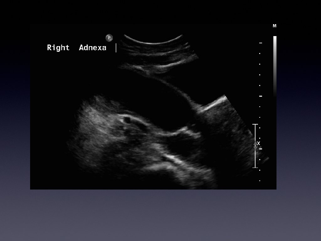
Presentation • • • 36 year old female Presented with chronic right iliac fossa pain Tenderness on palpation Bloods normal Investigated further with a pelvic ultrasound
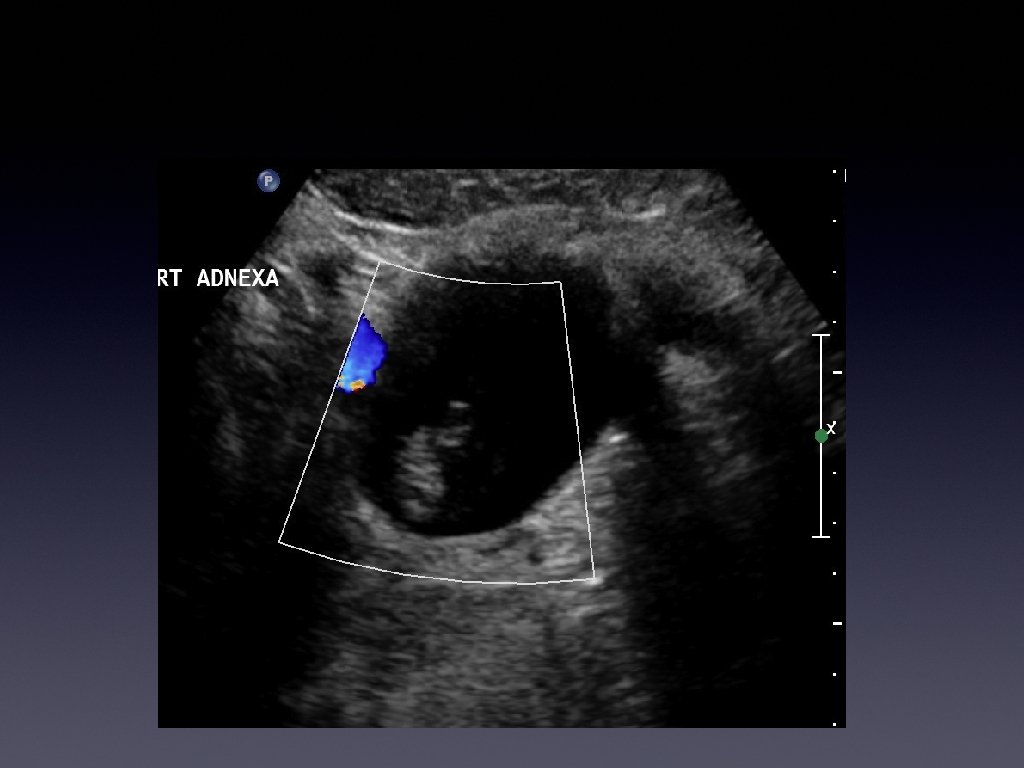
Few months later. . . • • • Patient continued to have pain Came back and this time tenderness was worse Bloods showed raised WBC count and CRP
Differentials • ? ? ? ?
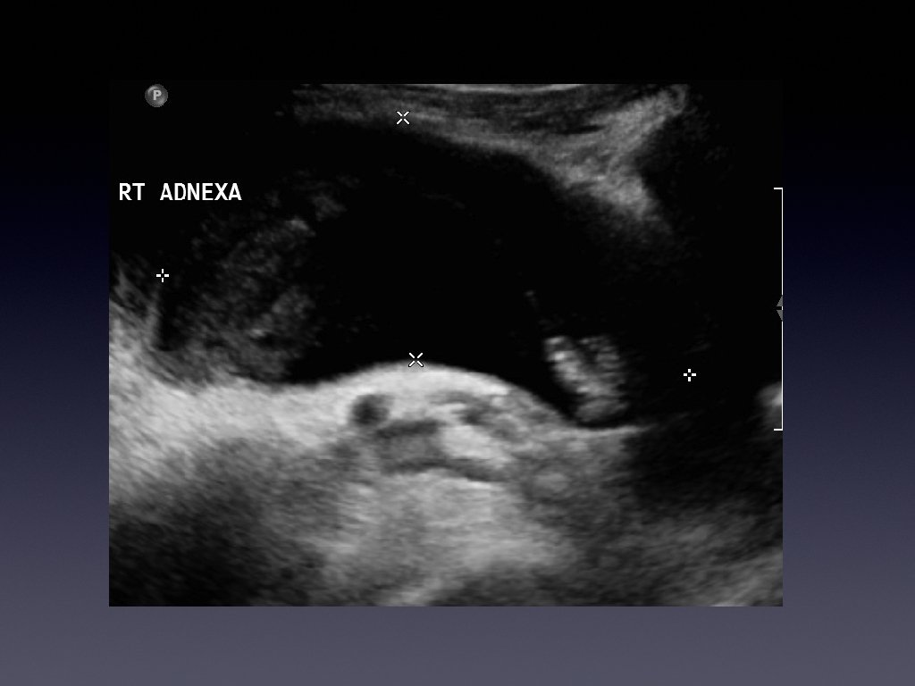
Management • • • Patient was diagnosed with an infected hydrosalpinx She was taken in for surgery Surgery revealed it was an appendiceal mucocoele
Learning Points • • • Hydrosalpinx is usually a tubular cystic mass However it has septations Septations are caused by the normal fold present in the fallopian tube which become thickened with inflammation.
Learning points • • • Usually a cystic mass Variable internal echogenicity Devoid of septations Can have debris Not every tubular mass in the iliac fossa is ovarian in origin!!!!
References • • Kim JS, Woo SK, Suh SJ, Morettin LB. Sonographic diagnosis of paraovarian cysts: value of detecting a separate ipsilateral ovary. AJR Am J Roentgenol 1995; 164(6): 1441– 1444 Madwed D, Mindelzun R, Jeffrey RB. Mucocele of the appendix: imaging findings. AJR Am J Roentgenol 1992; 159(1): 69– 72.