
MBBS MC MIRPUR m GROSS ANATOMY PROF DR
MBBS MC MIRPUR m GROSS ANATOMY

PROF DR ZUBAIR / DR KAMRAN. TTTTT
The Tibia
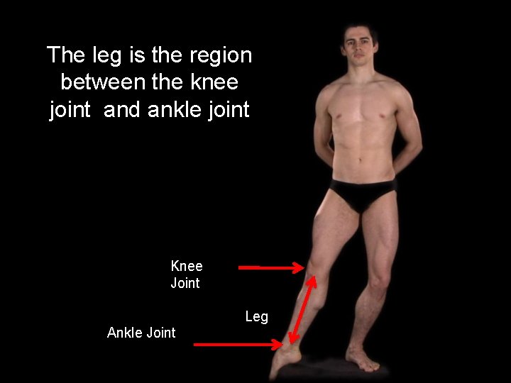
The leg is the region between the knee joint and ankle joint Knee Joint Leg Ankle Joint
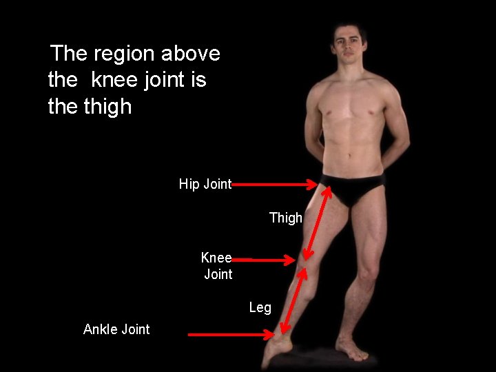
The region above the knee joint is the thigh Hip Joint Thigh Knee Joint Leg Ankle Joint

There are two bones in the leg
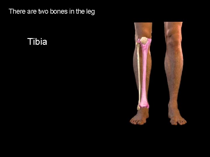
There are two bones in the leg Tibia
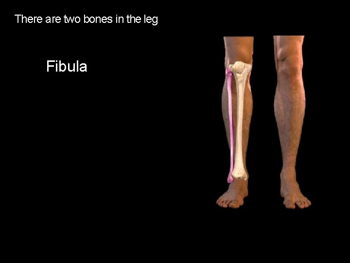
There are two bones in the leg Fibula
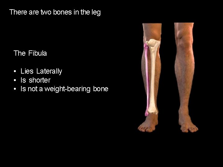
There are two bones in the leg The Fibula • Lies Laterally • Is shorter • Is not a weight-bearing bone
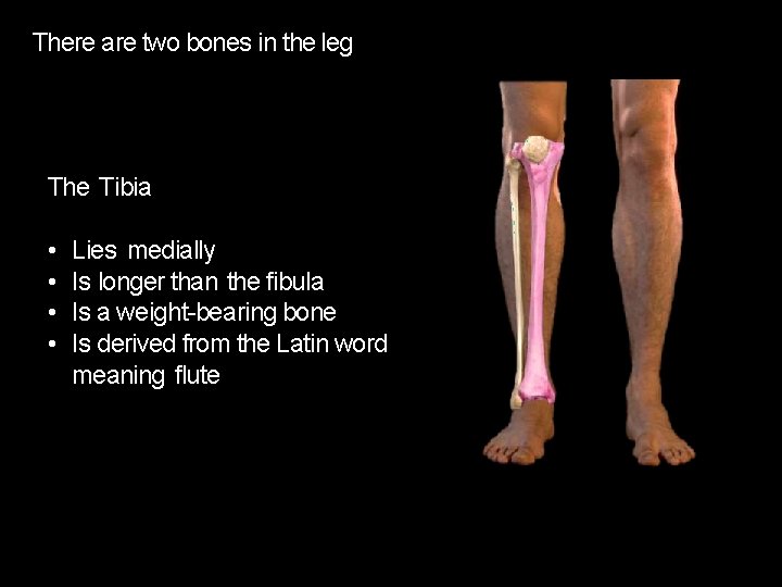
There are two bones in the leg The Tibia • • Lies medially Is longer than the fibula Is a weight-bearing bone Is derived from the Latin word meaning flute

The Fibula and Tibia are held together by the Interosseous Membrane

Proximal end The Tibia is a long bone with expanded proximal and distal ends with a shaft in between Shaft Distal end right Tibia, anterior
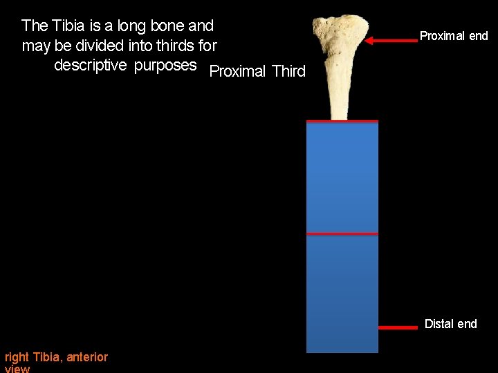
The Tibia is a long bone and may be divided into thirds for descriptive purposes Proximal Third Proximal end Distal end right Tibia, anterior

The Tibia is a long bone and may be divided into thirds for descriptive purposes Proximal Third Proximal end (Head of the Fibula) Middle Third Distal end right Tibia, anterior
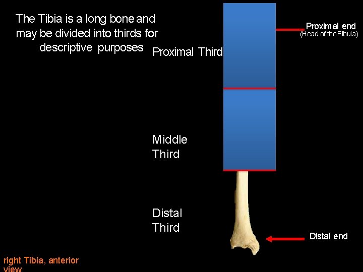
The Tibia is a long bone and may be divided into thirds for descriptive purposes Proximal Third Proximal end (Head of the Fibula) Middle Third Distal Third right Tibia, anterior Distal end

The Tibia articulates proximally at the Knee Joint Fibula Interosseous Membrane Tibia right Tibia, anterior
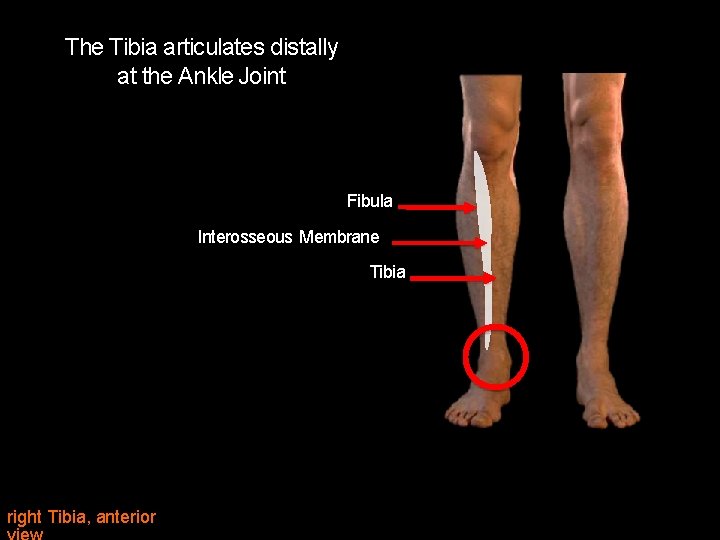
The Tibia articulates distally at the Ankle Joint Fibula Interosseous Membrane Tibia right Tibia, anterior
The Proximal Tibia
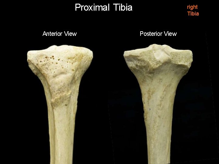
Proximal Tibia Anterior View right Tibia Posterior View
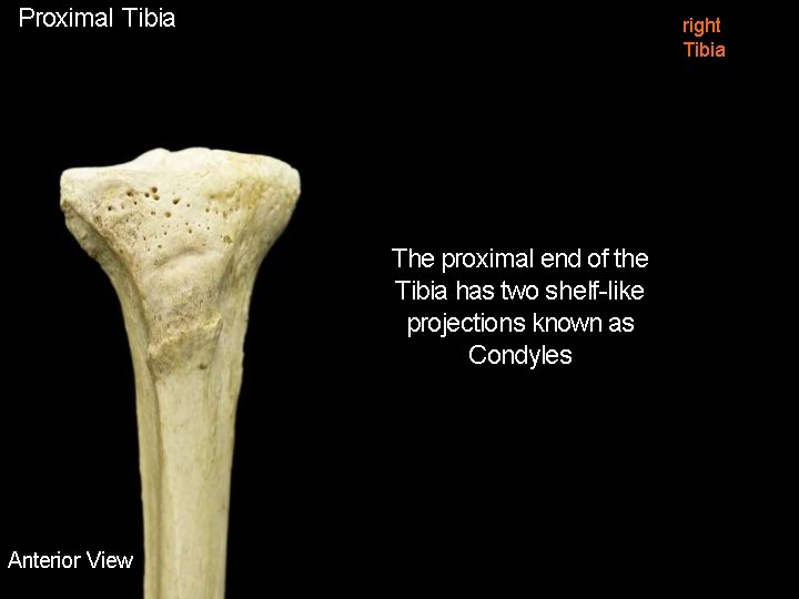
Proximal Tibia right Tibia The proximal end of the Tibia has two shelf-like projections known as Condyles Anterior View
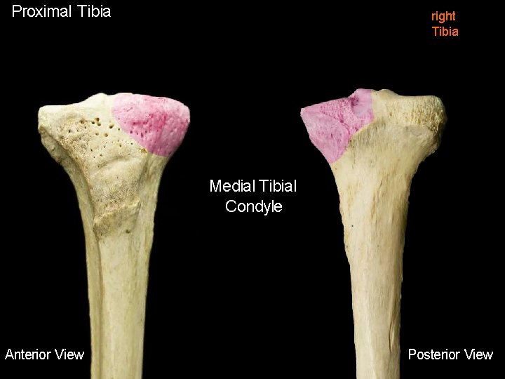
Proximal Tibia right Tibia Medial Tibial Condyle Anterior View Posterior View
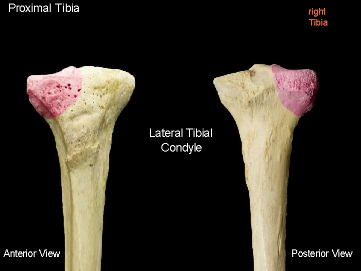
Proximal Tibia right Tibia Lateral Tibial Condyle Anterior View Posterior View
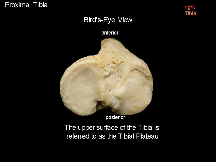
Proximal Tibia Bird’s-Eye View anterior posterior The upper surface of the Tibia is referred to as the Tibial Plateau right Tibia
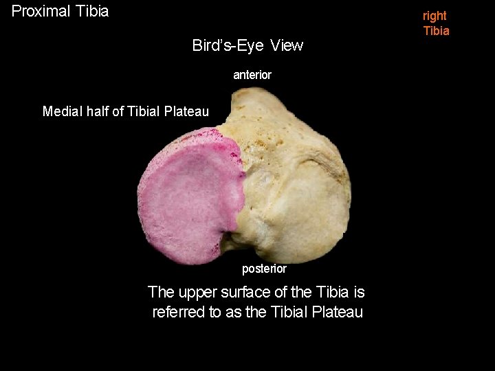
Proximal Tibia Bird’s-Eye View anterior Medial half of Tibial Plateau posterior The upper surface of the Tibia is referred to as the Tibial Plateau right Tibia
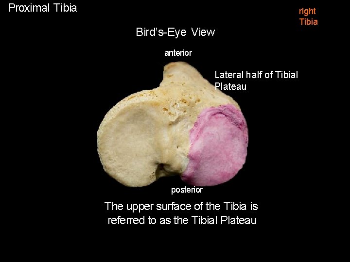
Proximal Tibia Bird’s-Eye View anterior Lateral half of Tibial Plateau posterior The upper surface of the Tibia is referred to as the Tibial Plateau right Tibia
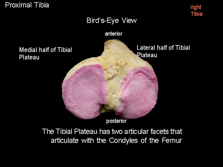
Proximal Tibia Bird’s-Eye View anterior Lateral half of Tibial Plateau Medial half of Tibial Plateau posterior The Tibial Plateau has two articular facets that articulate with the Condyles of the Femur right Tibia
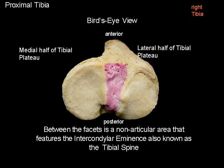
Proximal Tibia Bird’s-Eye View right Tibia anterior Lateral half of Tibial Plateau Medial half of Tibial Plateau posterior Between the facets is a non-articular area that features the Intercondylar Eminence also known as the Tibial Spine

right Tibia Menisci 2 crescent-shaped intra-capsular fibrocartilaginous structures situated one on each Tibial Facet anterior Medial Meniscus Lateral Meniscus posterior Superior view proximal Tibia
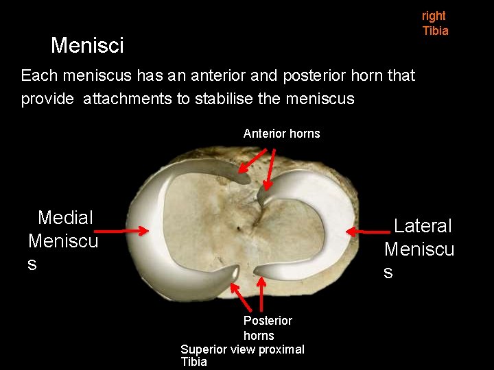
right Tibia Menisci Each meniscus has an anterior and posterior horn that provide attachments to stabilise the meniscus Anterior horns Medial Meniscu s Lateral Meniscu s Posterior horns Superior view proximal Tibia
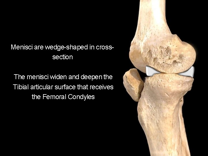
Menisci are wedge-shaped in crosssection The menisci widen and deepen the Tibial articular surface that receives the Femoral Condyles
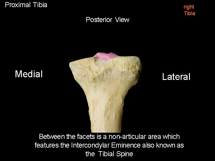
Proximal Tibia Posterior View Medial right Tibia Lateral Between the facets is a non-articular area which features the Intercondylar Eminence also known as the Tibial Spine
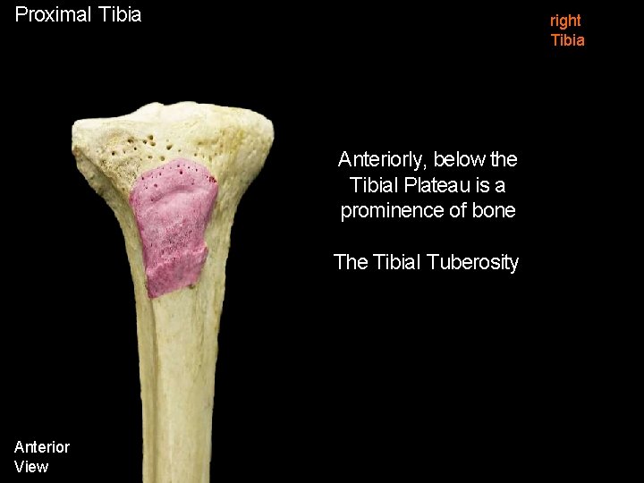
Proximal Tibia right Tibia Anteriorly, below the Tibial Plateau is a prominence of bone The Tibial Tuberosity Anterior View
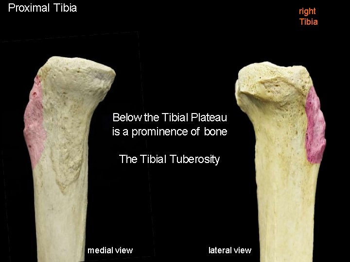
Proximal Tibia right Tibia Below the Tibial Plateau is a prominence of bone The Tibial Tuberosity medial view lateral view
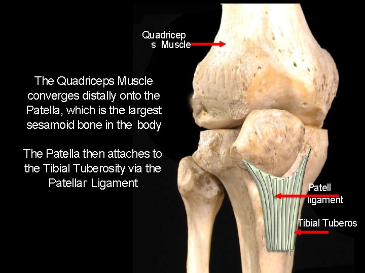
Quadricep s Muscle The Quadriceps Muscle converges distally onto the Patella, which is the largest sesamoid bone in the body The Patella then attaches to the Tibial Tuberosity via the Patellar Ligament Patell ligament Tibial Tuberos
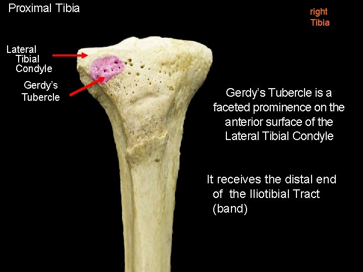
Proximal Tibia right Tibia Lateral Tibial Condyle Gerdy’s Tubercle is a faceted prominence on the anterior surface of the Lateral Tibial Condyle It receives the distal end of the Iliotibial Tract (band)

Iliotibial Tract Tensor Fasciae Latae Attaches to Gerdy’s Tubercle on the anterior aspect of the Lateral Tibial Condyle Proximally it receives two muscles • Tensor Fasciae Latae • Gluteus Maximus Iliotibial Tract Gerdy’s Tubercle right
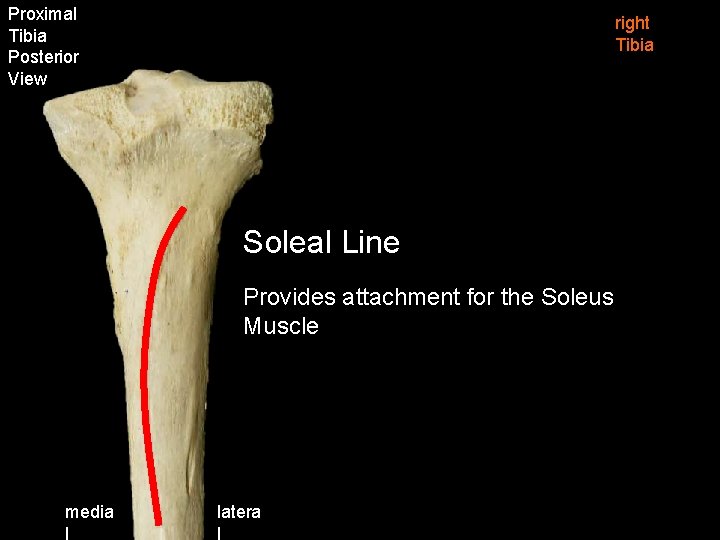
Proximal Tibia Posterior View right Tibia Soleal Line Provides attachment for the Soleus Muscle media l latera l
Tibial Shaft
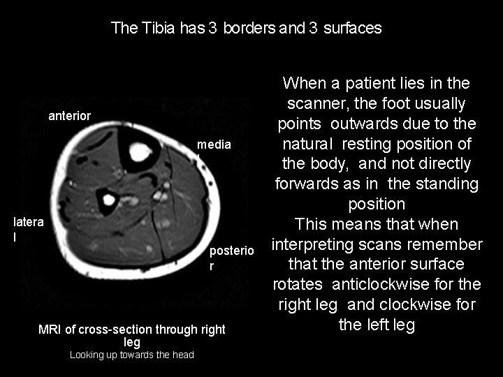
The Tibia has 3 borders and 3 surfaces anterior media l latera l posterio r MRI of cross-section through right leg Looking up towards the head When a patient lies in the scanner, the foot usually points outwards due to the natural resting position of the body, and not directly forwards as in the standing position This means that when interpreting scans remember that the anterior surface rotates anticlockwise for the right leg and clockwise for the left leg
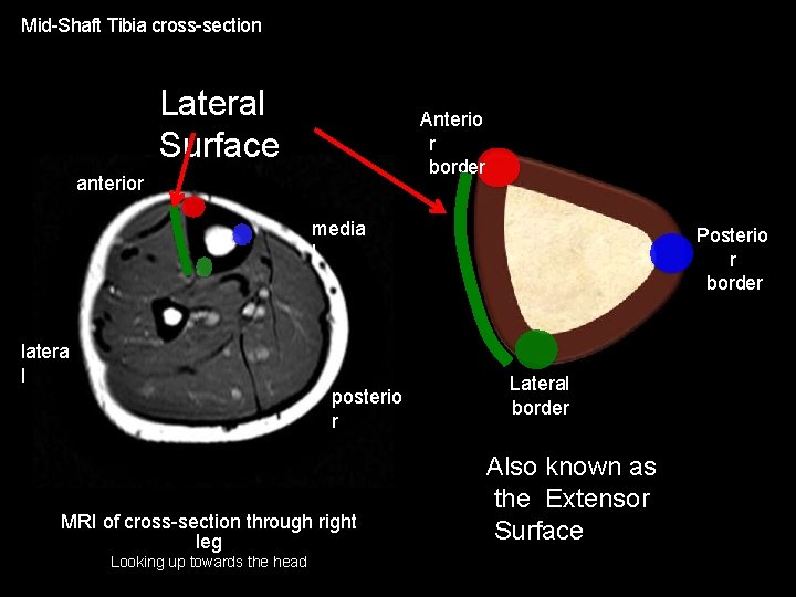
Mid-Shaft Tibia cross-section Lateral Surface Anterio r border anterior media l latera l posterio r MRI of cross-section through right leg Looking up towards the head Posterio r border Lateral border Also known as the Extensor Surface
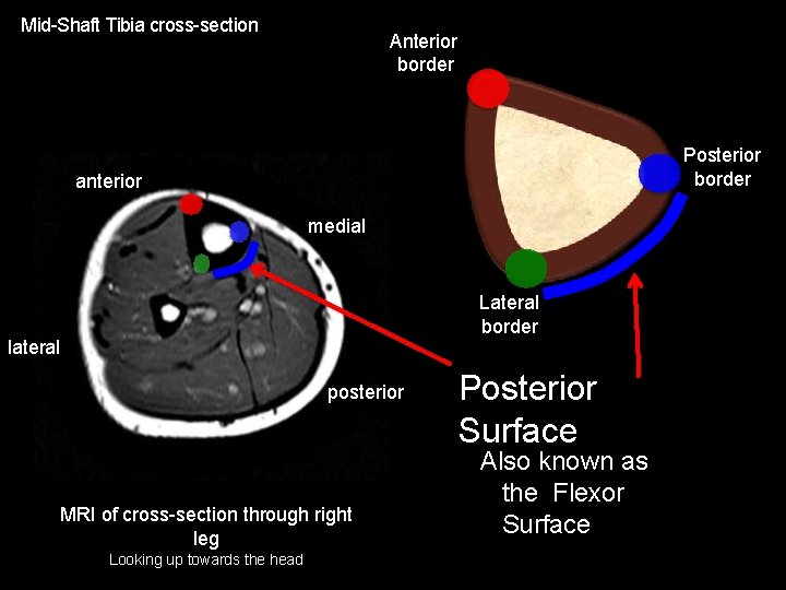
Mid-Shaft Tibia cross-section Anterior border Posterior border anterior medial Lateral border lateral posterior MRI of cross-section through right leg Looking up towards the head Posterior Surface Also known as the Flexor Surface
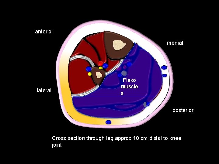
anterior medial lateral Flexo r muscle s posterior Cross section through leg approx 10 cm distal to knee joint
Distal Tibia
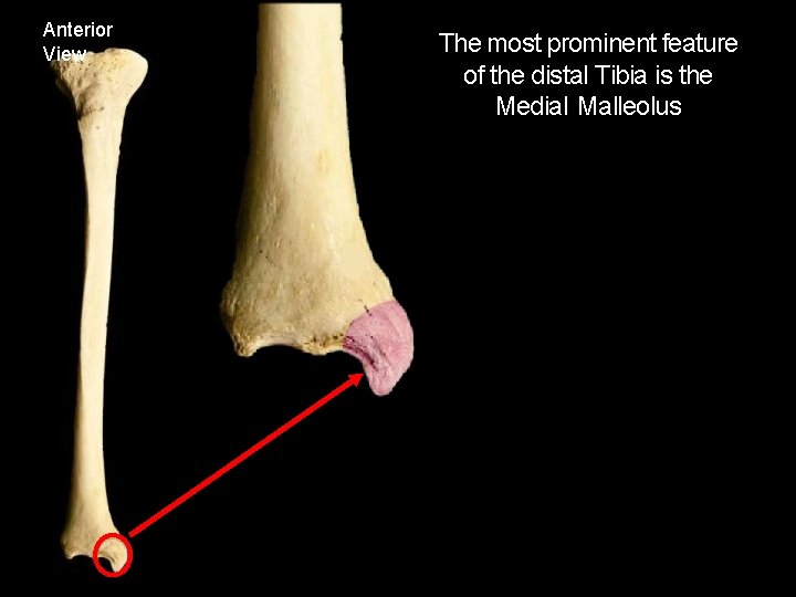
Anterior View The most prominent feature of the distal Tibia is the Medial Malleolus
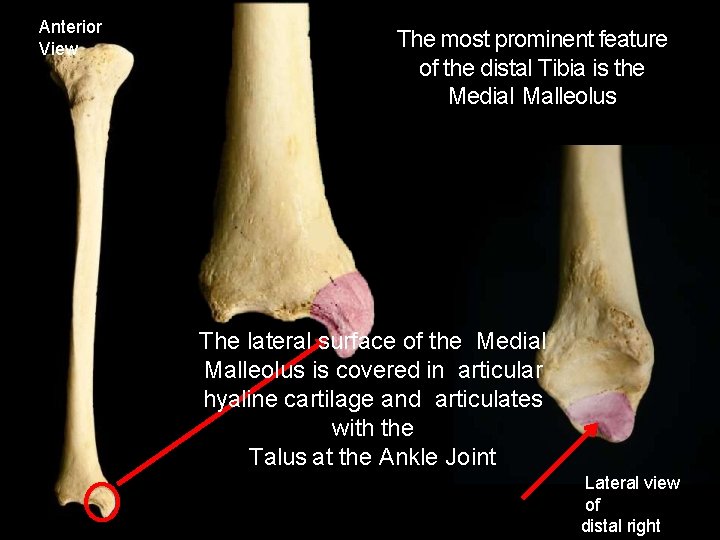
Anterior View The most prominent feature of the distal Tibia is the Medial Malleolus The lateral surface of the Medial Malleolus is covered in articular hyaline cartilage and articulates with the Talus at the Ankle Joint Lateral view of distal right
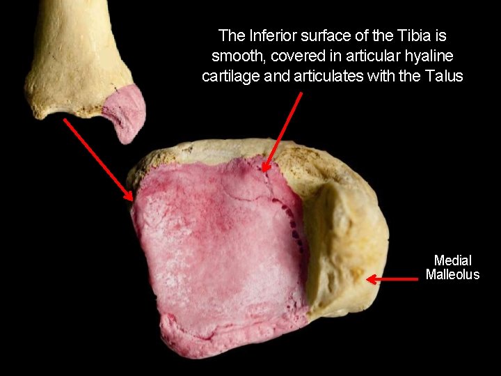
The Inferior surface of the Tibia is smooth, covered in articular hyaline cartilage and articulates with the Talus Lateral surface Medial Malleolus
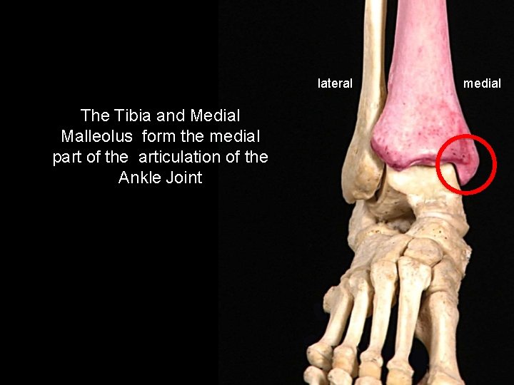
lateral The Tibia and Medial Malleolus form the medial part of the articulation of the Ankle Joint medial
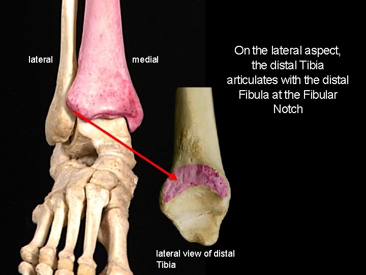
lateral medial On the lateral aspect, the distal Tibia articulates with the distal Fibula at the Fibular Notch lateral view of distal Tibia

Muscle Attachments
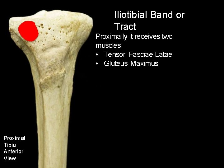
Iliotibial Band or Tract Proximally it receives two muscles • Tensor Fasciae Latae • Gluteus Maximus Proximal Tibia Anterior View
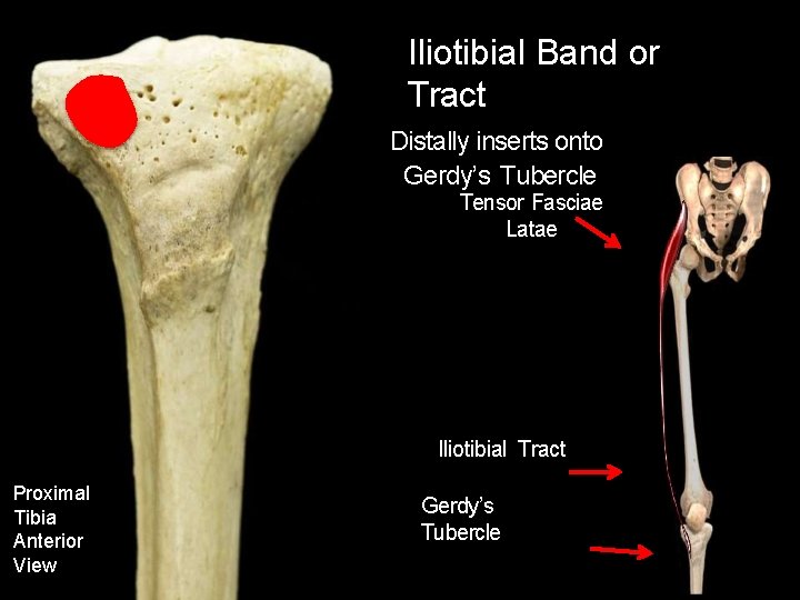
Iliotibial Band or Tract Distally inserts onto Gerdy’s Tubercle Tensor Fasciae Latae Iliotibial Tract Proximal Tibia Anterior View Gerdy’s Tubercle
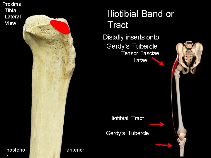
Proximal Tibia Lateral View Iliotibial Band or Tract Distally inserts onto Gerdy’s Tubercle Tensor Fasciae Latae Iliotibial Tract Gerdy’s Tubercle posterio r anterior
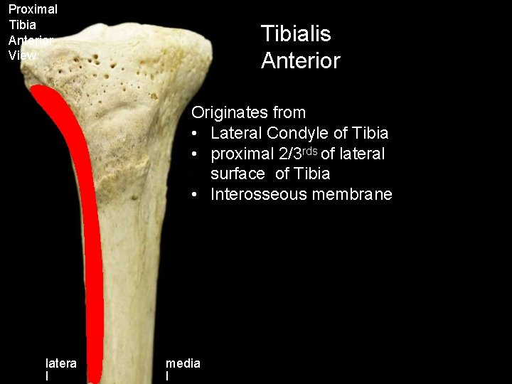
Proximal Tibia Anterior View Tibialis Anterior Originates from • Lateral Condyle of Tibia • proximal 2/3 rds of lateral surface of Tibia • Interosseous membrane latera l media l
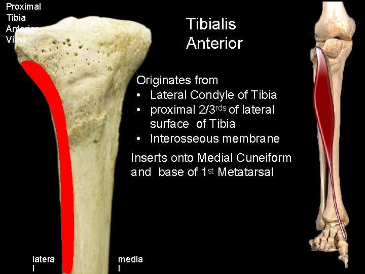
Proximal Tibia Anterior View Tibialis Anterior Originates from • Lateral Condyle of Tibia • proximal 2/3 rds of lateral surface of Tibia • Interosseous membrane Inserts onto Medial Cuneiform and base of 1 st Metatarsal latera l media l
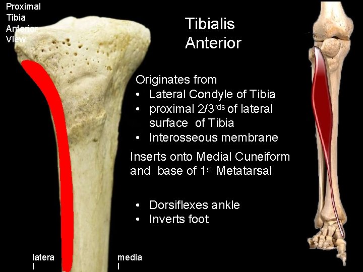
Proximal Tibia Anterior View Tibialis Anterior Originates from • Lateral Condyle of Tibia • proximal 2/3 rds of lateral surface of Tibia • Interosseous membrane Inserts onto Medial Cuneiform and base of 1 st Metatarsal • Dorsiflexes ankle • Inverts foot latera l media l
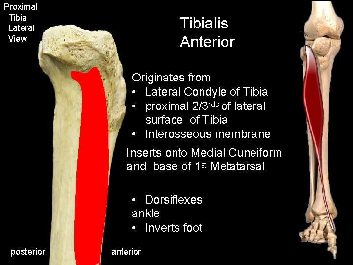
Proximal Tibia Lateral View Tibialis Anterior Originates from • Lateral Condyle of Tibia • proximal 2/3 rds of lateral surface of Tibia • Interosseous membrane Inserts onto Medial Cuneiform and base of 1 st Metatarsal • Dorsiflexes ankle • Inverts foot posterior anterior
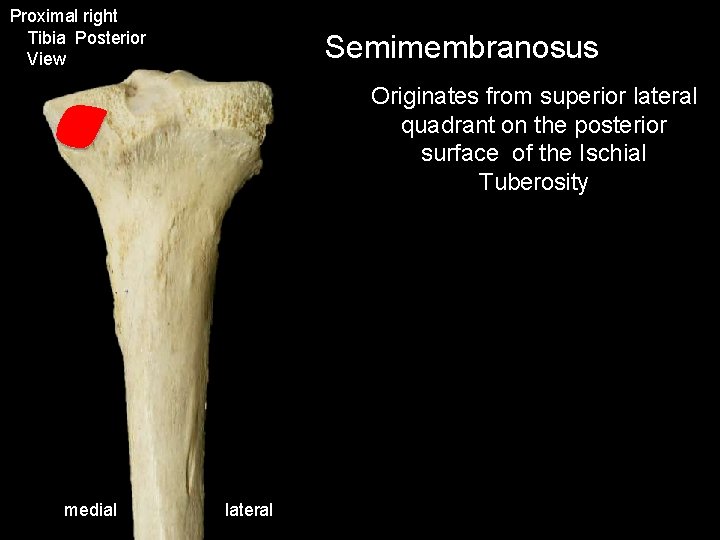
Proximal right Tibia Posterior View Semimembranosus Originates from superior lateral quadrant on the posterior surface of the Ischial Tuberosity medial lateral
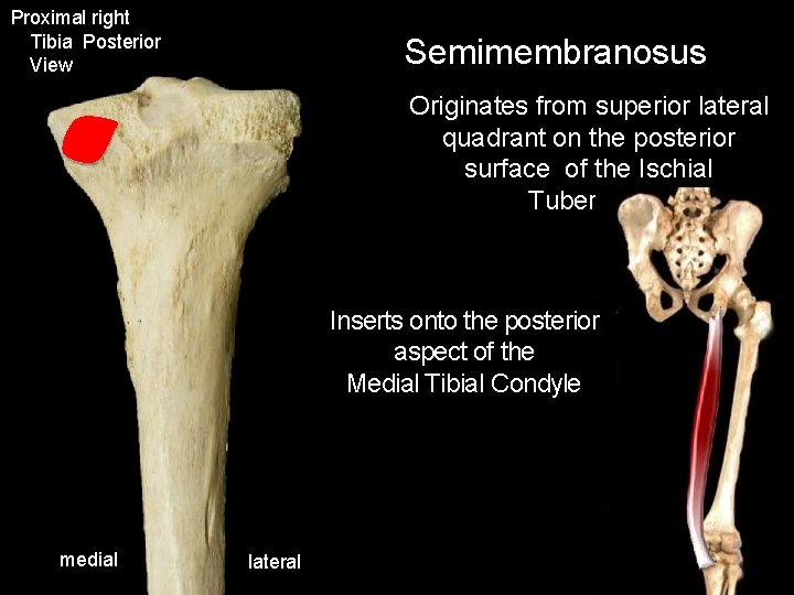
Proximal right Tibia Posterior View Semimembranosus Originates from superior lateral quadrant on the posterior surface of the Ischial Tuberosity Inserts onto the posterior aspect of the Medial Tibial Condyle medial lateral
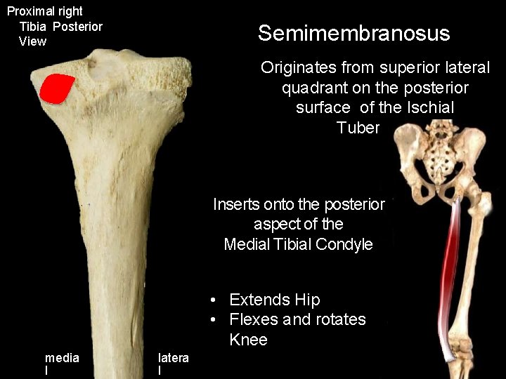
Proximal right Tibia Posterior View Semimembranosus Originates from superior lateral quadrant on the posterior surface of the Ischial Tuberosity Inserts onto the posterior aspect of the Medial Tibial Condyle • Extends Hip • Flexes and rotates Knee media l latera l
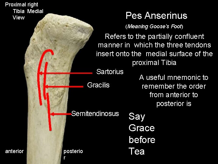
Proximal right Tibia Medial View Pes Anserinus (Meaning Goose’s Foot) Refers to the partially confluent manner in which the three tendons insert onto the medial surface of the proximal Tibia Sartorius A useful mnemonic to Gracilis remember the order from anterior to posterior is Semitendinosus anterior posterio r Say Grace before Tea
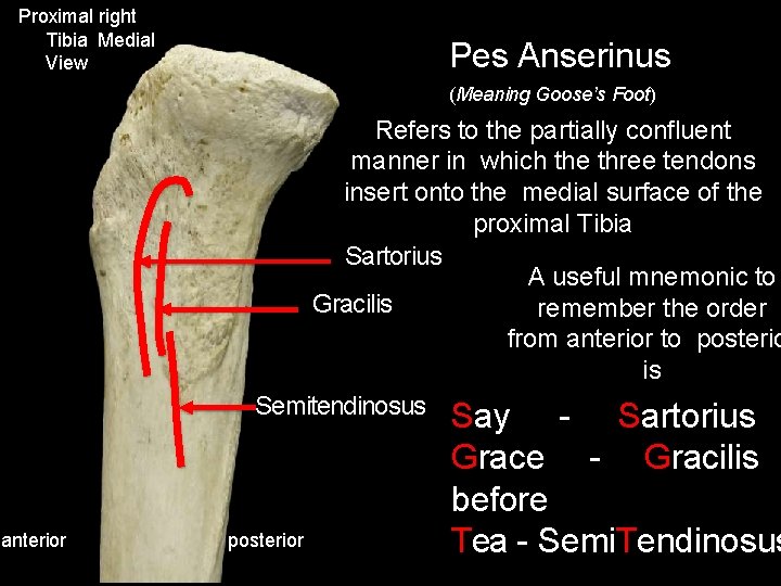
Proximal right Tibia Medial View Pes Anserinus (Meaning Goose’s Foot) Refers to the partially confluent manner in which the three tendons insert onto the medial surface of the proximal Tibia Sartorius A useful mnemonic to Gracilis remember the order from anterior to posterio is Semitendinosus anterior posterior Say - Sartorius Grace - Gracilis before Tea - Semi. Tendinosus
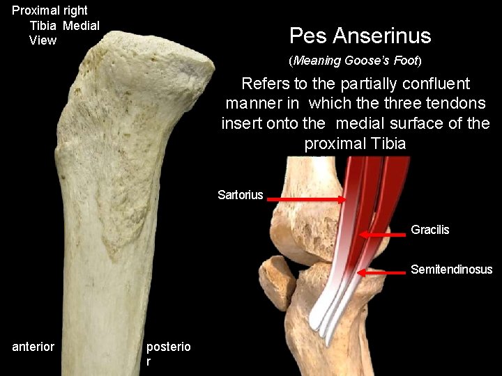
Proximal right Tibia Medial View Pes Anserinus (Meaning Goose’s Foot) Refers to the partially confluent manner in which the three tendons insert onto the medial surface of the proximal Tibia Sartorius Gracilis Semitendinosus anterior posterio r
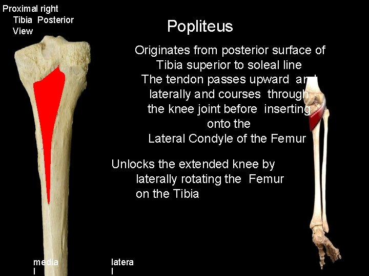
Proximal right Tibia Posterior View Popliteus Originates from posterior surface of Tibia superior to soleal line The tendon passes upward and laterally and courses through the knee joint before inserting onto the Lateral Condyle of the Femur Unlocks the extended knee by laterally rotating the Femur on the Tibia media l latera l
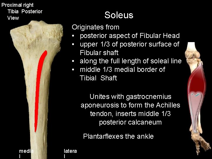
Proximal right Tibia Posterior View Soleus Originates from • posterior aspect of Fibular Head • upper 1/3 of posterior surface of Fibular shaft • along the full length of soleal line • middle 1/3 medial border of Tibial Shaft Unites with gastrocnemius aponeurosis to form the Achilles tendon, inserts middle 1/3 posterior calcaneum Plantarflexes the ankle media l latera l
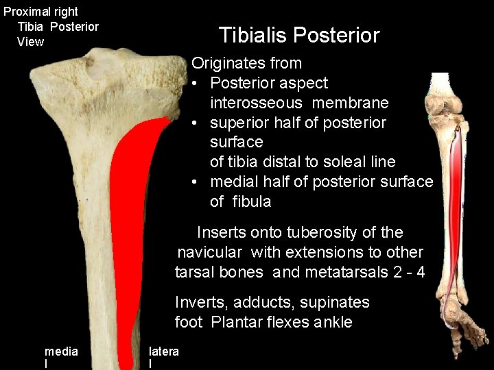
Proximal right Tibia Posterior View Tibialis Posterior Originates from • Posterior aspect interosseous membrane • superior half of posterior surface of tibia distal to soleal line • medial half of posterior surface of fibula Inserts onto tuberosity of the navicular with extensions to other tarsal bones and metatarsals 2 - 4 Inverts, adducts, supinates foot Plantar flexes ankle media l latera l
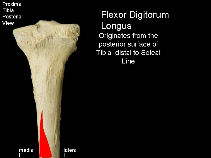
Proximal Tibia Posterior View Flexor Digitorum Longus Originates from the posterior surface of Tibia distal to Soleal Line media l latera l
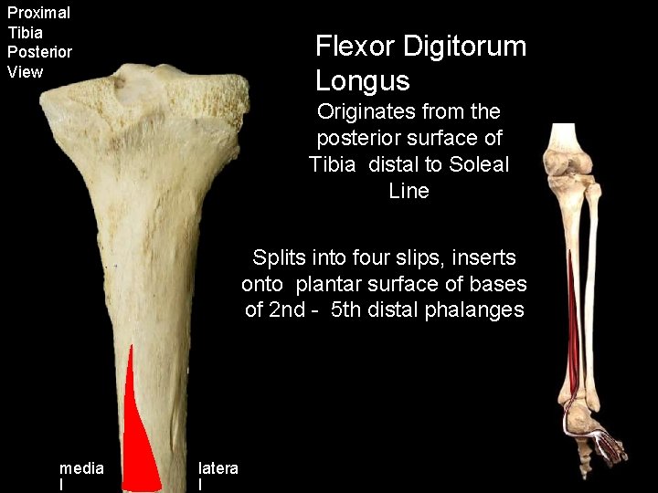
Proximal Tibia Posterior View Flexor Digitorum Longus Originates from the posterior surface of Tibia distal to Soleal Line Splits into four slips, inserts onto plantar surface of bases of 2 nd - 5 th distal phalanges media l latera l
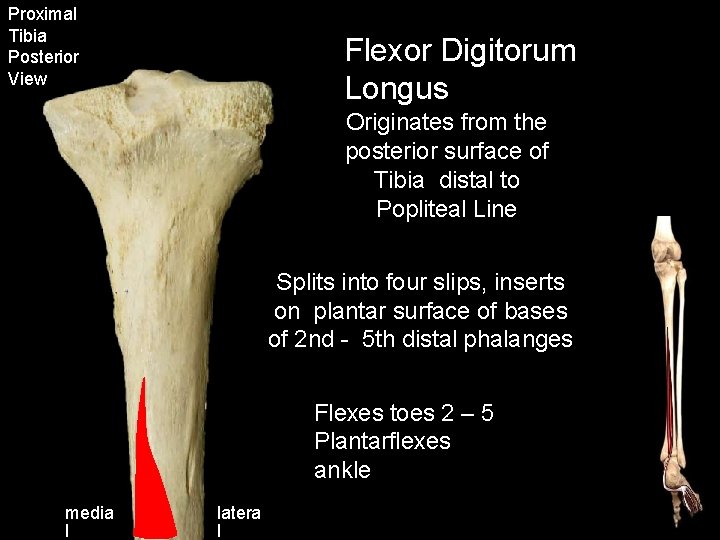
Proximal Tibia Posterior View Flexor Digitorum Longus Originates from the posterior surface of Tibia distal to Popliteal Line Splits into four slips, inserts on plantar surface of bases of 2 nd - 5 th distal phalanges Flexes toes 2 – 5 Plantarflexes ankle media l latera l

Ligament Attachments
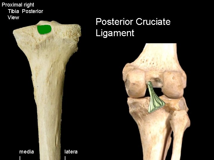
Proximal right Tibia Posterior View media l Posterior Cruciate Ligament latera l
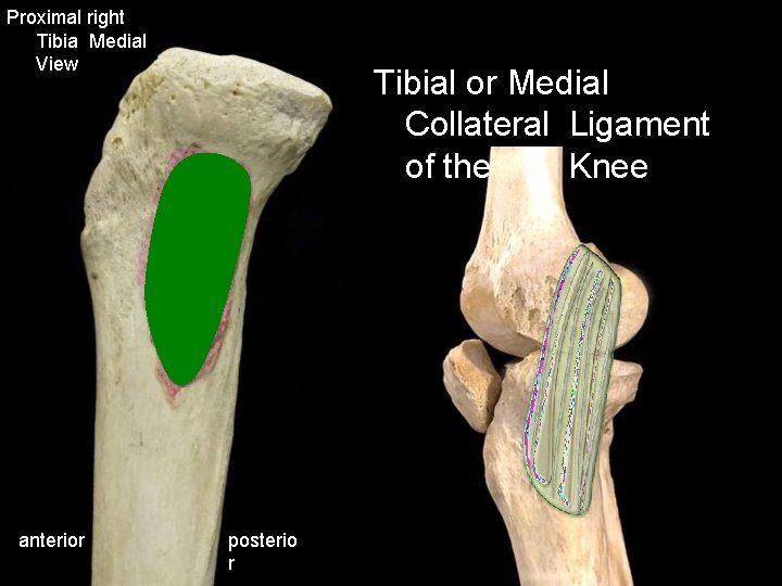
Proximal right Tibia Medial View anterior Tibial or Medial Collateral Ligament of the Knee posterio r
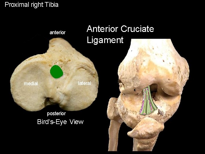
Proximal right Tibia Anterior Cruciate Ligament anterior lateral medial posterior Bird’s-Eye View
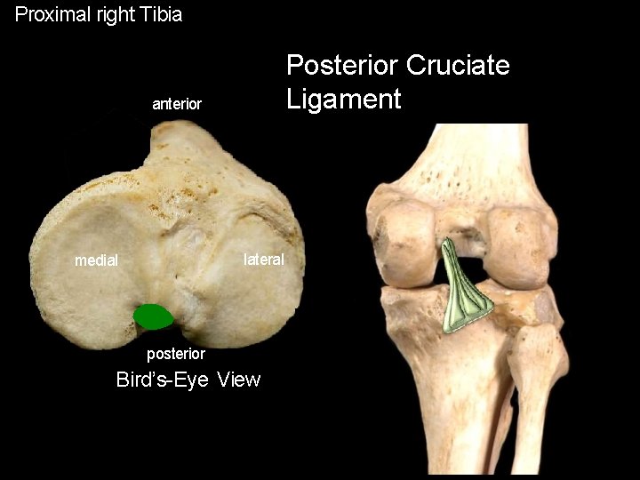
Proximal right Tibia Posterior Cruciate Ligament anterior lateral medial posterior Bird’s-Eye View
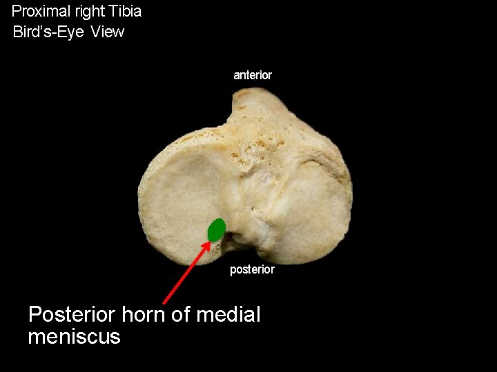
Proximal right Tibia Bird’s-Eye View anterior posterior Posterior horn of medial meniscus
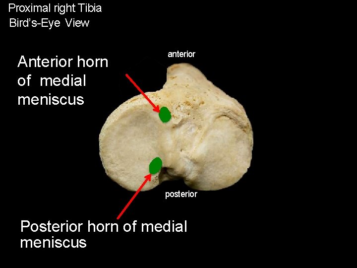
Proximal right Tibia Bird’s-Eye View Anterior horn of medial meniscus anterior posterior Posterior horn of medial meniscus
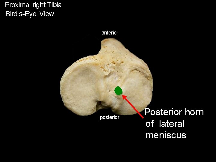
Proximal right Tibia Bird’s-Eye View anterior posterior Posterior horn of lateral meniscus
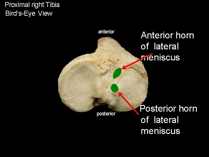
Proximal right Tibia Bird’s-Eye View anterior posterior Anterior horn of lateral meniscus Posterior horn of lateral meniscus
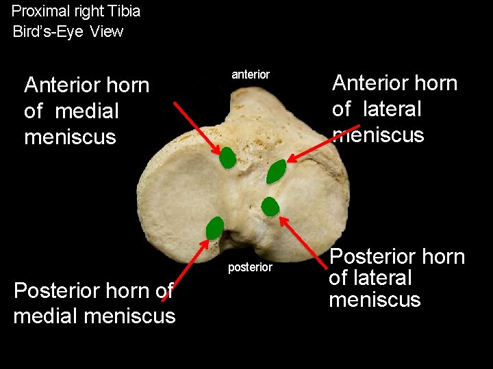
Proximal right Tibia Bird’s-Eye View Anterior horn of medial meniscus anterior posterior Posterior horn of medial meniscus Anterior horn of lateral meniscus Posterior horn of lateral meniscus

Muscle Summaries
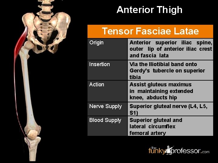
Anterior Thigh Tensor Fasciae Latae Origin Anterior superior iliac spine, outer lip of anterior iliac crest and fascia lata Insertion Via the Iliotibial band onto Gerdy’s tubercle on superior tibia Assist gluteus maximus in maintaining extended knee, abducts hip Action Nerve Supply Blood Supply Superior gluteal nerve (L 4, L 5, S 1) Superior gluteal and lateral circumflex femoral artery
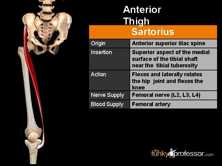
Anterior Thigh Sartorius Origin Anterior superior iliac spine Insertion Superior aspect of the medial surface of the tibial shaft near the tibial tuberosity Action Nerve Supply Flexes and laterally rotates the hip joint and flexes the knee Femoral nerve (L 2, L 3, L 4) Blood Supply Femoral artery
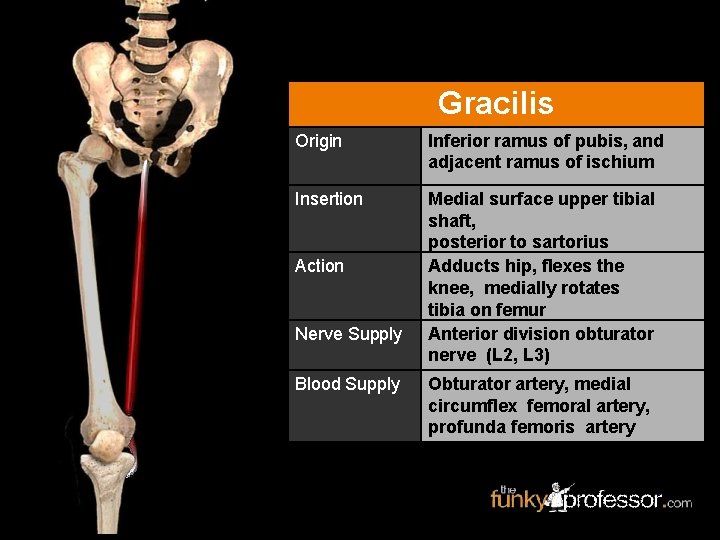
Gracilis Origin Inferior ramus of pubis, and adjacent ramus of ischium Insertion Medial surface upper tibial shaft, posterior to sartorius Adducts hip, flexes the knee, medially rotates tibia on femur Anterior division obturator nerve (L 2, L 3) Action Nerve Supply Blood Supply Obturator artery, medial circumflex femoral artery, profunda femoris artery
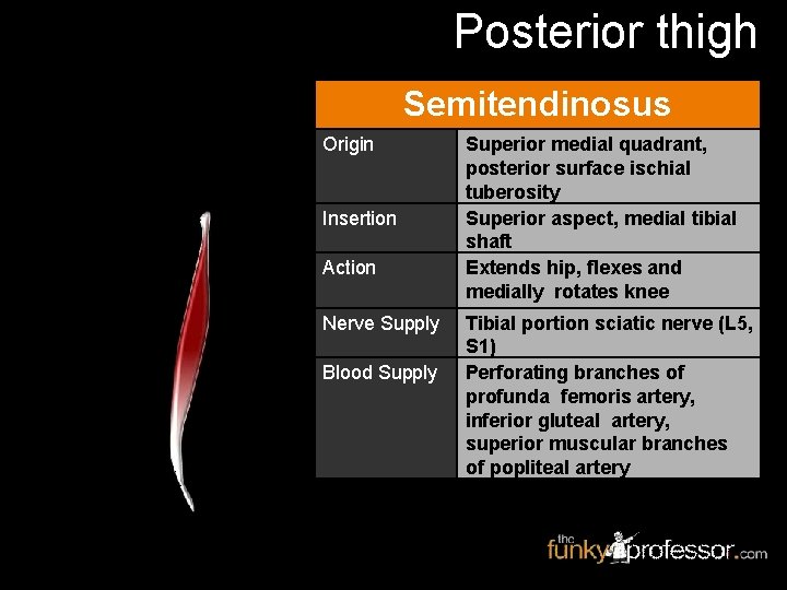
Posterior thigh Semitendinosus Origin Insertion Action Nerve Supply Blood Supply Superior medial quadrant, posterior surface ischial tuberosity Superior aspect, medial tibial shaft Extends hip, flexes and medially rotates knee Tibial portion sciatic nerve (L 5, S 1) Perforating branches of profunda femoris artery, inferior gluteal artery, superior muscular branches of popliteal artery
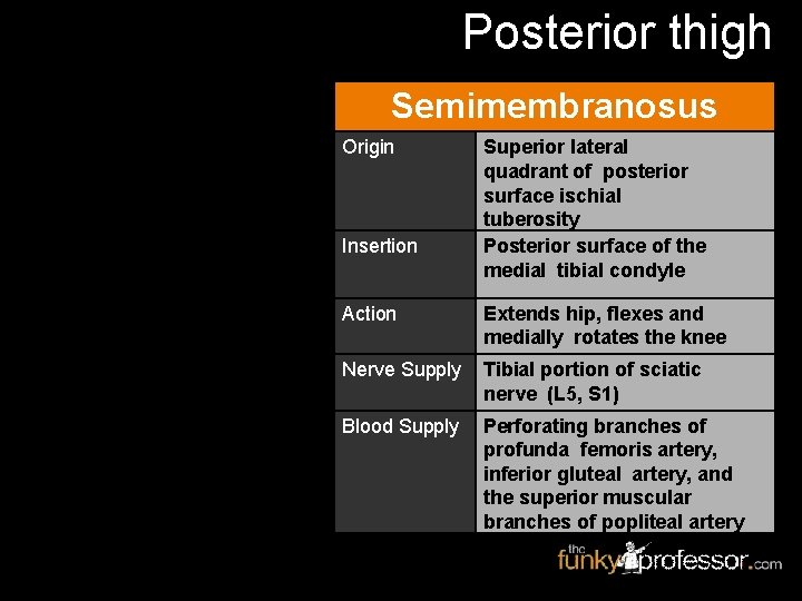
Posterior thigh Semimembranosus Origin Insertion Superior lateral quadrant of posterior surface ischial tuberosity Posterior surface of the medial tibial condyle Action Extends hip, flexes and medially rotates the knee Nerve Supply Tibial portion of sciatic nerve (L 5, S 1) Blood Supply Perforating branches of profunda femoris artery, inferior gluteal artery, and the superior muscular branches of popliteal artery
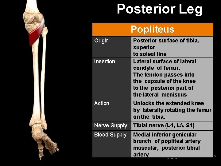
Posterior Leg Popliteus Origin Insertion Posterior surface of tibia, superior to soleal line Lateral surface of lateral condyle of femur. The tendon passes into the capsule of the knee to the posterior part of the lateral meniscus Action Unlocks the extended knee by laterally rotating the femur on the tibia. Nerve Supply Tibial nerve (L 4, L 5, S 1) Blood Supply Medial inferior genicular branch of popliteal artery muscular, posterior tibial artery
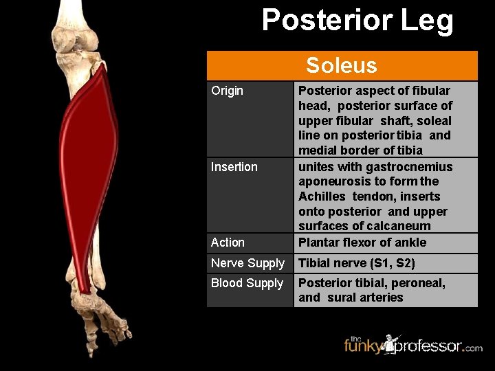
Posterior Leg Soleus Origin Action Posterior aspect of fibular head, posterior surface of upper fibular shaft, soleal line on posterior tibia and medial border of tibia unites with gastrocnemius aponeurosis to form the Achilles tendon, inserts onto posterior and upper surfaces of calcaneum Plantar flexor of ankle Nerve Supply Tibial nerve (S 1, S 2) Blood Supply Posterior tibial, peroneal, and sural arteries Insertion

Posterior Leg Tibialis Posterior Origin Insertion Action Posterior aspect interosseous membrane, superior half of tibia and medial posterior surface of fibula Inserts onto navicular tuberosity and sends extensions to other tarsal bones and metatarsals Inverts, adducts, supinates foot, plantar flexes ankle Nerve Supply Tibial nerve (L 4, L 5) Blood Supply sural, peroneal and posterior tibial arteries
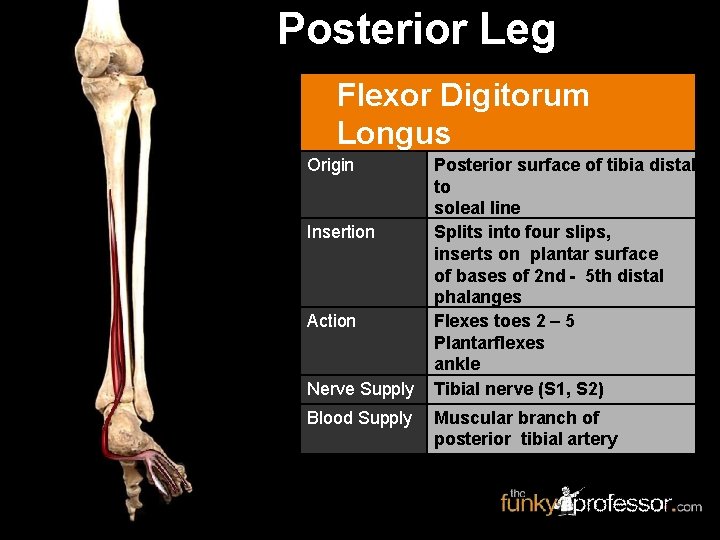
Posterior Leg Flexor Digitorum Longus Origin Insertion Action Nerve Supply Blood Supply Posterior surface of tibia distal to soleal line Splits into four slips, inserts on plantar surface of bases of 2 nd - 5 th distal phalanges Flexes toes 2 – 5 Plantarflexes ankle Tibial nerve (S 1, S 2) Muscular branch of posterior tibial artery
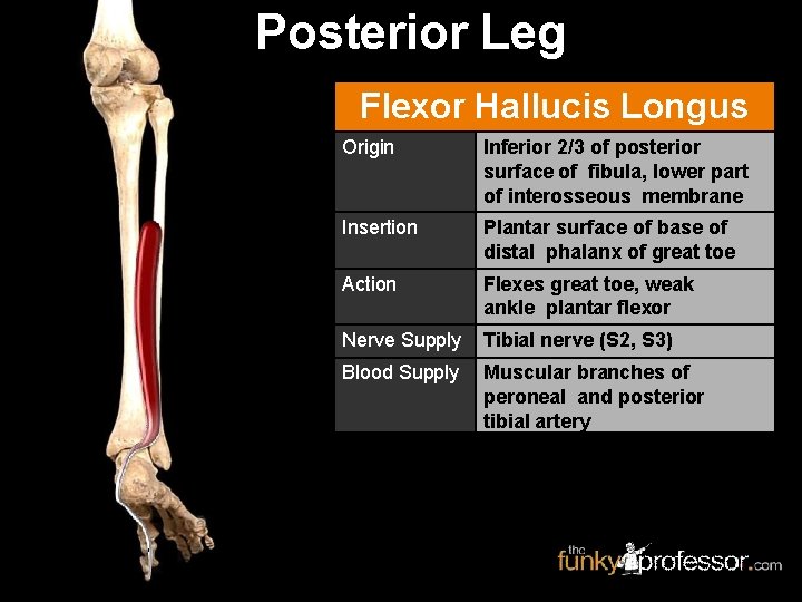
Posterior Leg Flexor Hallucis Longus Origin Inferior 2/3 of posterior surface of fibula, lower part of interosseous membrane Insertion Plantar surface of base of distal phalanx of great toe Action Flexes great toe, weak ankle plantar flexor Nerve Supply Tibial nerve (S 2, S 3) Blood Supply Muscular branches of peroneal and posterior tibial artery
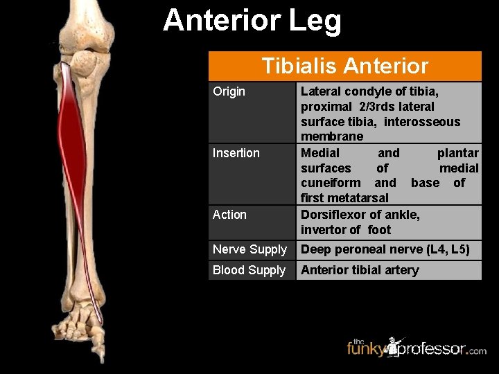
Anterior Leg Tibialis Anterior Origin Insertion Action Lateral condyle of tibia, proximal 2/3 rds lateral surface tibia, interosseous membrane Medial and plantar surfaces of medial cuneiform and base of first metatarsal Dorsiflexor of ankle, invertor of foot Nerve Supply Deep peroneal nerve (L 4, L 5) Blood Supply Anterior tibial artery

- Slides: 91