THE HEMIPORTOCAVAL SHUNT R Troisi MD Ph D

- Slides: 36
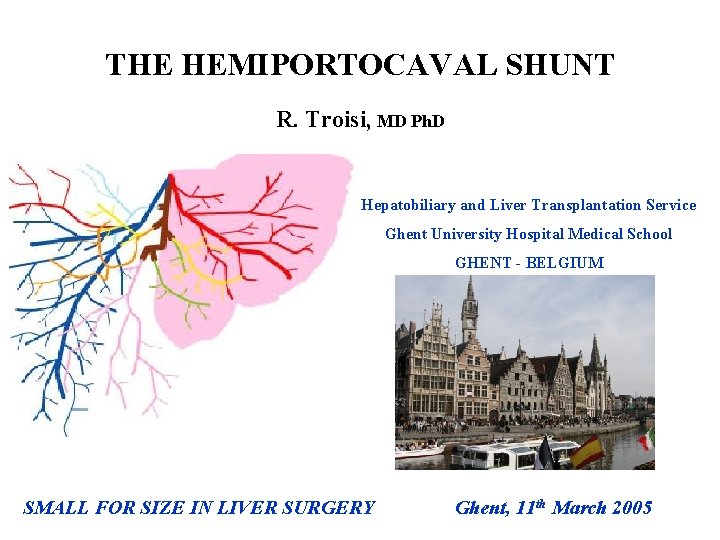
THE HEMIPORTOCAVAL SHUNT R. Troisi, MD Ph. D Hepatobiliary and Liver Transplantation Service Ghent University Hospital Medical School GHENT - BELGIUM SMALL FOR SIZE IN LIVER SURGERY Ghent, 11 th March 2005
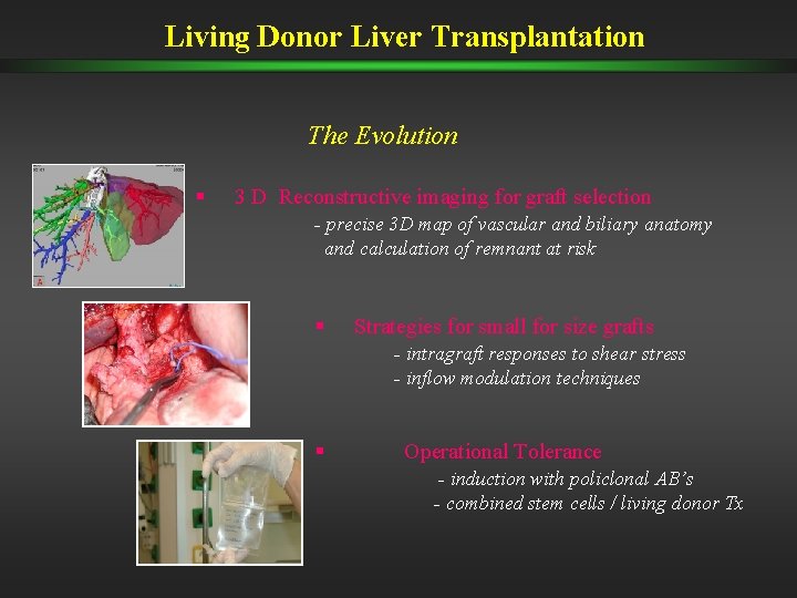
Living Donor Liver Transplantation The Evolution § 3 D Reconstructive imaging for graft selection - precise 3 D map of vascular and biliary anatomy and calculation of remnant at risk § Strategies for small for size grafts - intragraft responses to shear stress - inflow modulation techniques § Operational Tolerance - induction with policlonal AB’s - combined stem cells / living donor Tx
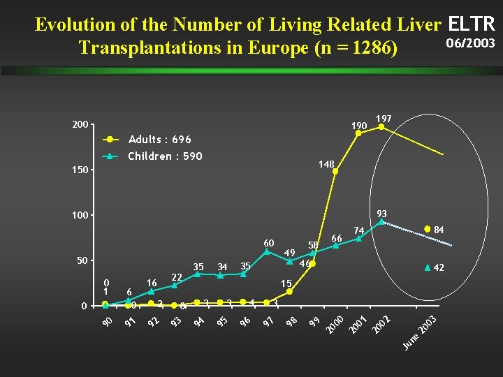
Evolution of the Number of Living Related Liver ELTR 06/2003 Transplantations in Europe (n = 1286) 200 197 Adults : 696 Children : 590 148 150 93 100 60 35 35 22 46 42 03 20 Ju ne 20 02 20 01 20 00 3 99 4 97 3 96 3 95 0 58 84 74 15 94 2 93 16 92 90 0 6 0 91 0 1 34 98 50 49 66
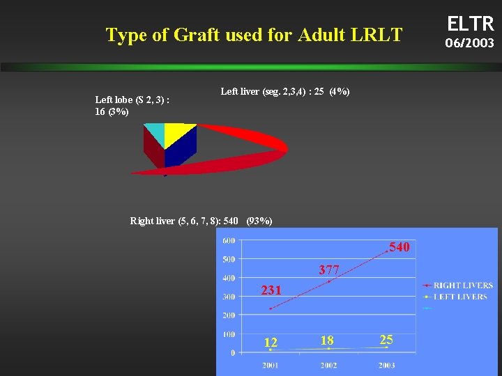
Type of Graft used for Adult LRLT Left lobe (S 2, 3) : 16 (3%) Left liver (seg. 2, 3, 4) : 25 (4%) Right liver (5, 6, 7, 8): 540 (93%) 540 377 231 12 18 25 ELTR 06/2003
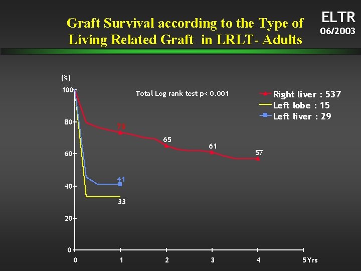
Graft Survival according to the Type of Living Related Graft in LRLT- Adults ELTR 06/2003 (%) 100 80 Right liver : 537 Left lobe : 15 Left liver : 29 Total Log rank test p< 0. 001 73 65 60 61 57 41 40 33 20 0 0 1 2 3 4 5 Yrs
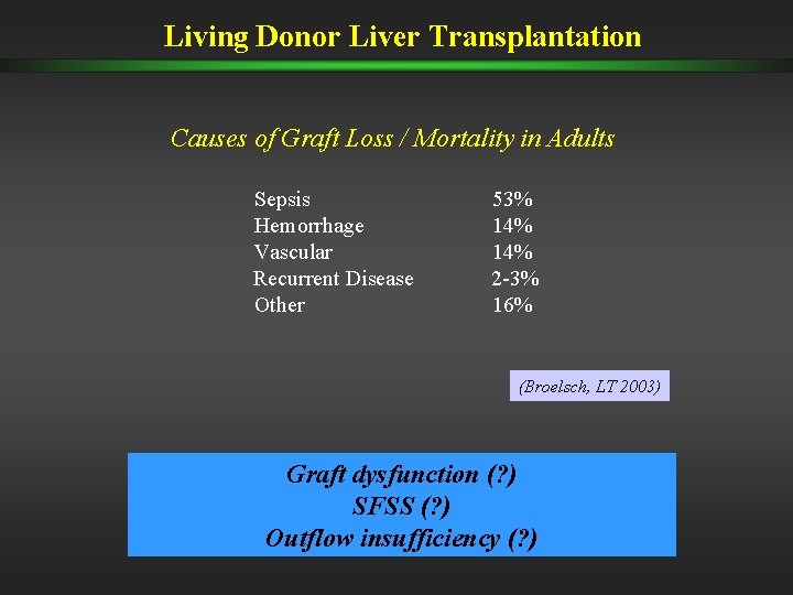
Living Donor Liver Transplantation Causes of Graft Loss / Mortality in Adults Sepsis Hemorrhage Vascular Recurrent Disease Other 53% 14% 2 -3% 16% (Broelsch, LT 2003) Graft dysfunction (? ) SFSS (? ) Outflow insufficiency (? )
Living Donor Liver Transplantation Relationships between Techniques and Complications Small for Size Syndrome New surgical techniques Vascular thrombosis Bleeding Biliary complications • leaks • stenosis Venous outflow insufficiency
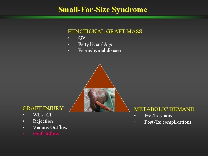
Small-For-Size Syndrome FUNCTIONAL GRAFT MASS • • • GRAFT INJURY • • WI / CI Rejection Venous Outflow Graft Inflow GV Fatty liver / Age Parenchymal disease METABOLIC DEMAND • • Pre-Tx status Post-Tx complications
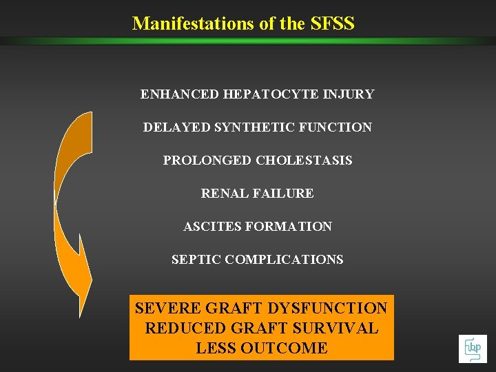
Manifestations of the SFSS ENHANCED HEPATOCYTE INJURY DELAYED SYNTHETIC FUNCTION PROLONGED CHOLESTASIS RENAL FAILURE ASCITES FORMATION SEPTIC COMPLICATIONS SEVERE GRAFT DYSFUNCTION REDUCED GRAFT SURVIVAL LESS OUTCOME
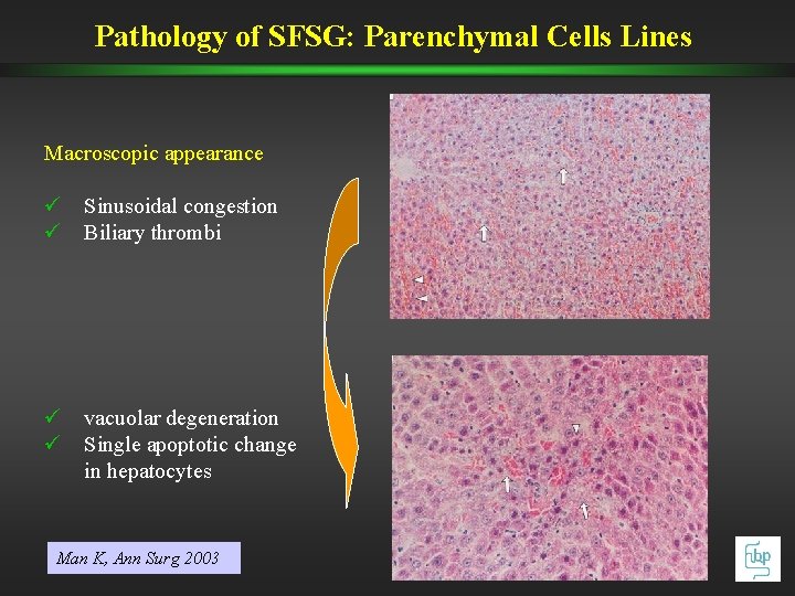
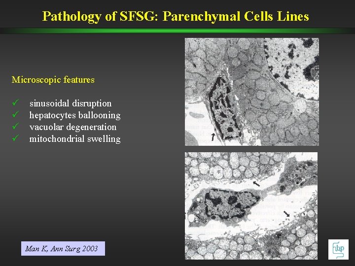
Pathology of SFSG: Parenchymal Cells Lines Macroscopic appearance ü Sinusoidal congestion ü Biliary thrombi ü vacuolar degeneration ü Single apoptotic change in hepatocytes Man K, Ann Surg 2003
Pathology of SFSG: Parenchymal Cells Lines Microscopic features ü ü sinusoidal disruption hepatocytes ballooning vacuolar degeneration mitochondrial swelling Man K, Ann Surg 2003
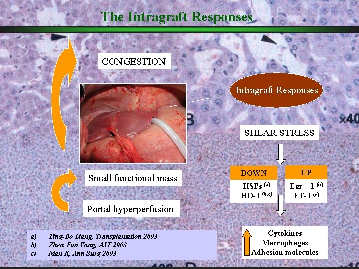
The Intragraft Responses CONGESTION Intragraft Responses SHEAR STRESS Small functional mass DOWN HSPs (a) HO-1 (b, c) UP Egr – 1 (a) ET-1 (c) Portal hyperperfusion a) b) c) Ting-Bo Liang, Transplantation 2003 Zhen-Fan Yang, AJT 2003 Man K, Ann Surg 2003 Cytokines Macrophages Adhesion molecules
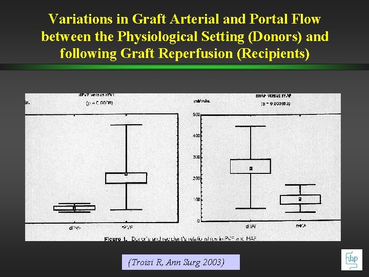
Variations in Graft Arterial and Portal Flow between the Physiological Setting (Donors) and following Graft Reperfusion (Recipients) (Troisi R, Ann Surg 2003)
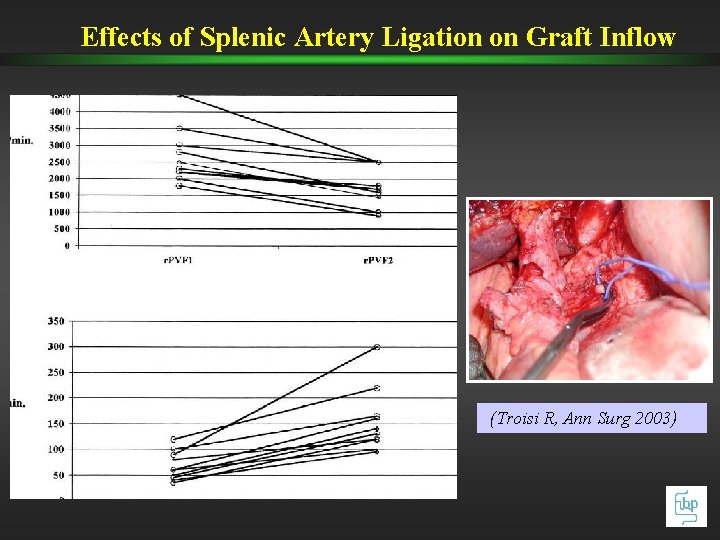
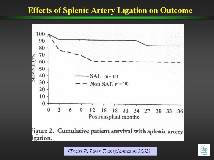
Effects of Splenic Artery Ligation on Graft Inflow (Troisi R, Ann Surg 2003)
Effects of Splenic Artery Ligation on Outcome (n = 14) (n = 10) (Troisi R, Liver Transplantation 2003)
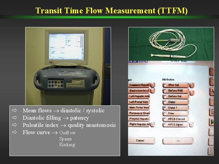
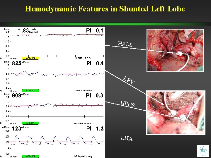
Transit Time Flow Measurement (TTFM) ð ð Mean flows diastolic / systolic Diastolic filling patency Pulsatile index quality anastomosis Flow curve Outflow Spasm Kinking
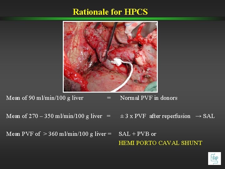
Rationale for HPCS Mean of 90 ml/min/100 g liver = Normal PVF in donors Mean of 270 – 350 ml/min/100 g liver = ± 3 x PVF after reperfusion → SAL Mean PVF of > 360 ml/min/100 g liver = SAL + PVB or HEMI PORTO CAVAL SHUNT
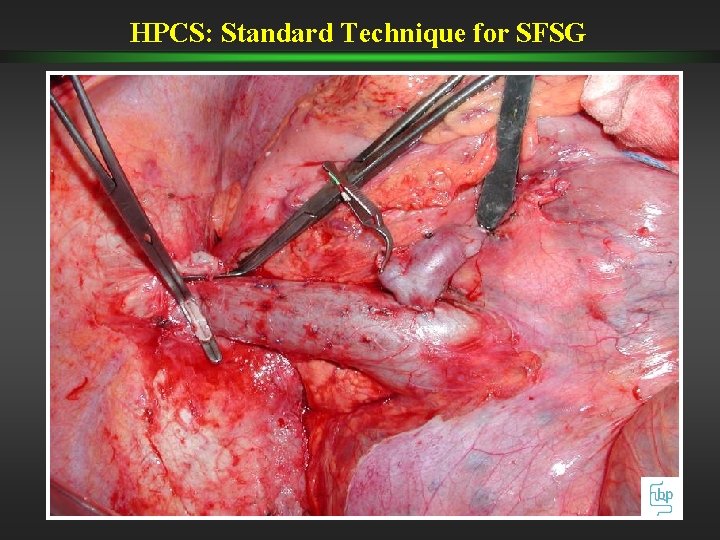
HPCS: Standard Technique for SFSG
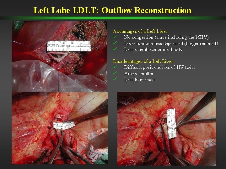
Left Lobe LDLT: Outflow Reconstruction Advantages of a Left Liver ü No congestion (since including the MHV) ü Liver function less depressed (bigger remnant) ü Less overall donor morbidity Disadvantages of a Left Liver ü Difficult position/risks of HV twist ü Artery smaller ü Less liver mass
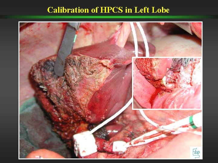
Calibration of HPCS in Left Lobe
Hemodynamic Features in Shunted Left Lobe HPCS LP V HPCS LHA
Effects of HPCS for Inflow Modulation on the Outcome of SFSG in LDLT üGraft hemodynamics in SFSG üFeatures of non-parenchymal cell lines following portal shunt üClinical outcome (Troisi R, Am J Transplantation 2005)
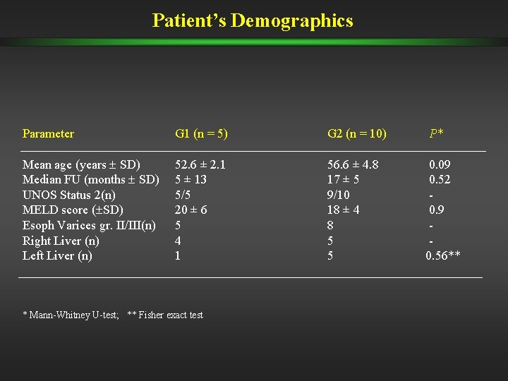
Patient’s Demographics Parameter G 1 (n = 5) G 2 (n = 10) P* Mean age (years SD) Median FU (months SD) UNOS Status 2(n) MELD score ( SD) Esoph Varices gr. II/III(n) Right Liver (n) Left Liver (n) 52. 6 ± 2. 1 5 ± 13 5/5 20 ± 6 5 4 1 56. 6 ± 4. 8 17 ± 5 9/10 18 ± 4 8 5 5 0. 09 0. 52 0. 9 0. 56** * Mann-Whitney U-test; ** Fisher exact test
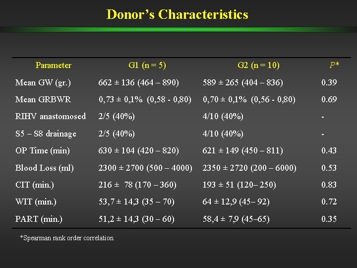
Donor’s Characteristics Parameter G 1 (n = 5) G 2 (n = 10) P* Mean GW (gr. ) 662 ± 136 (464 – 890) 589 ± 265 (404 – 836) 0. 39 Mean GRBWR 0, 73 ± 0, 1% (0, 58 - 0, 80) 0, 70 ± 0, 1% (0, 56 - 0, 80) 0. 69 RIHV anastomosed 2/5 (40%) 4/10 (40%) - S 5 – S 8 drainage 2/5 (40%) 4/10 (40%) - OP Time (min) 630 ± 104 (420 – 820) 621 ± 149 (450 – 811) 0. 43 Blood Loss (ml) 2300 ± 2700 (500 – 4000) 2350 ± 2720 (200 – 6000) 0. 53 CIT (min. ) 216 ± 78 (170 – 360) 193 ± 51 (120– 250) 0. 83 WIT (min. ) 53, 7 ± 14, 3 (35 – 70) 64 ± 12, 9 (45– 92) 0. 72 PART (min. ) 51, 2 ± 14, 3 (30 – 60) 58, 4 ± 7, 9 (45– 65) 0. 35 *Spearman rank order correlation
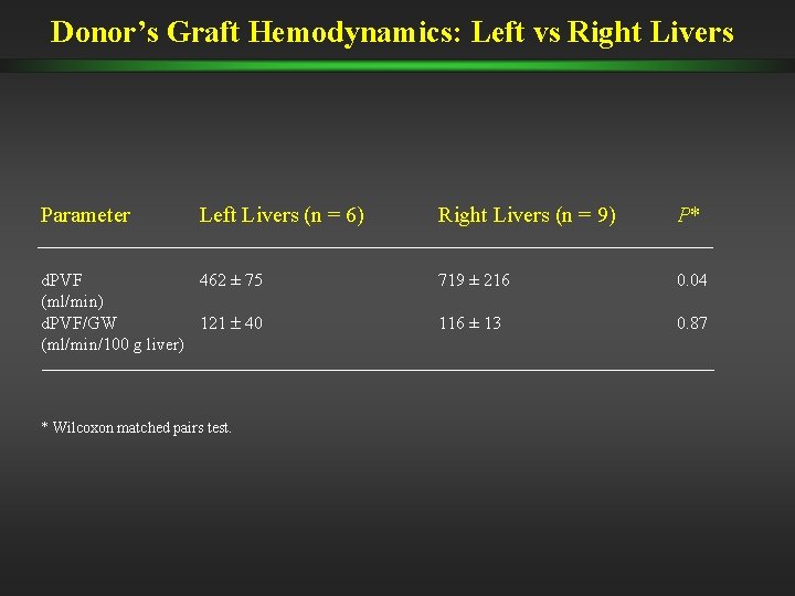
Donor’s Graft Hemodynamics: Left vs Right Livers Parameter Left Livers (n = 6) d. PVF 462 ± 75 (ml/min) d. PVF/GW 121 40 (ml/min/100 g liver) * Wilcoxon matched pairs test. Right Livers (n = 9) P* 719 ± 216 0. 04 116 ± 13 0. 87
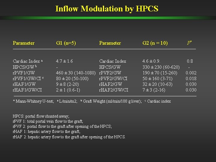
Inflow Modulation by HPCS Parameter G 1 (n=5) Parameter G 2 (n = 10) P* Cardiac Index a HPCS/GW b r. PVF 1/GW/CI c r. HAF 1/GW/CI 4. 7 ± 1. 6 460 ± 30 (140 -1080) 80 ± 20 (50 -100) 9 ± 8 (2 -20) 2 ± 1 (0. 6 -1) Cardiac Index HPCS/GW r. PVF 2/GW/CI r. HAF 2/GW/CI 4. 6 ± 0. 9. 330 ± 230 (60 -620) 190 ± 70 (15 -260) 50 ± 160 (3 -71) 32 ± 20 (10 -63) 7 ± 3 (2 -16) 0. 8 0. 002 0. 018 0. 030 * Mann-Whitney U-test; a L/min/m 2; b Graft Weight (ml/min/100 g liver); HPCS: portal flow shunted away; r. PVF 1: total portal vein flow to the graft; r. PVF 2: portal flow to the graft after opening of the HPCS; r. HAF 1: hepatic artery flow to the graft; r. HAF 2: hepatic artery flow to the graft after opening of the HPCS. c Cardiac index
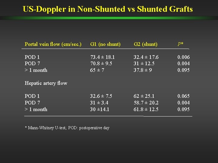
US-Doppler in Non-Shunted vs Shunted Grafts Portal vein flow (cm/sec. ) G 1 (no shunt) G 2 (shunt) P* POD 1 POD 7 > 1 month 73. 4 ± 18. 1 70. 8 ± 9. 5 65 ± 7 32. 4 ± 17. 6 31 ± 12. 5 37. 8 ± 9 0. 006 0. 004 0. 095 32. 6 ± 7. 5 31 ± 3. 4 30 ± 14. 1 62 ± 25. 1 58. 7 ± 20. 2 61. 8 ± 12. 5 0. 065 0. 004 0. 095 Hepatic artery flow POD 1 POD 7 > 1 month * Mann-Whitney U-test; POD: postoperative day
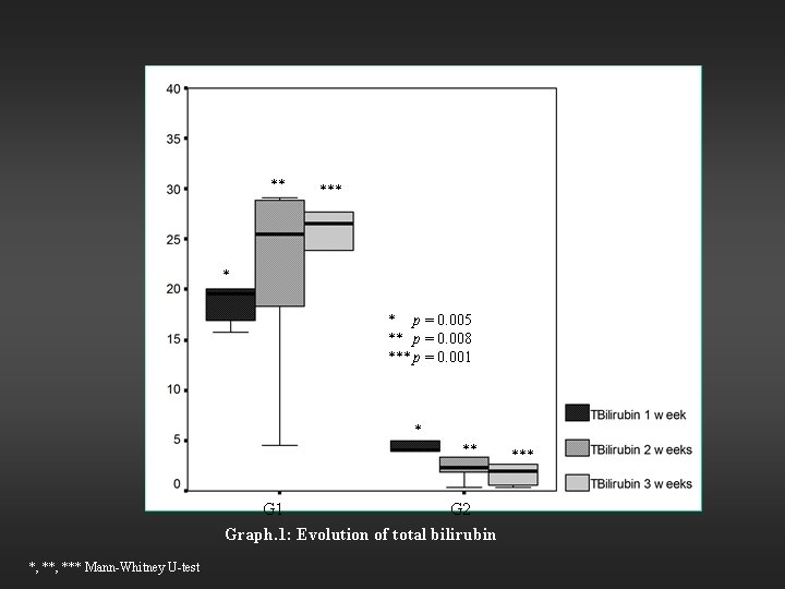
** * * p = 0. 005 ** p = 0. 008 *** p = 0. 001 * ** G 1 G 2 Graph. 1: Evolution of total bilirubin *, *** Mann-Whitney U-test ***
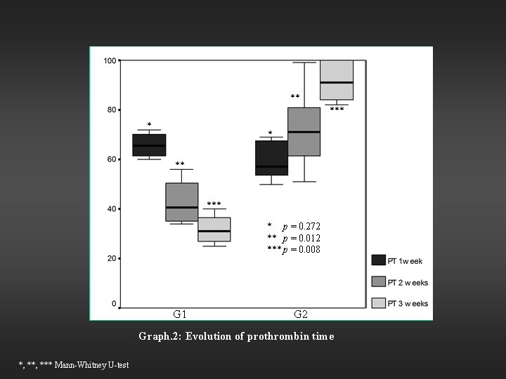
** * * ** * p = 0. 272 ** p = 0. 012 *** p = 0. 008 G 1 G 2 Graph. 2: Evolution of prothrombin time *, *** Mann-Whitney U-test
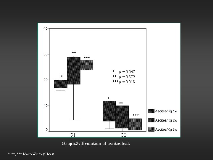
** * p = 0. 067 ** p = 0. 372 *** p = 0. 018 * * ** *** G 1 G 2 Graph. 3: Evolution of ascites leak *, *** Mann-Whitney U-test
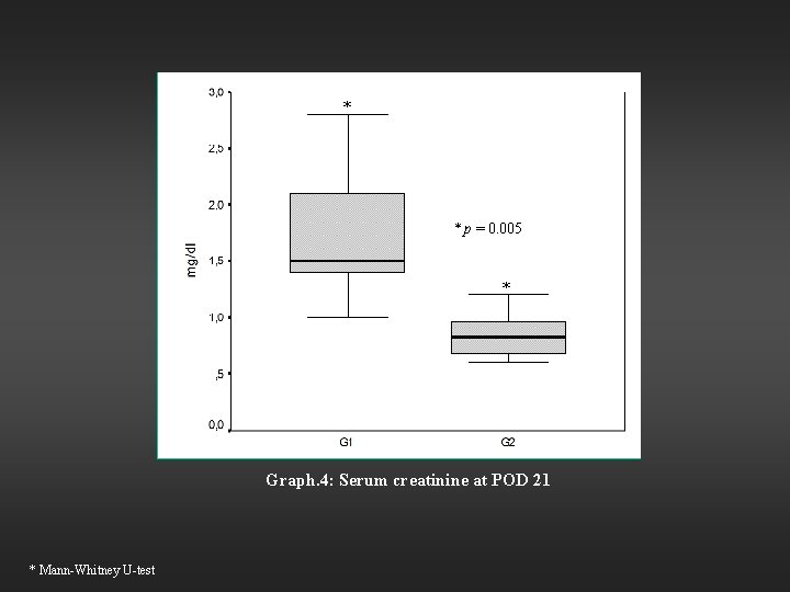
* * p = 0. 005 * Graph. 4: Serum creatinine at POD 21 * Mann-Whitney U-test
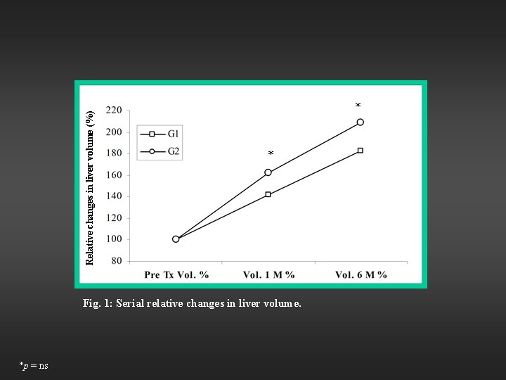
Relative changes in liver volume (%) * * Fig. 1: Serial relative changes in liver volume. *p = ns
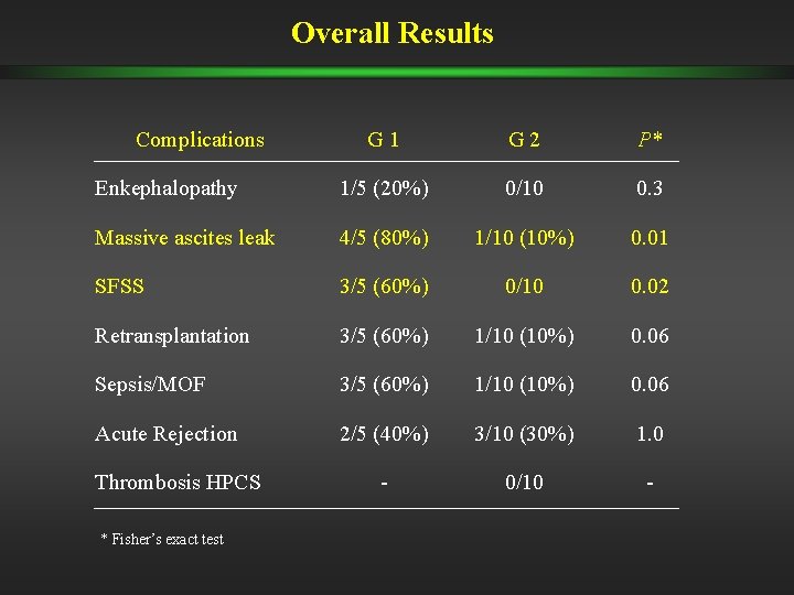
Overall Results Complications G 1 G 2 P* Enkephalopathy 1/5 (20%) 0/10 0. 3 Massive ascites leak 4/5 (80%) 1/10 (10%) 0. 01 SFSS 3/5 (60%) 0/10 0. 02 Retransplantation 3/5 (60%) 1/10 (10%) 0. 06 Sepsis/MOF 3/5 (60%) 1/10 (10%) 0. 06 Acute Rejection 2/5 (40%) 3/10 (30%) 1. 0 - 0/10 - Thrombosis HPCS * Fisher’s exact test
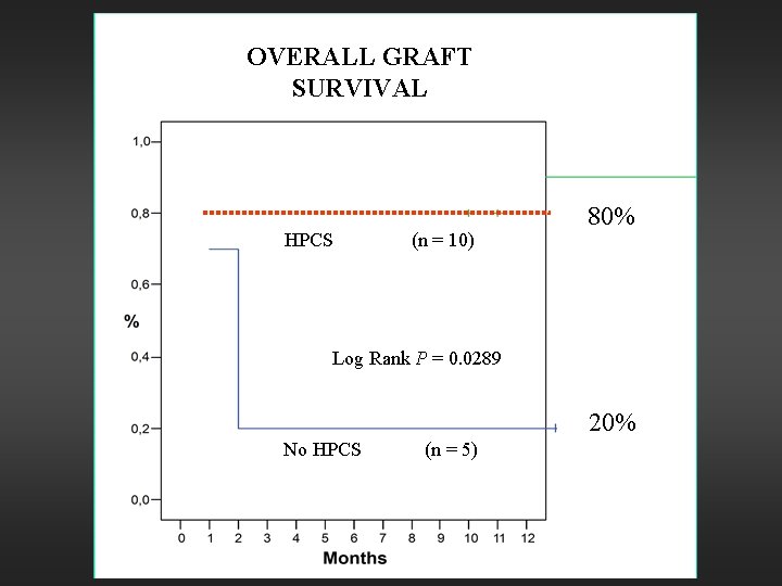
OVERALL GRAFT SURVIVAL HPCS (n = 10) 80% Log Rank P = 0. 0289 20% No HPCS (n = 5)
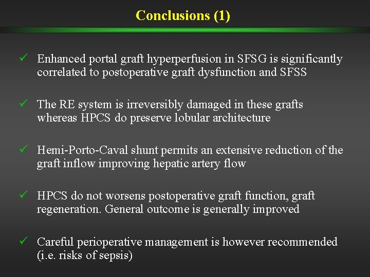
Conclusions (1) ü Enhanced portal graft hyperperfusion in SFSG is significantly correlated to postoperative graft dysfunction and SFSS ü The RE system is irreversibly damaged in these grafts whereas HPCS do preserve lobular architecture ü Hemi-Porto-Caval shunt permits an extensive reduction of the graft inflow improving hepatic artery flow ü HPCS do not worsens postoperative graft function, graft regeneration. General outcome is generally improved ü Careful perioperative management is however recommended (i. e. risks of sepsis)
Conclusions (2) ü Basing on the Asian data on reduced donor morbidity with the use of the left livers, upon this experience could we implement the use of left liver grafts for adult recipients?