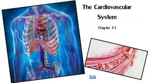
The Cardiovascular System A closed system of the

- Slides: 95
The Cardiovascular System • A closed system of the heart and blood vessels • The functions of the cardiovascular system © 2012 Pearson Education, Inc.
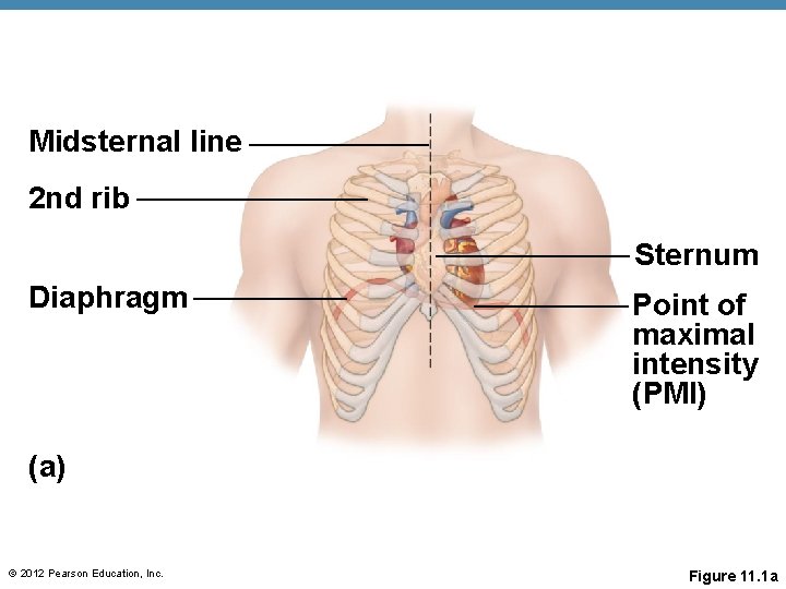
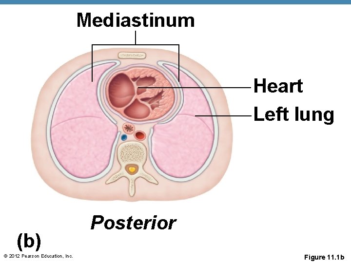
The Heart • Location • Orientation • About the size of your fist © 2012 Pearson Education, Inc.
Midsternal line 2 nd rib Sternum Diaphragm Point of maximal intensity (PMI) (a) © 2012 Pearson Education, Inc. Figure 11. 1 a
Mediastinum Heart Left lung (b) © 2012 Pearson Education, Inc. Posterior Figure 11. 1 b
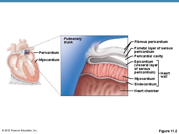
The Heart: Coverings • Pericardium— • Fibrous pericardium is loose and superficial • Serous membrane is deep to the fibrous pericardium and composed of two layers • Serous fluid fills the space between the layers of pericardium © 2012 Pearson Education, Inc.
Pulmonary trunk Pericardium Myocardium Fibrous pericardium Parietal layer of serous pericardium Pericardial cavity Epicardium (visceral layer of serous pericardium) Heart wall Myocardium Endocardium Heart chamber © 2012 Pearson Education, Inc. Figure 11. 2
The Heart: Heart Wall • Three layers • Epicardium • Myocardium • Endocardium © 2012 Pearson Education, Inc.
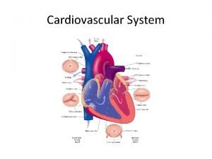
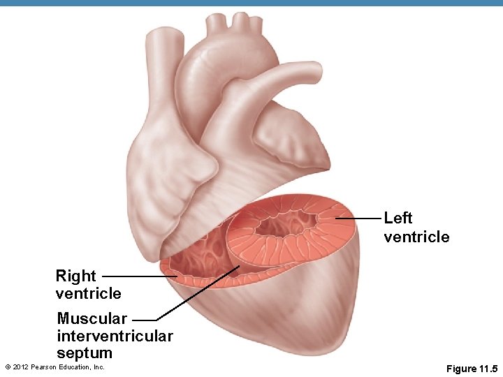
The Heart: Chambers • Right and left side act as separate pumps • Four chambers • Atria • Receiving chambers • Right atrium • Left atrium • Ventricles • Discharging chambers • Right ventricle • Left ventricle © 2012 Pearson Education, Inc.
Left ventricle Right ventricle Muscular interventricular septum © 2012 Pearson Education, Inc. Figure 11. 5
The Heart: Septa • Interventricular septum • Interatrial septum © 2012 Pearson Education, Inc.
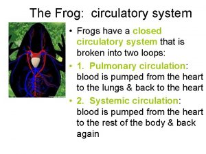
The Heart’s Role in Blood Circulation • Systemic circulation • Pulmonary circulation © 2012 Pearson Education, Inc.
The Heart: Valves • Allow blood to flow in only one direction to prevent backflow • Four valves © 2012 Pearson Education, Inc.
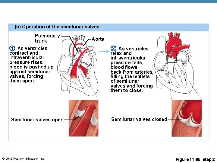
The Heart: Valves • AV valves • Semilunar valves © 2012 Pearson Education, Inc.
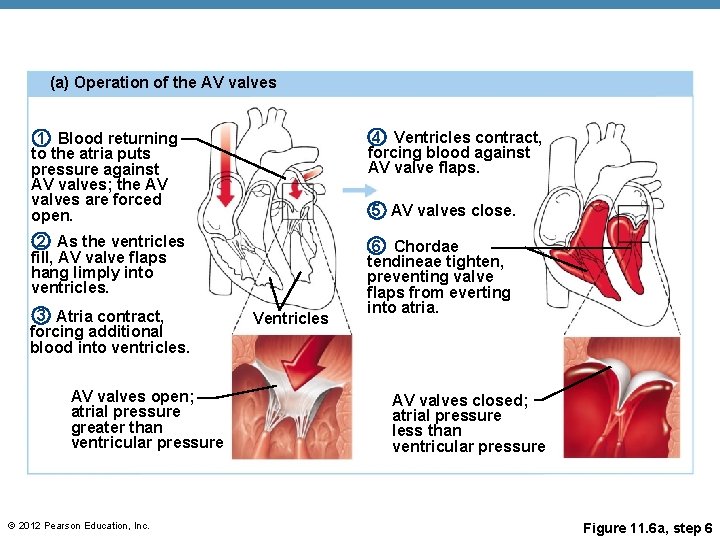
(a) Operation of the AV valves 1 Blood returning to the atria puts pressure against AV valves; the AV valves are forced open. 4 Ventricles contract, forcing blood against AV valve flaps. 2 As the ventricles fill, AV valve flaps hang limply into ventricles. 6 Chordae tendineae tighten, preventing valve flaps from everting into atria. 3 Atria contract, forcing additional blood into ventricles. AV valves open; atrial pressure greater than ventricular pressure © 2012 Pearson Education, Inc. 5 AV valves close. Ventricles AV valves closed; atrial pressure less than ventricular pressure Figure 11. 6 a, step 6
(b) Operation of the semilunar valves Pulmonary trunk 1 As ventricles contract and intraventricular pressure rises, blood is pushed up against semilunar valves, forcing them open. Semilunar valves open © 2012 Pearson Education, Inc. Aorta 2 As ventricles relax and intraventricular pressure falls, blood flows back from arteries, filling the leaflets of semilunar valves and forcing them to close. Semilunar valves closed Figure 11. 6 b, step 2
Cardiac Circulation • Blood in the heart chambers does not nourish the myocardium • The heart has its own nourishing circulatory system consisting of • Coronary • Cardiac • Coronary • Blood empties into the right atrium via the coronary sinus © 2012 Pearson Education, Inc.
The Heart: Associated Great Vessels • Arteries • Aorta • Pulmonary arteries © 2012 Pearson Education, Inc.
The Heart: Associated Great Vessels • Veins © 2012 Pearson Education, Inc.
Blood Flow Through the Heart • Superior and inferior venae cavae dump blood into the right atrium • From right atrium, through the tricuspid valve, blood travels to the right ventricle • From the right ventricle, blood leaves the heart as it passes through the pulmonary semilunar valve into the pulmonary trunk • Pulmonary trunk splits into right and left pulmonary arteries that carry blood to the lungs © 2012 Pearson Education, Inc.
Blood Flow Through the Heart • Oxygen is picked up and carbon dioxide is dropped off by blood in the lungs • Oxygen-rich blood returns to the heart through the four pulmonary veins • Blood enters the left atrium and travels through the bicuspid valve into the left ventricle • From the left ventricle, blood leaves the heart via the aortic semilunar valve and aorta © 2012 Pearson Education, Inc.
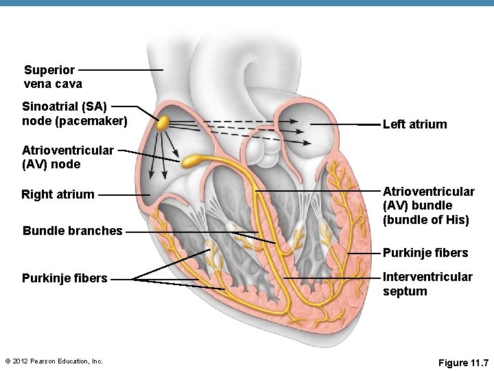
The Heart: Conduction System • Intrinsic conduction system (nodal system) © 2012 Pearson Education, Inc.
The Heart: Conduction System • Special tissue sets the pace • Sinoatrial node = • Atrioventricular bundle = • Bundle branches are in the interventricular septum • Purkinje fibers spread within the ventricle wall © 2012 Pearson Education, Inc.
Superior vena cava Sinoatrial (SA) node (pacemaker) Left atrium Atrioventricular (AV) node Right atrium Bundle branches Atrioventricular (AV) bundle (bundle of His) Purkinje fibers © 2012 Pearson Education, Inc. Interventricular septum Figure 11. 7
Heart Contractions • Contraction is initiated by the sinoatrial node (SA node) • Force cardiac muscle depolarization in one direction— © 2012 Pearson Education, Inc.
Heart Contractions • Once SA node starts the heartbeat • At the AV node, the impulse passes through the AV bundle, bundle branches, and Purkinje fibers • Blood is ejected from the ventricles to the aorta and pulmonary trunk as the ventricles contract © 2012 Pearson Education, Inc.
Heart Contractions • Homeostatic imbalance • Heart block— • Ischemia— • Fibrillation— © 2012 Pearson Education, Inc.
Heart Contractions • Homeostatic imbalance (continued) • Tachycardia— • Bradycardia— © 2012 Pearson Education, Inc.
The Heart: Cardiac Cycle & Heart Sounds • Atria contract simultaneously • Atria relax, then ventricles contract © 2012 Pearson Education, Inc.
The Heart: Cardiac Cycle & Heart Sounds • Cardiac cycle—events of one complete heart beat © 2012 Pearson Education, Inc.
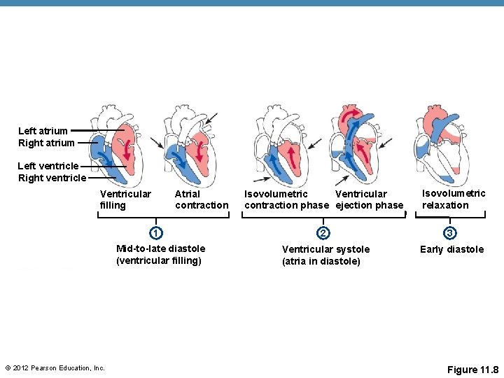
The Heart: Cardiac Cycle & Heart Sounds • Cardiac cycle—events of one complete heart beat • Ventricular systole • Atrioventricular valves close causes first heart sound, “lub” • Semilunar valves open as blood pushes against them • Blood travels out of the ventricles through pulmonary trunk and aorta • Atria are relaxed © 2012 Pearson Education, Inc.
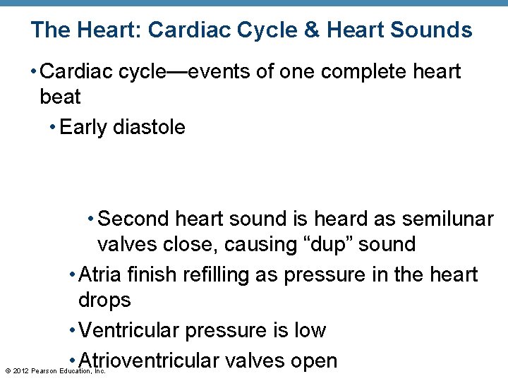
The Heart: Cardiac Cycle & Heart Sounds • Cardiac cycle—events of one complete heart beat • Early diastole • Second heart sound is heard as semilunar valves close, causing “dup” sound • Atria finish refilling as pressure in the heart drops • Ventricular pressure is low • Atrioventricular valves open © 2012 Pearson Education, Inc.
Left atrium Right atrium Left ventricle Right ventricle Ventricular filling Atrial contraction 1 Mid-to-late diastole (ventricular filling) © 2012 Pearson Education, Inc. Ventricular Isovolumetric contraction phase ejection phase Isovolumetric relaxation 2 3 Ventricular systole (atria in diastole) Early diastole Figure 11. 8
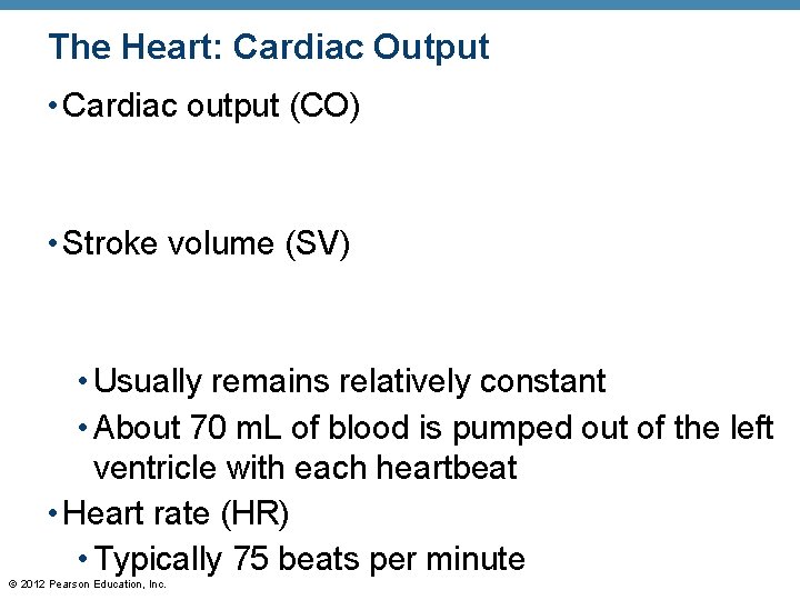
The Heart: Cardiac Output • Cardiac output (CO) • Stroke volume (SV) • Usually remains relatively constant • About 70 m. L of blood is pumped out of the left ventricle with each heartbeat • Heart rate (HR) • Typically 75 beats per minute © 2012 Pearson Education, Inc.
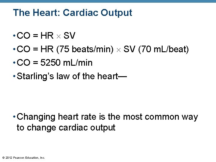
The Heart: Cardiac Output • CO = HR SV • CO = HR (75 beats/min) SV (70 m. L/beat) • CO = 5250 m. L/min • Starling’s law of the heart— • Changing heart rate is the most common way to change cardiac output © 2012 Pearson Education, Inc.
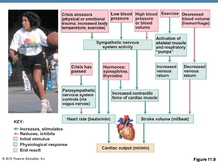
The Heart: Regulation of Heart Rate • Increased heart rate • Hormones • Exercise • Decreased blood volume © 2012 Pearson Education, Inc.
The Heart: Regulation of Heart Rate • Decreased heart rate © 2012 Pearson Education, Inc.
© 2012 Pearson Education, Inc. Figure 11. 9
Blood Vessels: The Vascular System • Transport blood to the tissues and back • Exchanges between tissues and blood • Capillary beds • Return blood toward the heart © 2012 Pearson Education, Inc.
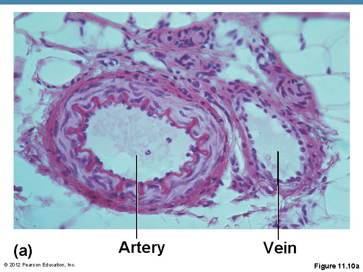
(a) © 2012 Pearson Education, Inc. Artery Vein Figure 11. 10 a
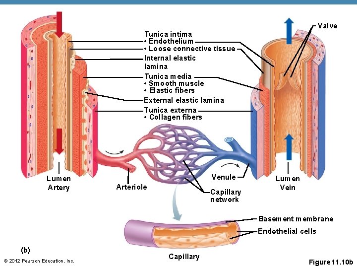
Blood Vessels: Microscopic Anatomy • Three layers (tunics) • Endothelium • Smooth muscle • Controlled by sympathetic nervous system • Mostly fibrous connective tissue © 2012 Pearson Education, Inc.
Valve Tunica intima • Endothelium • Loose connective tissue Internal elastic lamina Tunica media • Smooth muscle • Elastic fibers External elastic lamina Tunica externa • Collagen fibers Lumen Artery Venule Arteriole Capillary network Lumen Vein Basement membrane Endothelial cells (b) © 2012 Pearson Education, Inc. Capillary Figure 11. 10 b
Structural Differences Among Blood Vessels • Arteries have a thicker tunica media than veins • Capillaries are only one cell layer (tunica intima) to allow for exchanges between blood and tissue • Veins have a thinner tunica media than arteries © 2012 Pearson Education, Inc.
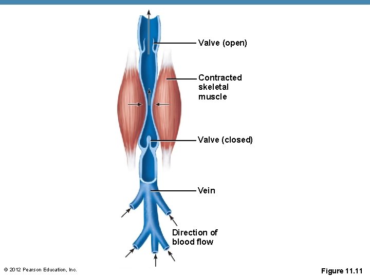
Venous Aids for the Return of Blood to the Heart • Veins: To assist in the movement of blood back to the heart: • Larger veins have valves to prevent backflow • Skeletal muscle “milks” blood in veins toward the heart © 2012 Pearson Education, Inc.
Valve (open) Contracted skeletal muscle Valve (closed) Vein Direction of blood flow © 2012 Pearson Education, Inc. Figure 11. 11
Movement of Blood Through Vessels • Most arterial blood is pumped by the heart • Veins use the milking action of muscles to help move blood © 2012 Pearson Education, Inc.
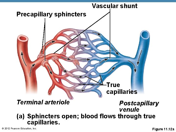
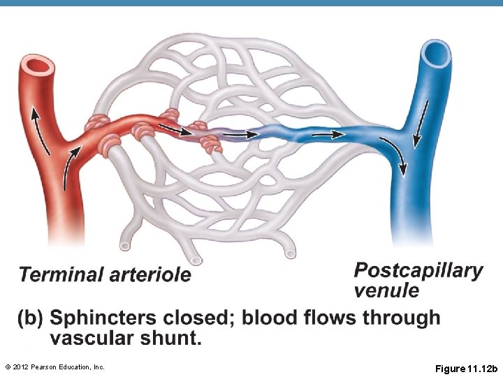
Capillary Beds • Capillary beds consist of two types of vessels • Vascular shunt—vessel directly connecting an arteriole to a venule • True capillaries— © 2012 Pearson Education, Inc.
Vascular shunt Precapillary sphincters True capillaries Terminal arteriole Postcapillary venule (a) Sphincters open; blood flows through true capillaries. © 2012 Pearson Education, Inc. Figure 11. 12 a
© 2012 Pearson Education, Inc. Figure 11. 12 b
Major Arteries of System Circulation • Aorta • Leaves from the left ventricle of the heart • Regions • Ascending aorta— • Aortic arch— • Thoracic • Abdominal © 2012 Pearson Education, Inc.
Major Arteries of System Circulation • Arterial branches of the ascending aorta • Right and left coronary arteries serve the heart © 2012 Pearson Education, Inc.
Major Arteries of Systemic Circulation • Arterial branches of the aortia arch (BCS) • Brachiocephalic trunk splits into the • Left common carotid artery splits into the • Left subclavian artery branches into the • Vertebral artery • In the axilla, the subclavian artery becomes the axillary artery brachial artery radial and ulnar arteries © 2012 Pearson Education, Inc.
Major Arteries of Systemic Circulation • Arterial branches of the thoracic aorta • Intercostal arteries supply the muscles of the thorax wall • Other branches of the thoracic aorta supply the © 2012 Pearson Education, Inc.
Major Arteries of Systemic Circulation • Arterial branches of the abdominal aorta • Celiac trunk is the first branch of the abdominal aorta. Three branches are • Superior mesenteric artery supplies most of the small intestine and first half of the large intestine © 2012 Pearson Education, Inc.
Major Arteries of Systemic Circulation • Arterial branches of the abdominal aorta • Left and right renal arteries (kidney) • Left and right gonadal arteries • Lumbar arteries serve muscles of the abdomen and trunk © 2012 Pearson Education, Inc.
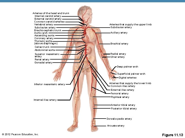
Major Arteries of Systemic Circulation • Arterial branches of the abdominal aorta • Inferior mesenteric artery serves the second half of the large intestine • Left and right common iliac arteries are the final branches of the aorta • Internal iliac arteries serve the pelvic organs • External iliac arteries enter the thigh femoral artery popliteal artery anterior and posterior tibial arteries © 2012 Pearson Education, Inc.
Arteries of the head and trunk Internal carotid artery External carotid artery Common carotid arteries Vertebral artery Subclavian artery Brachiocephalic trunk Aortic arch Ascending aorta Coronary artery Thoracic aorta (above diaphragm) Celiac trunk Abdominal aorta Superior mesenteric artery Renal artery Gonadal artery Arteries that supply the upper limb Subclavian artery Axillary artery Brachial artery Radial artery Ulnar artery Deep palmar arch Superficial palmar arch Digital arteries Inferior mesenteric artery Arteries that supply the lower limb Common iliac artery External iliac artery Femoral artery Popliteal artery Internal iliac artery Anterior tibial artery Posterior tibial artery Dorsalis pedis artery Arcuate artery © 2012 Pearson Education, Inc. Figure 11. 13
Major Veins of Systemic Circulation • Superior and inferior vena cava enter the right atrium of the heart © 2012 Pearson Education, Inc.
Major Veins of Systemic Circulation • Veins draining into the superior vena cava • Radial and ulnar veins brachial vein axillary vein • These veins drain the arms © 2012 Pearson Education, Inc.
Major Veins of Systemic Circulation • Veins draining into the superior vena cava • Subclavian vein receives © 2012 Pearson Education, Inc.
Major Veins of Systemic Circulation • Veins draining into the superior vena cava • Left and right brachiocephalic veins receive venous blood from the • Brachiocephalic veins join to form the superior vena cava right atrium of heart • Azygous vein drains the thorax © 2012 Pearson Education, Inc.
Major Veins of Systemic Circulation • Veins draining into the inferior vena cava • Anterior and posterior tibial veins and fibial veins drain the legs • Posterior tibial vein popliteal vein femoral vein external iliac vein • Great saphenous veins (longest veins of the body) receive superficial drainage of the legs © 2012 Pearson Education, Inc.
Major Veins of Systemic Circulation • Veins draining into the inferior vena cava • Right gonadal vein drains the right ovary in females and right testicle in males • Left gonadal vein empties into the left renal vein • Left and right renal veins drain the kidneys © 2012 Pearson Education, Inc.
Major Veins of Systemic Circulation • Veins draining into the inferior vena cava • Left and right hepatic veins drain the liver © 2012 Pearson Education, Inc.
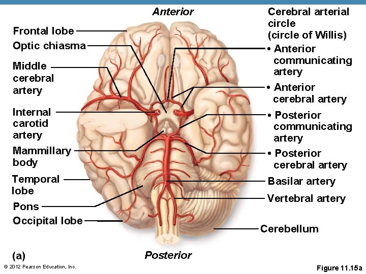
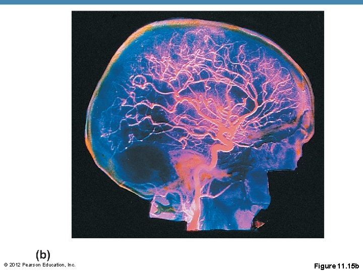
Arterial Supply of the Brain • Internal carotid arteries divide into • Vertebral arteries join once within the skull to form the basilar artery © 2012 Pearson Education, Inc.
Arterial Supply of the Brain • Posterior cerebral arteries form from the division of the basilar artery © 2012 Pearson Education, Inc.
Circle of Willis • Anterior and posterior blood supplies are united by small communicating arterial branches • Result— © 2012 Pearson Education, Inc.
Anterior Frontal lobe Optic chiasma Middle cerebral artery Cerebral arterial circle (circle of Willis) • Anterior communicating artery • Anterior cerebral artery Internal carotid artery Mammillary body • Posterior communicating artery • Posterior cerebral artery Temporal lobe Basilar artery Vertebral artery Pons Occipital lobe (a) © 2012 Pearson Education, Inc. Cerebellum Posterior Figure 11. 15 a
© 2012 Pearson Education, Inc. Figure 11. 15 b
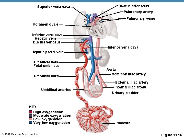
Fetal Circulation • Fetus receives exchanges of gases, nutrients, and wastes through the placenta • Umbilical cord contains three vessels © 2012 Pearson Education, Inc.
Superior vena cava Ductus arteriosus Pulmonary artery Pulmonary veins Foramen ovale Inferior vena cava Hepatic vein Ductus venosus Inferior vena cava Hepatic portal vein Umbilical vein Fetal umbilicus Umbilical cord Umbilical arteries KEY: High oxygenation Moderate oxygenation Low oxygenation Very low oxygenation © 2012 Pearson Education, Inc. Aorta Common iliac artery External iliac artery Internal iliac artery Urinary bladder Placenta Figure 11. 16
Fetal Circulation • Blood flow bypasses the liver through the ductus venosus and enters the inferior vena cava right atrium of heart • Blood flow bypasses the lungs © 2012 Pearson Education, Inc.
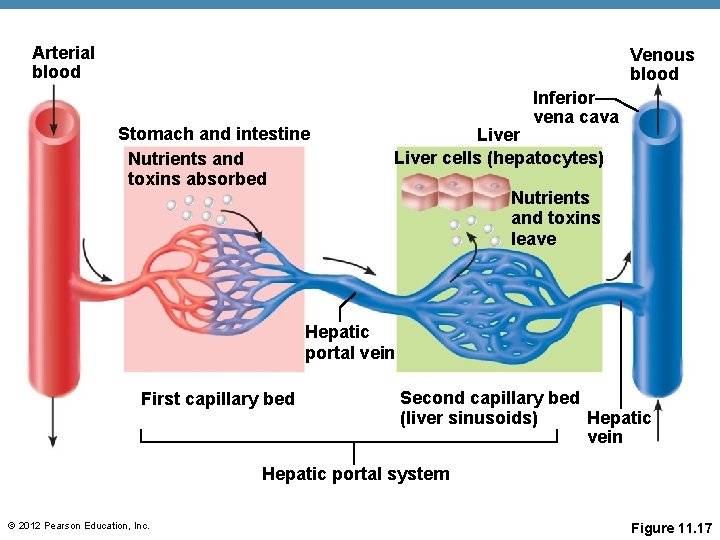
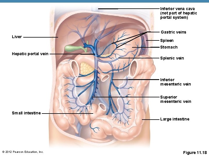
Hepatic Portal Circulation • Veins of hepatic portal circulation drain • Hepatic portal vein carries this blood to the liver • Liver helps maintain proper glucose, fat, and protein concentrations in blood © 2012 Pearson Education, Inc.
Hepatic Portal Circulation • Major vessels of hepatic portal circulation © 2012 Pearson Education, Inc.
Arterial blood Venous blood Stomach and intestine Nutrients and toxins absorbed Inferior vena cava Liver cells (hepatocytes) Nutrients and toxins leave Hepatic portal vein First capillary bed Second capillary bed (liver sinusoids) Hepatic vein Hepatic portal system © 2012 Pearson Education, Inc. Figure 11. 17
Inferior vena cava (not part of hepatic portal system) Gastric veins Liver Spleen Stomach Hepatic portal vein Splenic vein Inferior mesenteric vein Superior mesenteric vein Small intestine Large intestine © 2012 Pearson Education, Inc. Figure 11. 18
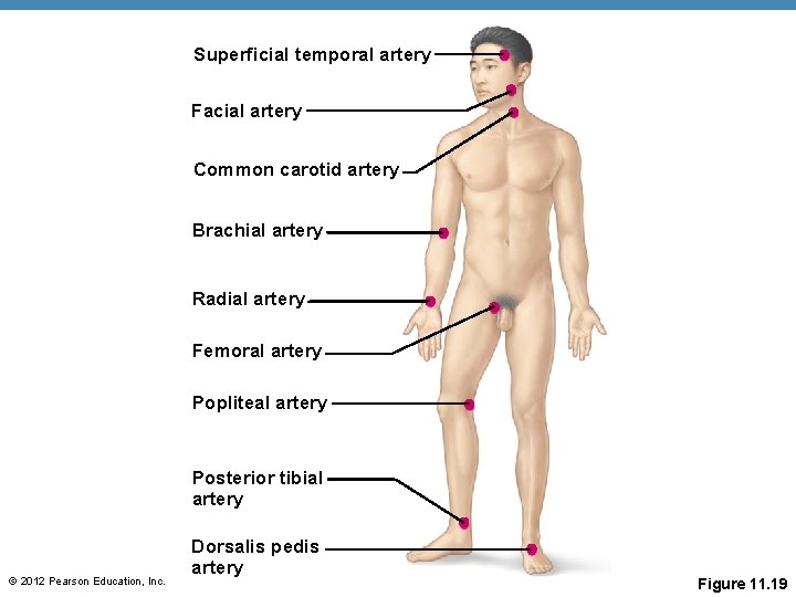
Pulse • Pressure wave of blood © 2012 Pearson Education, Inc.
Superficial temporal artery Facial artery Common carotid artery Brachial artery Radial artery Femoral artery Popliteal artery Posterior tibial artery © 2012 Pearson Education, Inc. Dorsalis pedis artery Figure 11. 19
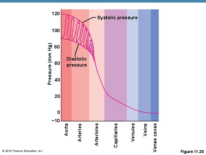
Blood Pressure • Measurements by health professionals are made on the pressure in large arteries • Pressure in blood vessels decreases as distance from the heart increases © 2012 Pearson Education, Inc.
120 Systolic pressure Pressure (mm Hg) 100 80 60 Diastolic pressure 40 20 Veins Venules Capillaries Venae cavae © 2012 Pearson Education, Inc. Arterioles Aorta − 10 Arteries 0 Figure 11. 20
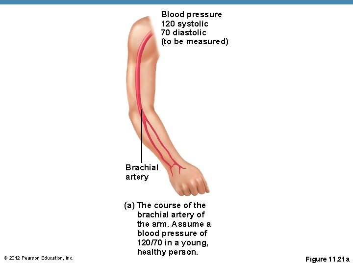
Blood pressure 120 systolic 70 diastolic (to be measured) Brachial artery © 2012 Pearson Education, Inc. (a) The course of the brachial artery of the arm. Assume a blood pressure of 120/70 in a young, healthy person. Figure 11. 21 a
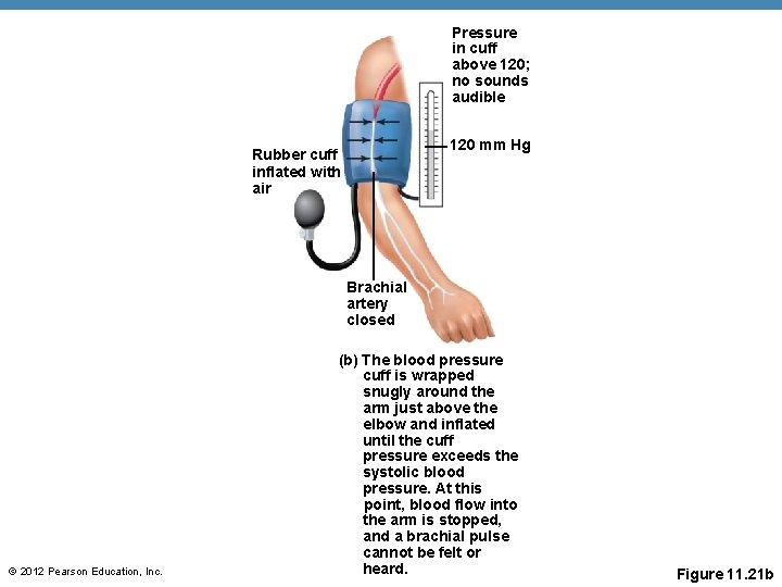
Pressure in cuff above 120; no sounds audible 120 mm Hg Rubber cuff inflated with air Brachial artery closed © 2012 Pearson Education, Inc. (b) The blood pressure cuff is wrapped snugly around the arm just above the elbow and inflated until the cuff pressure exceeds the systolic blood pressure. At this point, blood flow into the arm is stopped, and a brachial pulse cannot be felt or heard. Figure 11. 21 b
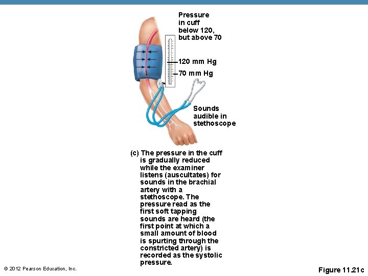
Pressure in cuff below 120, but above 70 120 mm Hg 70 mm Hg Sounds audible in stethoscope © 2012 Pearson Education, Inc. (c) The pressure in the cuff is gradually reduced while the examiner listens (auscultates) for sounds in the brachial artery with a stethoscope. The pressure read as the first soft tapping sounds are heard (the first point at which a small amount of blood is spurting through the constricted artery) is recorded as the systolic pressure. Figure 11. 21 c
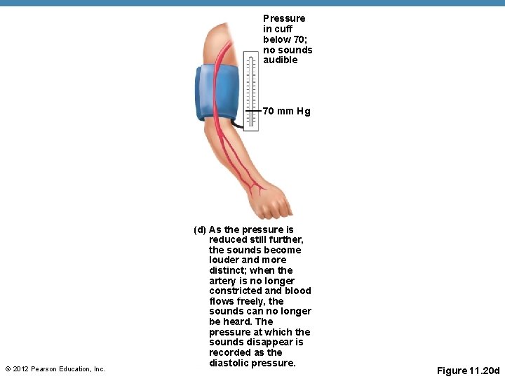
Pressure in cuff below 70; no sounds audible 70 mm Hg © 2012 Pearson Education, Inc. (d) As the pressure is reduced still further, the sounds become louder and more distinct; when the artery is no longer constricted and blood flows freely, the sounds can no longer be heard. The pressure at which the sounds disappear is recorded as the diastolic pressure. Figure 11. 20 d
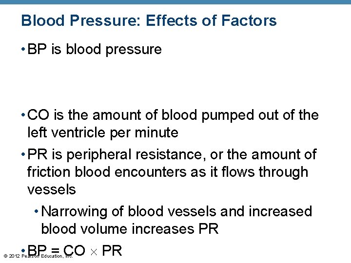
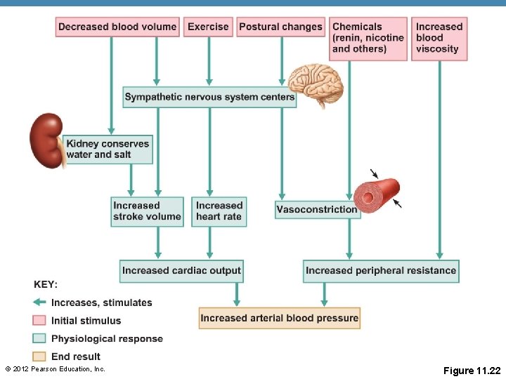
Blood Pressure: Effects of Factors • BP is blood pressure • CO is the amount of blood pumped out of the left ventricle per minute • PR is peripheral resistance, or the amount of friction blood encounters as it flows through vessels • Narrowing of blood vessels and increased blood volume increases PR • BP = CO PR © 2012 Pearson Education, Inc.
Blood Pressure: Effects of Factors • Neural factors • Renal factors © 2012 Pearson Education, Inc.
Blood Pressure: Effects of Factors • Temperature • Heat has a vasodilating effect • Cold has a vasoconstricting effect • Chemicals • Various substances can cause increases or decreases • Diet © 2012 Pearson Education, Inc.
© 2012 Pearson Education, Inc. Figure 11. 22
Variations in Blood Pressure • Normal human range is variable • Normal • 140 to 110 mm Hg systolic • 80 to 75 mm Hg diastolic • Hypotension • Low systolic (below 110 mm Hg) • Often associated with illness • Hypertension • High systolic (above 140 mm Hg) • Can be dangerous if it is chronic © 2012 Pearson Education, Inc.
Capillary Exchange • Substances exchanged due to concentration gradients © 2012 Pearson Education, Inc.
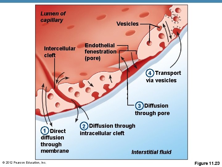
Capillary Exchange: Mechanisms • Direct diffusion across plasma membranes • Endocytosis or exocytosis • Some capillaries have gaps (intercellular clefts) • Fenestrations (pores) of some capillaries © 2012 Pearson Education, Inc.
Lumen of capillary Intercellular cleft Vesicles Endothelial fenestration (pore) 4 Transport via vesicles 3 Diffusion through pore 1 Direct diffusion through membrane © 2012 Pearson Education, Inc. 2 Diffusion through intracellular cleft Interstitial fluid Figure 11. 23
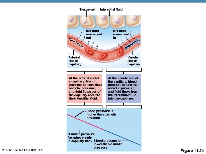
Fluid Movements at Capillary Beds • Blood pressure forces fluid and solutes out of capillaries • Osmotic pressure draws fluid into capillaries © 2012 Pearson Education, Inc.
Tissue cell Bl oo d Interstitial fluid Net fluid movement out flo Net fluid movement in w low f od o Bl Arterial end of capillary Venule end of capillary At the arterial end of a capillary, blood pressure is more than osmotic pressure, and fluid flows out of the capillary and into the interstitial fluid. At the venule end of the capillary, blood pressure is less than osmotic pressure, and fluid flows from the interstitial fluid into the capillary. Blood pressure is higher than osmotic pressure © 2012 Pearson Education, Inc. Osmotic pressure (remains steady in capillary bed) Blood pressure is lower than osmotic pressure Figure 11. 24
Developmental Aspects of the Cardiovascular System • A simple “tube heart” develops in the embryo and pumps by the fourth week • Few structural changes occur after the seventh week © 2012 Pearson Education, Inc.
Developmental Aspects of the Cardiovascular System • Aging problems associated with the cardiovascular system include • Progressive atherosclerosis • Loss of elasticity of vessels leads to hypertension • Coronary artery disease results from vessels filled with fatty, calcified deposits © 2012 Pearson Education, Inc.