Spinal Cord Injury Partial or complete disruption of


, cervical (4 -7), and T 12")
(45%) • Domestic / Industrial Accidents (34%) • Sport")


 • Pathway for control of voluntary motion")


 vs. Lower Motor Neuron (LMN) Syndrome UMN syndrome LMN Syndrome")


 Frontal Lobe, Micturation Centre Pontine Micturation Centre")

 Pontine Micturation Centre A lesion affecting S 2 -S")


: an emergency situation in which a noxious stimulus precipitates a pathological")


")


- Slides: 87
Spinal Cord Injury
• Partial or complete disruption of spinal cord resulting in paralysis, sensory loss, altered autonomic and reflex activities.
Complete Lesion Incomplete Lesion • A lesion to the spinal cord where there is no preserved motor or sensory function below the level of lesion • A lesion to the spinal cord with incomplete damage to the cord. There may be scattered motor function, sensory function or both below the level of lesion Introduction : 2 General Classifications
SCI COMMON TERMINOLOGY
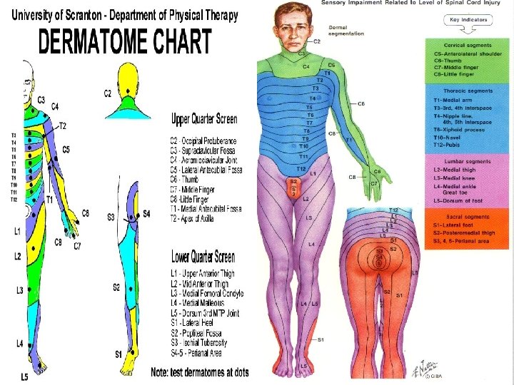
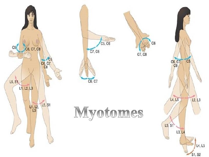
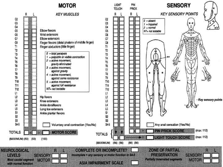
• Cauda Equina Injuries: a term used to describe injuries that • occur below the L 1 level of the spine (LMN) • Dermatome: Designated sensory areas based on spinal segment innervation • Myotome: Designated motor areas based on spinal segment innervation • Neurologic level: the lowest segment of the spinal cord with intact strength and sensation. Muscle groups at this level must receive a grade of 3 Paraplegia: a term used to describe injuries that occur at the level of the thoracic lumber or sacral spine
EPIDEMIOLOGY/ETIOLOGY
Epidemiology • Vertebral column injuries are reported to occur in 6% of trauma patients • Half of these patients sustain spinal cord or nerve root injury
Spinal Cord Injuries �Most common locations: cervical (1&2), cervical (4 -7), and T 12 – L 2 vertebrae �Cervical and lumbar spine injuries are often associated with incomplete cord injury while thoracic injuries often have complete cord injury.
Mode of Injury • Accidents (RTA)(45%) • Domestic / Industrial Accidents (34%) • Sport Injury 15% • Assault 6%
PATHOPHYSIOLOGY OF SPINAL CORD INJURY Neural tissue injuries • Primary injury – refers to physical tissue disruption caused by mechanical forces • Secondary injury – refers to additional neural tissue damage resulting from the biological response initiated by the physical tissue disruption e. g. edema
ANATOMY OF THE SPINAL CORD
• Gross Anatomy • • Sagittal Diameter: 7 -12 mm Cervical Enlargement: C 3 -T 2 level Lumbar Enlargement: T 10 -T 12 level Length: approximately 45 cm. From foramen magnum to L 1 -L 2 vertebrae • Vascular Supply • 1 anterior spinal artery which supplies the anterior 2/3 of the cord • 2 posterior spinal arteries which supply the posterior 1/3 of the cord • Both anterior and posterior arteries receive reinforcement from the Radicular Artery • Spinal vertebral venous plexus to the azygous vein
• Internal Anatomy • Gray Matter – neuronal cell bodies & synapses • Anterior Horn – motor neurons • Posterior Horn – sensory neurons • White Matter – ascending & descending fiber pathways • Ascending – relays sensory information to the brain • Descending – relays motor information down to the cord
ASCENDING TRACTS Consists of axons that conduct action potentials or impulses towards the brain (afferent)
• Pain and Temperature • Lateral Spinothalamic Tract – Main Pathway • Ventral Spinothalamic Tract • Both tracts cross to the contralateral side of the cord about 2 -3 cord levels above as it travels up the cerebral cortex (Area 3, 1, 2) • Lesion of this tract presents with loss of contralateral pain & temperature below the level of injury
• Proprioception and Stereognosis • Posterior Column – ascends up the spinal cord and crosses to the contralateral side at the level of the medulla to the cerebral cortex (Area 3, 1, 2) • • Fasciculus Cuneatus – laterally located; UE proprioception Fasciculus Gracilis – medially located; LE proprioception • Lesion of Posterior Column: Loss of ipsilateral proprioception & stereognosis below the level of injury
• Lateral & Anterior Spinothalamic Tracts • Pain & temperature sensation and crude touch • Dorsal Column • Fine touch, proprioception, two-point discrimination • Dorsal & Ventral Spinocerebellar Tracts • Proprioceptive and exteroceptive stimuli for movement and position sense • Spinoreticular Tract • Deep and chronic pain Summary Ascending Tracts
DESCENDING TRACTS Consists of axons that conduct action potentials or impulses away from the brain (efferent)
• Lateral Corticospinal Tract – main pathway • Ventral Corticospinal Tract • Both tracts decussate from the cerebral cortex (area 4, 6) at the level of the medulla as it descends to the cord ipsilaterally • Lesion of corticospinal tract: loss of ipsilateral motor function below the level of injury Motor Pathway
• Rubrospinal Tract • Serves as motor junction • For large muscle movement & fine motor control • Facilitates flexion & inhibits extension of upper extremities • Reticulospinal Tract • For modulation of sensory transmission esp. pain; spinal reflexes • Tectospinal Tract • For reflex head turning • Mediate reflex postural movements of the head in response to visual & auditory stimuli • Medial Longitudinal Fissure • For coordination of head and eye movements Summary Descending Tracts
• Anterior Corticospinal (Direct Pyramidal Tract) • Pathway for control of voluntary motion • Conduct voluntary motor impulses from the precentral gyrus to the motor centers of the cord. • Lateral Corticospinal (Crossed Pyramidal Tract) • Pathway for control of voluntary motion • Provides fine motor control of limbs and digits • Vestibulospinal Tract • For postural reflexes • Facilitates extensor muscle tone & equilibrium • Spino-olivary Tract • Proprioception from muscles & tendons and cutaneous impulses to the olivary nucleus Summary Descending Tracts
CLASSIFICATION
• Lesion level indicates most distal uninvolved nerve root segment with normal function; muscles must have a grade of at least 3+/5 or fair + function • Tertraplegia (quadreplegia): injury occurs between C 1 and C 8, involves all extremities and trunk • Paraplegia: injury occurs between T 1 and T 12 L 1; involves both lower extremities and trunk (varying levels) UMN Injury
Upper Motor Neuron (UMN) vs. Lower Motor Neuron (LMN) Syndrome UMN syndrome LMN Syndrome Type of Paralysis Spastic Paresis Flaccid Paralysis Atrophy No (Disuse) Atrophy Severe Atrophy Deep Tendon Reflex Increase Absent DTR Pathological Reflex Positive Babinski Sign Absent Superficial Reflex Absent Present Fasciculation and Fibrillation Absent Could be Present
Functional consequences of spinal cord injury • Complete injury • Incomplete injury • Transient spinal cord dysfunction Grades of severity of neurological injury • Anterior cord syndrome • Central cord syndrome • Brown-Sequard syndrome Pattern of neurological dysfunction observed during clinical evaluation
Assessment of spinal cord injury • Level of cord injury • Spinal shock is over or not • Injury is complete or incomplete
• A= complete: no motor or sensory function is preserved in the sacral segments S 4 -S 5 • B= incomplete: sensory but not motor function is preserved below the neurological level and includes the sacral segments S 4 -S 5 • C= incomplete: motor functional is preserved below the neurological level, and most key muscles below the neurological level have a muscle grade <3 • D= incomplete: motor function is preserved below the neurological level, and most key muscles below the neurological level have a muscle grade of ≤ 3 • E= normal: motor and sensory function in normal American Spinal Injury Association
SPECIFIC INCOMPLETE LESIONS
• An incomplete lesion that results from compression and damage to the anterior part of the spinal cord or anterior spinal artery. The mechanism of injury is usually cervical flexion. There is loss of motor function and pain and temperature sense below the lesion due to damage of the corticospinal and spinothalamic tracts Anterior Cord Syndrome
• A relatively rare syndrome that is caused by compression of the posterior spinal artery and is characterized by loss of pain perception, proprioception, two point discrimination, and stereognosis. Motor function is preserved. Posterior Cord Syndrome
• An incomplete lesion usually caused by a stab wound, which produces hemisection of the spinal cord. There is paralysis and loss of vibratory and position sense on the same side as the lesion due to the damage to the CST and dorsal columns. There is a loss of pain and temp sense on the opposite side of the lesion from damage to the lateral spinothalamic tract. Brown Sequard Syndrome
• An incomplete lesion that results from compression and damage to the central portion of the spinal cord. The mechanism of injury is usually cervical hyperextension that damages the spinothalamic tract, CST and dorsal columns. The upper extremities present with greater involvement than the lower extremities and greater motor deficits exist as compared to sensory deficits. Central Cord Syndrome
Conus Medullaris Syndrome– Most of these injuries occur b/w T 11 and L 2 vertebrae – – Sphincter disturbances Saddle anesthesia(S 3 -S 5) Absence of lower extremity reflexes Usually it is associated With involvement of lumbar roots Cauda Equina Syndrome It is bilateral involvement of multiple lumbosacral nerve roots with in the spinal canal, characterized by Absent reflexes in lower limbs Bladder and bowel involvement Loss of perineal sensation
• sparing of tracts to sacral segments with preservation of perianal sensation, rectal sphincter tone or active toe flexion. Sacral Sparing
COMMON COMPLICATIONS IN SCI AND THEIR INTERVENTIONS
• Spinal Shock • Respiratory complication • Cardiovascular complication • Renal and Bladder complication • Bed sores • Deep vein thrombosis • Musculoskeletal complication
• Spinal shock: A physiologic response that occurs between 30 and 60 minutes after trauma to the spinal cord and can last up to several weeks. Spinal shock presents with total flaccid paralysis and loss of all reflexes below the level of injury.
Two reflexes are important in acute spinal cord injury assessment • Anal wink reflex • Bulbocavernous reflex Return of bulbocavernous reflex marks the end of spinal shock
Innervation of the bladder Parasympethetic, S 2, 3, 4 Sympethetic, T 11 -L 2 Somatic S 2, 3, 4 Stimulates the internal The afferent fibers carry sensations from the bladder. parasympathetic nerves stimulate the detrusor to contract urinary The sphincter pudendal tonerve originates remain tightly from closed. the nucleus of Onuf in the activity anteriolateral The situated sympathetic also horns of the sacral segments inhibits parasympathetic 2, 3 and 4. stimulation. It regulates the voluntary actions of the external urinary sphincter and the pelvic diaphragm. Nucleus of Onuf Hypogastric nerve Pudendal Nerve Primitive voiding centre S 2, 3, 4 Preganglionic fibers Pelvic Nerve Postganglio nic fibers Pelvic Plexus Internal urinary sphincter Detrusor Muscle
Neuroanatomy of Voiding Frontal Lobe, Micturation Centre Pontine Micturation Centre Sends inhibitory signals PMC until a socially acceptable time and place are found. Primitive voiding centre Major relay center/ inherent excitatory nature. Coordinates urinary sphincters and the Bladder Independent until 3 -4 yrs of age.
Neuroanatomy of Voiding Frontal Lobe, Micturation Centre Pontine Micturation Centre There is first a voluntary relaxation of the perineum, followed sequentially by an increase tension of abdominal wall, a slow contraction of the detrusor, and an associated opening of internal sphincter; finally there is relaxation of the external sphincter resulting in urinary flow. Primitive voiding centre Sympethetic Parasympethetic Pudendal Nerve Relaxation of IUS External sphincter opens Detrusor Contraction Facilitation of voluntary urination
Neurogenic dysfunction of bladder Frontal Lobe, Micturation Centre Uninhibited Neurogenic Bladder Pontine Micturati on Centre If a problem occurs within the nervous system, the entire voiding cycle is affected. Any part of the nervous system may be affected, including the brain, pons, spinal cord, sacral cord, and peripheral nerves. Pudendal Nerve Reflex Neurogenic Bladder (Automatic bladder, Spastic bladder) Autonomic Neurogenic Bladder Primitive voiding centre Motor paralytic bladder (atonic detrusor) Sensory paralytic bladder
The Neurogenic Bladder Lapides Classification Five Types – • Two upper motor neuron type (reflex and uninhibited neurogenic bladder) and • Three lower motor neuron type (autonomous, sensory paralytic, and motor paralytic bladder).
Uninhibited Neurogenic Bladder Frontal Lobe, Micturation Centre Pontine Micturation Centre A hyper-reflexic detrusor with a normal sphincter. Lesions of the brain above the pons destroy the master control center, causing a complete loss of voiding control. Urgency, frequency, hesitancy, urge incontinence. The bladder empties too quickly and too often, with relatively low quantities. Cerebro-vascular accident Brain tumors Parkinson disease Shy-drager syndtrome Cerebral palsy Dementia Primitive voiding centre
Reflex Neurogenic Bladder (Automatic bladder, Spastic bladder) Frontal Lobe, Micturation Centre Pontine Micturation Centre During spinal shock, all sensory and motor innervations are depressed. When intravesical pressure rises above sphincter pressure some amount of urine dribbles out until again intravesical pressure comes below urethral pressure. Motor vehicle and diving accidents leading to spinal cord injury Myelitis Cervical Spondylosis Multiple sclerosis Arterio-venous malformation Syrigomyelia Other spinal cord disorder Children born with myelomeningocele Primitive voiding centre
Autonomous neurogenic bladder The bladder is paralyzed and there is no awareness of the state of fullness. voluntary initiation of micturition is impossible Overflow incontinence, painless retention. saddle anesthesia with absence of bulbocavernosus and superficial anal reflex. Typical causes are • A sacral cord tumor, • Herniated disc(conus medullaris), • Injuries that crush the pelvis, lumbar laminectomy, radical hysterectomy, or abdominoperineal resection. • Tethered cord syndrome. Frontal Lobe, Micturation Centre Pontine Micturation Centre Primitive voiding centre
Motor paralytic bladder (atonic detrusor) Pontine Micturation Centre A lesion affecting S 2 -S 4 motor neurons (detrusor motor neurons), Painful urinary retention with overflow incontinence and inability to initiate the urination. The saddle and bladder sensations are normal, but anal and bulbocavernosus reflexes are usually absent. Develops when the motor supply is interrupted, as in – • Poliomyelitis, • Polyradiculoneuritis, • Trauma, • Some pt may present this in association with Lumber canal stenosis and with lumbosacral meningo-myelocoele, • Pelvic surgery Frontal Lobe, Micturation Centre Primitive voiding centre
Sensory paralytic bladder ØAfferent limb of the micturition spinal reflex is compromised, posterior root ganglion of sacral nerve or the posterior column of spinal cord. ØBladder sensations are absent and there is no desire to void. ØPainless retention with overflow incontinence is present, but pt can initiate the urination with difficulty. ØSaddle anesthesia is present and anal & bulbocavernosus reflexes may or may not be present. Frontal Lobe, Micturation Centre Pontine Micturation Centre Primitive voiding centre Causes - • Multiple sclerosis, • Diabetic autonomic neuropathy, • Tabes dorsalis
Bladder Management for Supra Sacral SCI Frontal Lobe, Micturation Centre Pontine Micturati on Centre Four Important Goals � Prevent complications Most common methods of the kidneys Intermittent catheterization � Prevent complications Reflex voiding (for men) of the bladder Indwelling catheter � Tofrequent have a bladder Less methods management program to Bladder augmentation that best fits lifestyle surgically increase bladder of the patient Pudendal Nerve size � To keep skin and Surgical diversion clothing free of urine Neurostimulation Uninhibited Neurogenic Bladder Reflex Neurogenic Bladder (Automatic bladder, Spastic bladder) Primitive voiding centre
Frontal Lobe, Micturation Centre Bladder Management for Supra Sacral SCI b-Antispasmodic drugs – Anticholinergic drugs Medication They inhibit involuntary bladder Pontine Micturati on Centre These relax the smooth muscles of the contractions. urinary bladder. By exerting a direct spasmolytic action Propantheline bromide – 30 -60 mg qid. Oxybutynin chloride Dicyclomine hydrochloride – 10 mg tds Tolterodine L-tartrate Primitive Darifenacin voiding Trospium centre § Imipramine hydrochloride Amitriptyline hydrochloride Urge incontinence § The 3 main categories of • § • drugs used to treat § • include § Solifenacin succinate Anticholinergic drugs – c-Tricyclic antidepressant They function to increase Antispasmodics, norepinephrine and serotonin levels. In addition, they exhibit anticholinergic Tricyclic antidepressant and direct muscle relaxant effects on agents. the urinary bladder. § Pudendal Nerve Uninhibited Neurogenic Bladder Reflex Neurogenic Bladder (Automatic bladder, Spastic bladder)
Frontal Lobe, Micturation Centre Management Techniques for Sacral SCI Pontine Micturati on Centre 1. Intermittent Catheterization Easier than for those with Supra SCI because the bladder is flaccid and underactive and generally upper body strength and mobility are relatively better 2. Intra-abdominal Pressure Voiding • Valsalva (bearing down) • Credé (pushing inward on the bladder with fist) Autonomic Neurogenic Bladder Primitive voiding centre Motor paralytic bladder (atonic detrusor) Sensory paralytic bladder
Intermittent Catheterization • There are two general catheterization techniques. • Sterile technique (catheters put in using sterile gloves and a sterile catheter or a catheter contained in a sterile bag) • “Clean” technique (catheters are washed, dried, stored and reused) • Generally done every 4 -6 hours to prevent the bladder from getting too full
Indwelling Catheters: Two Types • Urethral Catheter – : Placed in the bladder through the urethra – Held in place by an inflated balloon – Does not require fluid restriction, good hand function or transfer/dressing skill. – 30% become coated with stones that prevents drainage and contributes to UTIs • Suprapubic Catheter �Requires minor surgery for insertion �Does not require fluid restriction, good hand function or transfer/dressing skill. �Clinically preferable over an indwelling catheter (less likely to get plugged with stones, easier to change) �Preferable by men because sexual activity is not limited (Women are not limited by either catheter type)
Concern with Credé Voiding �Can have negative side effects such as hemorrhoids, hernias, rectal prolapse, and reflux of urine up the ureters to the kidney �Can increase the risk for infections �Best not to do unless you have a weak sphincter
• Can occasionally be useful to a patient with a SCI however more often serves to interfere with functional activities. Spasticity can be enhanced by both internal and external sources such as stress, decubiti, urinary tract infections, bowel or bladder obstruction, temperature changes or touch. • Increased involuntary contraction of muscle groups, increased tonic stretch reflexes, excessive deep tendon reflexes. • Treatment: medications are usually administered in an attempt to reduce the degree of spasticity (Dantrium, Baclofen, Lioresal). Aggressive treatment includes rhizotomies, myelotomies, and other surgical intervention. Physical therapy intervention includes positioning, aquatic therapy, weight bearing, FES, ROM, resting splints and inhibitive casting. Spasticity
• (hyperreflexia): an emergency situation in which a noxious stimulus precipitates a pathological autonomic reflex with symptoms of paraoxysmal hypertension, bradycardia, headache, diaphoresis, flushing, diplopia, or convulsions; examine for irritating stimuli; treat as a medical emergency, elevate head, check and empty catheter first. • Treatment: the first reaction to this medical crisis is to transfer the patient to a sitting position and then immediately check the catheter for blockage. The bowel should also be checked for impaction. A patient should remain in sitting position. Lying a patient down is contraindicated and will only assist to further elevate blood pressure. The patient should be examined for any other irritating stimuli. If the cause remains unknown the patient should receive immediate medical intervention. Autonomic Dysreflexia
• Abnormal bone growth in soft tissues; examine for early changessoft tissue swelling, pain, erythema, generally near large joint; late changes- calcification, initial signs of ankylosis • Treatment: Drug intervention usually involves diphosphates that inhibit ectopic bone formation. Physical therapy and surgery are often incorporated into treatment. Physical therapy must focus on maintaining functional range of motion and allowing the patient the most independent functional outcome possible. Heterotopic Bone Formation
• Results from the formation of a blood clot that becomes dislodged and is termed an embolus. This is considered a serious medical condition since the embolus may obstruct a selected artery. A pateint with a spinal cord injury has a greater risk of developing a DVT due to the absence or decrease in the normal pumping action by active contractions of muscles in the lower extremities. • Treatment: once a DVT is suspected there should be no active or passive movement performed to the involved lower extremity. Bed rest and anticoagulant drug therapy are usually indicated. Surgical procedures can be performed if necessary. Deep Vein Thrombosis
• Occurs due to a loss of sympathetic control of vasoconstriction in combination with absent or severely reduced muscle tone. Venous pooling is fairly common during the early stages of rehab. A decrease in systolic blood pressure greater than 20 mm. HG after moving from supine to sitting is typically indicative of this. • Treatment: Monitoring vital signs assists with minimizing the effects of orthostatic hypotension. The use of elastic stockings, ace wraps to the lower extremities, and abdominal binders are common. Gradual progression to a vertical position using a tilt table is often indicated. During intervention may be indicated in order to increase blood pressure Orthostatic Hypotension
• Caused by sustained pressure, friction, and/or shearing to a surface. The most common areas susceptible to pressure ulcers are the coccyx, sacrum, ischium, trochanters, elbows, buttocks, malleoli, scapulae, and prominent vertebrae. Pressure ulcers require immediate medical intervention and can often significantly delay the rehab process. • Prevention is of greatest importance. A patient should change position frequently, maintain proper skin care, sit on an appropriate cushion, consistently weight shift, and maintain proper nutrition and hydration. Surgical intervention is often necessary with advanced pressure ulcers. Pressure Ulcers
PHYSICAL THERAPY GOALS, OUTCOMES AND INTERVENTIONS
• Deep breathing exercises, strengthening exercises to respiratory muscles; assisted coughing, respiratory hygiene (postural drainage, percussion, vibration, suctioning) as needed to keep airway clear; abdominal support Improve Respiratory Capacity
• Prevent contracture: PROM, positioning, splinting, selective stretching to preserve function (tenodesis grasp) Maintain ROM
• Free of pressure ulcers and other injury positioning program, pressure relieving devices (cushion, ankleboots) patient education: pressure relief activities (pushups) and skin inspection; provide prompt treatment of pressure sores Maintain Skin Integrity
• Strengthen all remaining innervated muscles use selective strengthening during acute phase to reduce stress on spinal segments; resistive training to hypertrophy muscles Improve Strength
• Tilt table, wheelchair, use of abdominal binder, elastic lower extremity wraps to decrease venous pooling; examine for signs and symptoms of orthostatic hypotension (light headedness, syncope, mental or visual blurring, sense of weakness Reorient Patient to Vertical Position
• Emphasis on independent rolling and bed mobility assumption of sitting, transfers, sit-to-stand, and ambulation as indicated • Tolerance, postural control, symmetry, and standing balance as indicated Promote early return of ADLs
• 1. Motorcyclist fracture is a. Ring fracture b. Comminuted fracture c. Fracture base of skull d. Separation of anterior and posterior half of skull
2. Most common cause of SCI in India is a. RTA b. Fall from height c. Fall in well d. House collapse 72 11/9/2020
• 3. Dislocation without fracture is seen in a. Sacral spine b. Lumbar spine c. Cervical Spine d. Thoracic spine 73 11/9/2020
• 4. Whip-lash injury is caused due to a. Fall from height b. Acute hyperextension of spine c. Blow on top of head d. Acute hyperflexion of spine
5. Jeffersons fracture is a. C 1 b. C 2 c. C 2 -C 1 d. C 2 -C 3 75 11/9/2020
6. Hangmans fracture is fracture of C 2 a. Dens b. Lamina c. Pars intercularis d. Spinous process 76 11/9/2020
• 7. Burst fracture of cervical spine is due to a. Whiplash injury b. Fall of weight on neck c. Car accident d. Vertebral compression injury
8. Tear drop fracture of lower cervical spine implies a. Wedge compression fracture b. Axial compression fracture c. Flexion compression failure of body d. Flexion rotation failure of body 78 11/9/2020
9. Dennis stability concept of spine is based on a. 1 column b. 2 column c. 3 column d. 5 column 79 11/9/2020
• 10. Spinal shock is associated with a. Increased spinal reflexes b. Absent spinal reflexes c. Loss of autonomic reflexes d. Bizzare reflexes
11. A 29 year old man following RTA presented with tenderness present at the thoracolumbar region in the back and there was a suspicion about posterior ligamentous complex disruption on local examination of spine. There was no visceral injury. On neurological examination, AIS was A. There was no prior history of spinal complaints. X- ray AP and lateral demonstrated a flexion compression injury of T 12. 81 11/9/2020
1. Most appropriate method for determining stability of the spine • Modified Denis 3 column classification • CT spine • MRI spine • TLICS score 82 11/9/2020
2. What is the TLICS score for the above case • 3 • 5 • 7 • 9 83 11/9/2020
. TLICS is summation of points on 3 categories: fracture morphology, neurologic status, and integrity of the posterior ligamentous complex. Fracture morphology: Compression Injuries (1 pt), Burst ( 2 pts), Translational/Rotational Injuries (3 pts), Distraction Injuries (4 pts); Neurologic status: Intact (0), Root Injury (2 pts), Complete injury (2 pts), Incomplete injury (3 pts), Cauda Equina injury (3 pts); Integrity of posterior ligamentous complex (PLC) Supraspinous ligament (SSL), interspinous ligament (ISL), capsular ligaments and ligamentum flavum: Intact (0), Injury suspected (2 pts), Injured (3 pts) In this patients it is 1 point for morphology + 2 points for complete neurological injury(AIS score A) + 2 points for suspected PLC injury. Total TLICS= 5. • Management according to summation of point values from 84 each category 3 or less - nonoperative 11/9/2020
Management for this case would be • Surgical stabilization • Spinal bracing • Complete bed rest only • Surgical or conservative. 85 11/9/2020
Which of the following is not a component of TLICS score • Injury morphology • Posterior ligamentous complex integrity • Dynamic X ray of spine • Neurological status. 86 11/9/2020
……. THANK YOU