Spinal Cord Injury SCI J Hardjono SPINAL CORD
 J. Hardjono")
. Car Crashes: 83% Motorcycle incidents: 10%")


 1. Paralysiss 1. Tetraplegia ( tingkat servical ) 2.")
 Kontaktur dan Nyeri Decubitis Infeksi saluran kencing")

 is a simple sensorimotor pathway that")


- Slides: 48
Spinal Cord Injury ( SCI ) J. Hardjono
SPINAL CORD INJURIES Caused by sudden trauma (USA). Car Crashes: 83% Motorcycle incidents: 10% Bicycle accidents: 3% Medical/Surgical Complications: 38% Hit by falling Object: 30% Pedestrian: 22% Gunshot: 92% Personal Contact: 6% Diving: 55% Snow skiing: 8% Surfing: 6% Approx. 10, 0000 new cases/year, 80% male. Age group most commonly injured: 16 -30 years (43%) and 31 -45 (28%). Although Vehicle is the leading cause overall, Falls become the leading cause in people over 60 years. Source: National Spinal Cord Injury Statistical Center
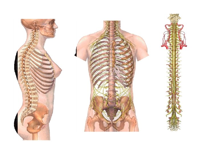
Functions of the Vertebral or Spinal Column Include: Protection • Spinal Cord and Nerve Roots • Many internal organs Base for Attachment • Ligaments • Tendons • Muscles
Structural Support • Head, shoulders, chest • Connects upper and lower body • Balance and weight distribution Flexibility and Mobility • Flexion (forward bending) • Extension (backward bending) • Side bending (left and right) • Rotation (left and right) • Combination of above Other • Bones produce red blood cells • Mineral storage
Spine Terdiri atas 33 tulang, • 24 tulang saling bersendi membentuk kolumna yang fleksibel: – 7 buah vertebra cervicalis – 12 buah vertebra thoracalis – 5 buah vertebra lumbalis – 1 buah (penyatuan 5 buah) os sacral • 1 buah (penyatuan 3 – 5 buah) os coccygeus
Stuktur yang mungkin cidera • Tulang belakang – Fraktur – Dilokasi • • Spinal Cord Paru – pleura Viseral abdomem Viseral Pelvis
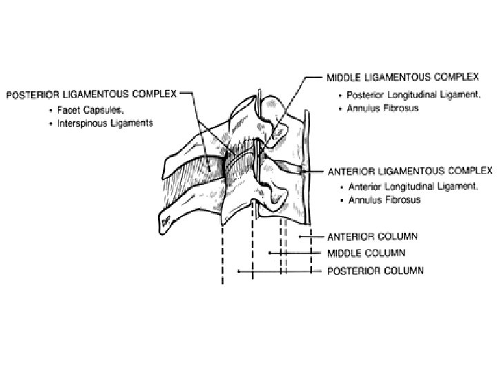
Fraktur pada tulang belakang Jenis • Dengan atau tanpa dislokasi • Kompresi raktur • Pecahnya corpus vertebra Kausa • Fleksi • Ekstensi ( whiplash) • Rotasi • Kompresi vertikal • Avulsi otot FR Patologis • Osteoporosis • Tumor • TBC Kondisi • Stable • instable
Classification of Neurologic Injury • • Root Injury Incomplete Spinal Cord Lesions Brown-Séquard Syndrome Central Cord Syndrome Anterior Cord Syndrome Posterior Cord Syndrome Complete Spinal Cord Injury
Root Injury • A nerve root may be damaged at the neural foramen by a fractured or dislocated facet or a fracture of the uncinate process of the vertebral body. • These are essentially peripheral nerve lesions and are expected to recover, at least partially. Root avulsion rarely occurs, except in a classic brachial plexus injury with a distraction force on the shoulder.
Incomplete Spinal Cord Lesions • Partial loss of cord function may be determined by the presence, although altered, of voluntary muscle power or sensory preservation distal to the zone of cord injury. The patterns of preserved sensory and motor function differentiate the various incomplete cord lesion syndromes. Each syndrome has a specific prognosis for the probability of recovery. In general, any sparing distal to the injury constitutes an incomplete lesion, and recovery, varying from minimal to full, is possible. The greater the motor and sensory sparing, and the more rapid the recovery during the first several days or weeks, the better the prognosis for full recovery.
Brown-Séquard Syndrome • An injury limited to either side of the spinal cord produces ipsilateral muscle paralysis and contralateral hypesthesia to pain and temperature. This syndrome has a good prognosis for recovery. More than 90% of the patients regain bladder and bowel control and the ability to walk. 104
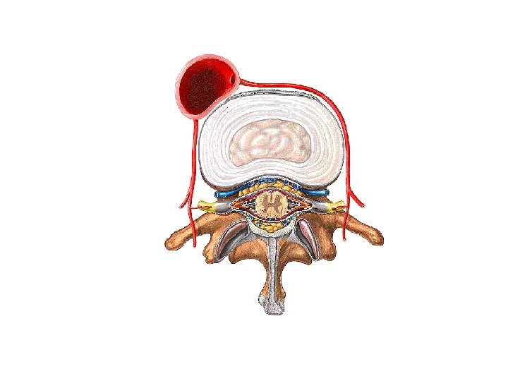
Central Cord Syndrome • Central cord syndrome is the most common incomplete cord syndrome. • It is frequently associated with an extension injury to an osteoarthritic spine in a middle-aged person. • X-ray examination may reveal no fracture or dislocation; however, the patient demonstrates almost complete flaccid quadriplegia. Impact damage to the central gray matter, produced by the pincer effect of the osteophytes anteriorly and the infolded ligamentum flavum posteriorly, produces severe flaccid lower motor neuron paralysis of the fingers, hands, and arms.
• Damage to the central portion of the corticospinal and spinothalamic long tracts in the white matter produces upper motor neuron spastic paralysis of the trunk and lower extremities. The sacral tracts are positioned on the periphery of the cord and are usually spared from the injury. The patient demonstrates gross quadriplegia, but careful examination reveals sacral sparing manifested by perianal sharp-dull sensation and an early return of sphincter and toe flexor control. Prognosis for this syndrome is fair. Fifty to 60% of the patients can be expected to have progressive return of motor and sensory power to the lower extremities and trunk, but they have poor recovery of hand function, owing to irreversible central gray matter destruction. These patients are likely to regain bowel and bladder control and to be able to walk with a spastic gait, but significant paralysis of the hands remains permanent.
Anterior Cord Syndrome • The anterior cord syndrome 118 is manifested by complete motor paralysis and sensory anesthesia, with the exception of dorsal column sparing providing deep pressure and proprioception as the only retained sensibility of the trunk and lower extremities. Prognosis is good if recovery is evident and progressive within the first 24 hours. After 24 hours, if no sign of sacral sensibility to pinprick or temperature is present, the prognosis for further functional recovery is poor. 104 Only 10% to 15% of patients demonstrate functional recovery. The rest remain with a permanent motor paralysis and only vague deep-pressure sensibility remaining in the trunk and lower extremities.
Posterior Cord Syndrome • The posterior cord syndrome consists of the loss of deep pressure, deep pain, and proprioceptive sensation only. The patient has full voluntary motor power, pain, and temperature sensation throughout the body; therefore, he or she will walk with a slapping gait similar to that of tabes dorsalis. This incomplete spinal cord injury syndrome is rare.
Complete Spinal Cord Injury • Complete anesthesia and complete absence of voluntary motor power distal to the level of injury on the first examination suggest a complete spinal cord injury. • Before making this diagnosis, sacral sparing must be examined for specificity. If the patient has immediate paralysis and no signs of sacral sparing, he or she is considered to have a complete cord lesion. As soon as spinal shock is over (ie, the bulbocavernosus reflex returns), a definite diagnosis of complete lesion can be made. This reflex is mediated through the spinal cord reflex centers and indicates that reflex activity has returned to the distal spinal cord segment and that spinal shock is over.
• Although the patient will not recover functional motor power in the lower extremities, there may be progressive return of cervical nerve root function with recovery of wrist and hand muscles. This must not be confused with regeneration or recovery of spinal cord function. Early determination of a diagnosis and prognosis allows the physician and rehabilitation team to make intelligent treatment choices suited to the individual patient and family.
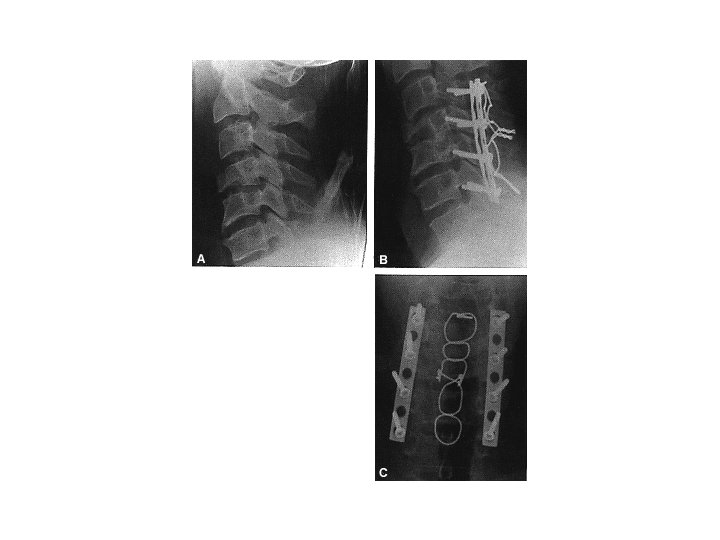
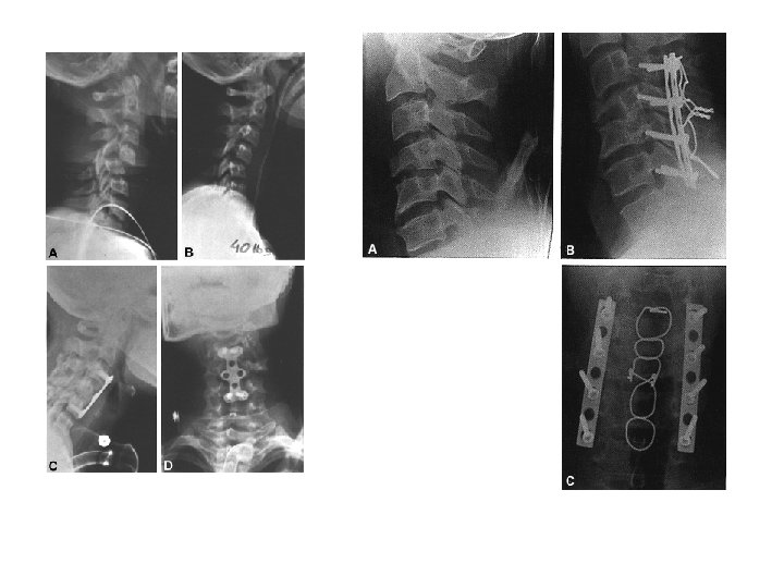
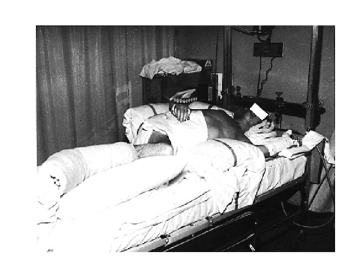
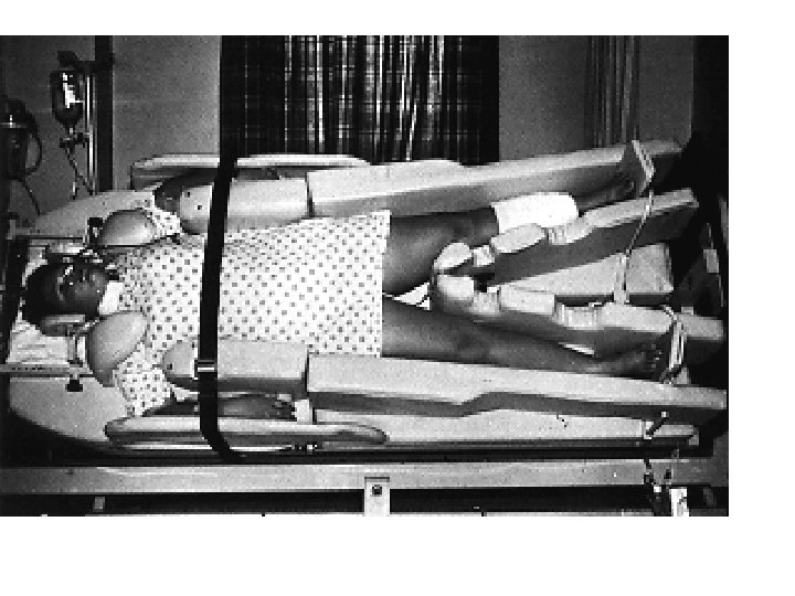
Terapi Prinsip • Konservative – bed rest 6 mg – 6 mg brace – 6 mg rehabiltation • Operatif – Screw and Plate dll • Reposisi • Immobilisasi – Eksternal – Internal
Lesi Sumsum tulang Belakang (SCI) 1. Paralysiss 1. Tetraplegia ( tingkat servical ) 2. Paraplegia ( thoracal – lumbal ) 2. 3. 4. 5. Kerusakan sensorik Inkontinensia Hilangnya respon vasomotor Fungsi Seksual
Komplikasi • • Gangguan pernafasan ( otot) Kontaktur dan Nyeri Decubitis Infeksi saluran kencing Osifikasi sendi Immobilitas Ketergantungan Depresi
Cauda Equina • Below the level of the conus medullaris (L 1– L 2 disk), the spinal canal is filled with the cauda equina (the motor and sensory roots of the lumbosacral myelomeres). • They exit caudally through their respective foramina. These roots are less likely to be injured in that they have more room in the canal and are not tethered to the same degree as the spinal cord. Furthermore, the motor nerve root is composed of lower motor neuron axons, which are more resilient to trauma than the brain and spinal cord.
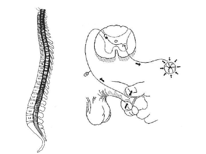
Reflex Arc • The reflex arc (eg, bulbocavernosus) is a simple sensorimotor pathway that can function without using either ascending or descending white -matter, long-tract axons (Fig. 23 -2). If the level of the reflex arc is both physiologically and anatomically intact, then the reflex will function despite dysfunction of the cephalad spinal cord.
Spinal Shock • With respect to spinal cord injury, spinal shock is defined as a spinal cord nervous tissue dysfunction based on physiologic rather than structural disruption. • Spinal shock has resolved when the reflex arcs below the level of the injury begin to function again (eg, bulbocavernosus reflex). • It generally lasts several hours to days.
Neurogenic Shock • Neurogenic shock is defined as vascular hypotension with bradycardia as a result of spinal cord injury. • The first few minutes after spinal cord injury are associated with hypertension and tachycardia, with a subsequent drop in pressure and pulse rate. • Neurogenic shock results from disruption of sympathetic outflow (T 1–L 2) and unopposed vagal tone (parasympathetic nervous system).
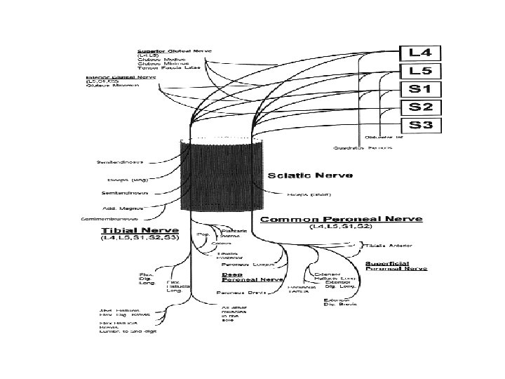
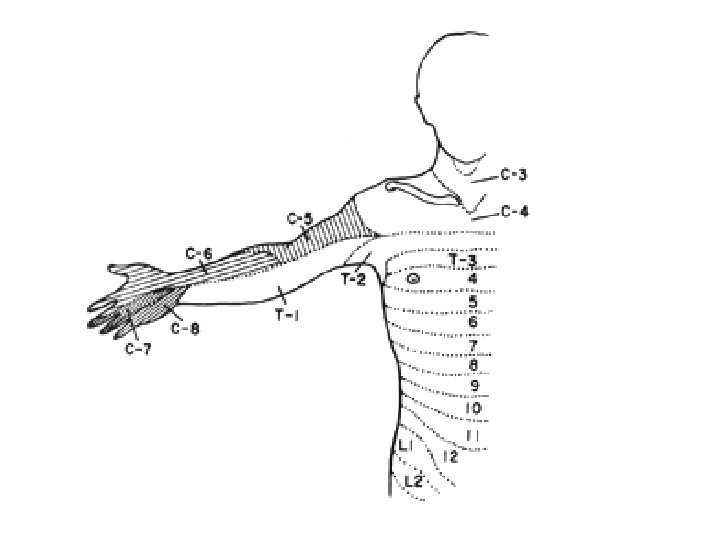
Sensory Examination The neurologic sensory examination should be done with a pinwheel and very light pressure. The patient is asked to identify sharp and dull discrimination of the face and then the cervical nerves are examined serially. • examining the back of the scalp (C 2); • the anterior aspect of the neck (C 3); • laterally and anteriorly over the clavicles down to the second rib space (C 4); • the lateral deltoid area of the arm (C 5); • the radial aspect of the thumb, index, and middle finger (C 6); • the ring and small fingers (C 7); • the ulnar border of the hand forearm (C 8); • the medial side of the upper arm (T 1); • and the anterior chest wall above the nipple line (T 2).
Motor Examination • The diagnosis of a complete or incomplete spinal cord injury syndrome is also documented. A careful motor function test is then carried out by evaluating sequential nerve root levels. Survival of persons with cervical spine injuries resulting in quadriplegia proximal to the C 4 level is rare, owing to the sudden paralysis of all respiratory muscles, which usually results in death at the scene of the accident. Modern resuscitation techniques, if applied rapidly, with mechanical ventilation consisting of mouth-to -mouth resuscitation or an Ambu bag can maintain ventilation, and therefore keep the patient alive until endotracheal intubation and mechanical respirator assistance are available.
• The number of surviving respiratory-dependent quadriplegics is growing, due to these rapid resuscitation techniques. If the patient is found breathing with diaphragm motion, function is present at least down to the C 4 nerve root; the patient will also have active control of the trapezius and sternocleidomastoid muscles. Voluntary control of the deltoid and biceps documents an intact C 5 level. Voluntary contraction of the extensor carpi radialis longus or brevis documents function of the C 6 level. Voluntary contractions of the pronator teres, flexor carpi radialis, and triceps or finger extensors document function at the C 7 level. Voluntary flexor digitorum sublimis or profundus control documents function at the C 8 level, and intrinsic function documents an intact T 1 level.
• After documentation of the lowest functioning muscles, and thus establishment of a functional neurologic level, the rest of the body must be examined for any evidence of voluntary muscle function indicating sparing of the corticospinal tract at the level of the injury. Specifically, the toe flexors, toe extensors, and rectal sphincter muscles must be tested. As in the case of sacral sensory sparing, sacral motor sparing (consisting of voluntary control of the sphincter or toe flexor muscles) may be the only evidence of an incomplete motor quadriplegia.
• When examining for toe flexor control, one should not touch the toes but should ask the patient to move the toes of either foot. Confusion with the flexor withdrawal reflex, which is commonly present after complete physiologic cervical transection, should be avoided. The patient must be able voluntarily to contract the toes of one foot, individually and independently, before he or she can be considered to have true voluntary control.
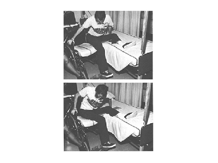
Physical Therapy • The physical therapist assesses the patient's sensory and motor power and evaluates him or her for spasticity. • Realistic goals of bed-to-wheelchair transfer activities, wheelchair propulsion, and ambulation for the incompletely injured patient are designated by the physical therapist. • The specific individualized wheelchair prescription for the appropriate level of disability is of primary importance.
• Lower extremity bracing prescriptions for the incompletely injured patient should be as small and light as possible. • The patient with a functional level only down to C 5 will have function of the deltoid and biceps and no wrist extensors. He or she is not capable of performing independent transfer, wheelchair propulsion, or dressing and will always require assistance in these activities.
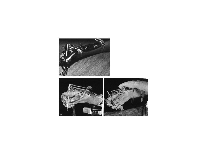
• The patient with the complete cord lesion and functioning at the C 6 level will have voluntary control of the wrist extensors and pectoral muscles. He or she can be taught independent transfer from bed-to-wheelchair and independent propulsion of a wheelchair with a plasticized hand rim or quad pegs on the hand rims and can be taught to dress himself or herself. • C 7 and C 8 functional level quadriplegics have more motor power in the wrist and hand muscles and are more likely to be independent with dressing, transfer, wheel chair propulsion, and automobile driving activities. In some instances, improvement of upper extremity function can be provided with tendon transfers.