INTRODUCTION TO NEUROANATOMY Kaan Ycel M D Ph


 Telencephalon Diencephalon 2. Midbrain (Mesencephalon) 3. Hindbrain")


. §")


 marrow Medulla spinalis –")


 12 pairs of cranial nerves 31 pairs of spinal nerves")
 in the skull. Entirely afferent")
 fibers SKELETAL MUSCLES CORTICOSPINAL TRACT")
 fibers Muscles of Pharynx Larynx Soft palate Facial")
 fibers Heart muscle (cardiomotor) Smooth muscles in the")
 fibers information from the skin, muscle, tendon and")
 EPICRITIC (Discriminative) “two-point discrimination")

 carry the visceral sensory information. Located in the blood vessels, walls of")


: tough, thick external fibrous layer Arachnoid mater (arachnoid): thin intermediate")
![CSF [Cerebrospinal fluid] Lateral ventricles Third ventricle Fourth ventricle](https://slidetodoc.com/presentation_image/2b24d9a0c1978e59844c874bc7ad8e28/image-52.jpg "CSF [Cerebrospinal fluid] Lateral ventricles Third ventricle Fourth ventricle")


- Slides: 70
INTRODUCTION TO NEUROANATOMY Kaan Yücel M. D. , Ph. D. 6. April. 2015 Monday
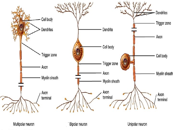
Neurite ONE Axon Dendirites NEUROGLIA
1 axon and a lot of dendirites Most neurons of the brain and spinal cord 1 neurite from each end @ retina, cochlear, vestibular ganglia, olfactory bulb 1 short neurite and two branches @ dorsal root ganglia 1 neurite; later branching @ ganglia of autonomic nervous system
PSEUDO-UNIPOLAR NEURONS FAKE
PSEUDO-UNIPOLAR NEURONS FAKE
NERVE FIBRE Dendirite or axon Bundles of nerve fibers Tract @ CNS Peripheral nerve @ PNS
NON-MYELINATED NERVE FIBERS Smaller axons of the central nervous system Postganglionic axons of the autonomic part of the nervous system Some fine sensory axons associated with the reception of pain
NEUROGLIA- GLIA
The nervous system can be separated into parts based on structure and on function: Structurally 1) Central nervous system (CNS) 2) Peripheral nervous system (PNS Functionally 1) Somatic nervous system 2) Visceral nervous system
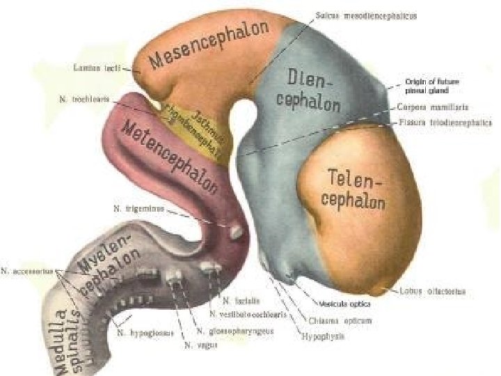
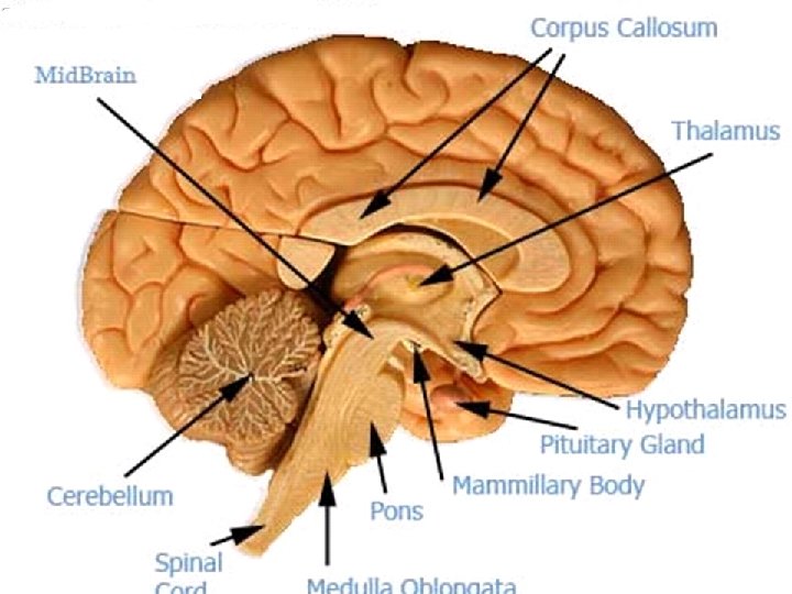
Brain in the cranium 1. Forebrain (Prosencephalon) Telencephalon Diencephalon 2. Midbrain (Mesencephalon) 3. Hindbrain (Rhombencephalon) Metencephalon (Pons & Cerebellum) Myelencephalon (Medulla oblongata-Medulla; Bulbus; Omurilik soğanı)
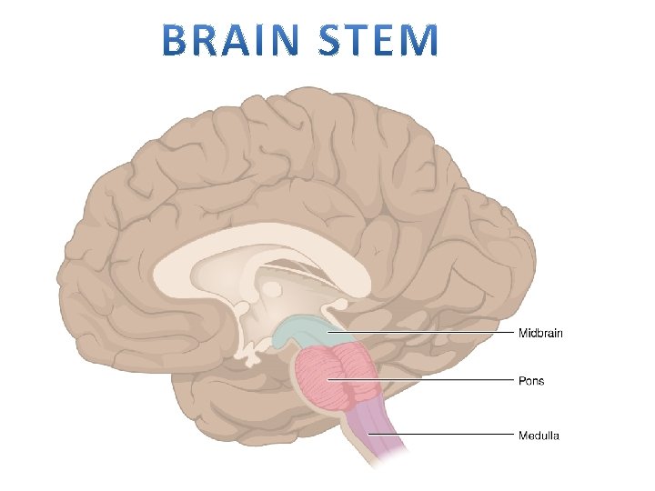
BRAIN STEM MIDBRAIN PONS MEDULLA OBLONGATA
Nuclei of 12 cranial nerves 10 of them in the brainstem midbrain Of the IV & III Of the other 4 pons VIII, VI, V the last 4 medulla Of XII, X, IX
SPINAL CORD IN THE VERTEBRAL COLUMN
Telencephalon Tele Far, end At a distance
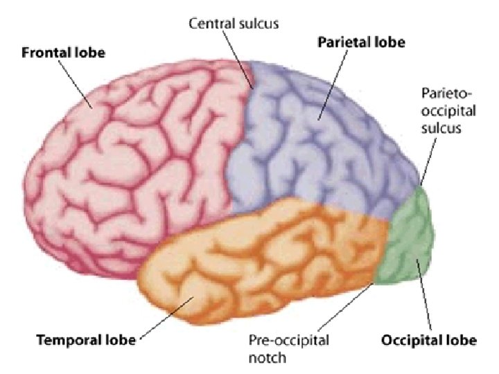
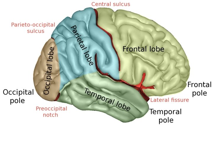
Gyrus & Sulcus & Fissure Greek gyros "ring, circle SULCI-depressions. DIVIDE THE BRAIN INTO GYRI -elevations. LOBES are MADE UP BY GYRI
BASAL GANGLIA EXTRAPYRAMIDAL SYSTEM
Diencephalon Epithalamus Subthalamus Basal ganglia structure
§ Important station that receives the main sensory tracts (except the olfactory pathway). § A station where much of the information is integrated and relayed to the cerebral cortex and many other subcortical regions. § A key role in the integration of visceral and somatic functions.
Adhesio interthalamica Septum pellicidum Pineal gland Anterior commissure Posterior commissure Corpora quadrigemina AC Thalamus Optic chiasm Hypothalamus Mamillary body Cerebral aqueduct Pituitary gland
Mesencephalon Midbrain
Hindbrain Rhombencephalon
Metencephalon meta, after, beyond, over Cerebellum Pons
Myelencephalon Medulla oblongata Medulla Bulbus Omurilik soğanı Myel—Spinal cord, (bone) marrow Medulla spinalis – Spinal cord
SUBCORTICAL NUCLEI GANGLIA
NUCLEI NUCLEUS v. s. GANGLIA GANGLION
Peripheral Nervous System (PNS) 12 pairs of cranial nerves 31 pairs of spinal nerves Associated ganglia Nerve fibers of the autonomic system
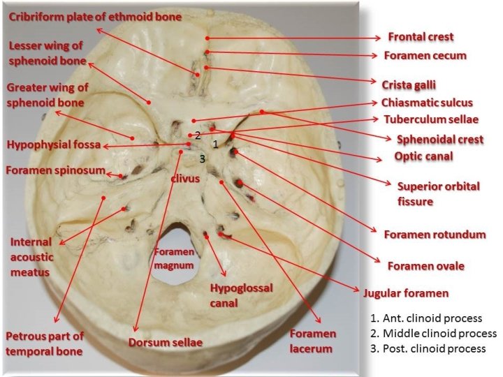
The cranial nerves pass through openings (foramine or fissures) in the skull. Entirely afferent fibers CNI; olfactory, CN II optic, and CN VIII; vestibulocochlear Entirely efferent fibers CN III; oculomotor, CN IV; trochlear, CN VI; abducent, CN XI; accessory, and CN XII; hypoglossal Both afferent and efferent fibers CN V; trigeminal, CN VII; facial, CN IX; glossopharyngeal, and CN X; vagus)
EFFERENT FIBERS General somatic efferent (GSE) fibers SKELETAL MUSCLES CORTICOSPINAL TRACT
EFFERENT FIBERS Special visceral efferent (SVE) fibers Muscles of Pharynx Larynx Soft palate Facial muscles Mastication Muscles in the middle ear
EFFERENT FIBERS General visceral efferent (GVE) fibers Heart muscle (cardiomotor) Smooth muscles in the organs (visceromotor) Smooth muscles in the walls of vessels (vasomotor) Smooth muscles in the roots of the hair follicles (pilomotor) Glands in the digestive system (secretomotor)
AFFERENT FIBERS General somatic afferent (GSA) fibers information from the skin, muscle, tendon and joints touch, pressure, pain, temperature and proprioception General visceral afferent (GVA) fibers information originates from the organs, glands and membrane touch, pressure, pain, temperature and proprioception Special visceral afferent (SVA) fibers gustatory (taste) and olfactory (smell) sensations
SENSORY INFORMATION SUPERFICIAL SENSATIONS PROTOPATHIC ( Touch, Pressure, Temperature, Pain) EPICRITIC (Discriminative) “two-point discrimination discriminative (fine) touch defines the type, intensity, localization of the touched site. Crude touch light touch on the skin. shirt touching the skin lightly. No discriminative feature in crude (nondiscriminative) touch.
DEEP SENSATIONS Proprioceptive Vibration Deep muscle pain Close your eyes Somebody moves your finger, let’s say abducts your thumb! You will know about the new position of your thumb! Proprioception via Proprioceptive receptors
Visceroreceptors (interoceptors) carry the visceral sensory information. Located in the blood vessels, walls of the organs, and in glands as free nerve endings. Carry sensory information such as pain, pressure, chemical changes, thirst, stetching, etc.
The sensations are carried by somatic afferent fibers in the spinal and cranial nerves from the abundant receptors in the skin. These receptors are simply the dendirites of the afferent neurons.
RECEP-TORS An individual receives impressions from the outside world and from within the body by special sensory nerve endings or receptors. Mechanoreceptors Thermoreceptors Nociceptors Photoreceptors Chemoreceptors Osmoreceptors
PERIPHERAL NERVE PLEXUSES
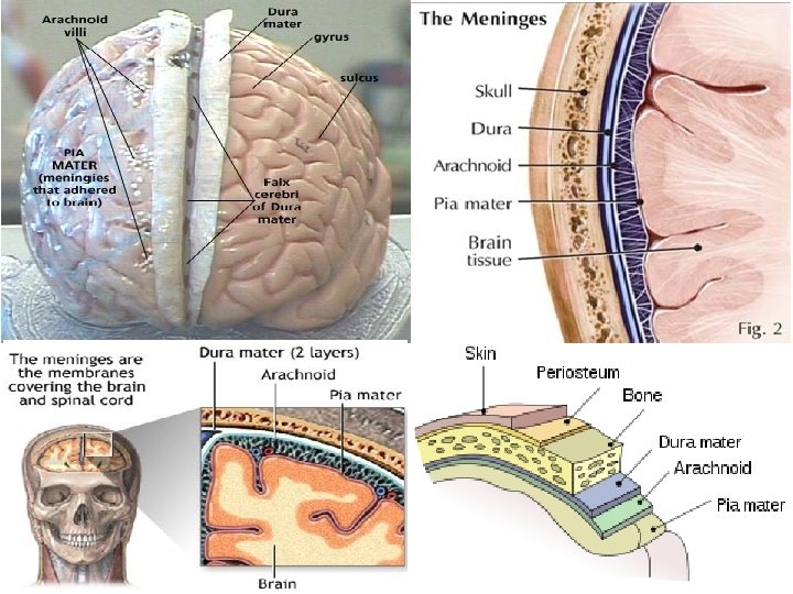
Meninges Dura mater (dura): tough, thick external fibrous layer Arachnoid mater (arachnoid): thin intermediate layer Pia mater (pia): delicate internal vasculated layer
CSF [Cerebrospinal fluid] Lateral ventricles Third ventricle Fourth ventricle
What is inside the skull? Meninges CSF Neuron Cortex Neuroglia Grey matter Axons White matter Tracts Hemispheres Lobes Sulcus & gyrus Arteries & Veins
SPINAL CORD
part of the CNS in the superior 2/3 of the vertebral canal It is roughly cylindrical in shape, and is circular to oval in cross-section. Internally, the cord has a small central canal surrounded by gray and white matter. The spinal cord extends from foramen magnum to lower border of L 1 (first lumbar vertebra). .
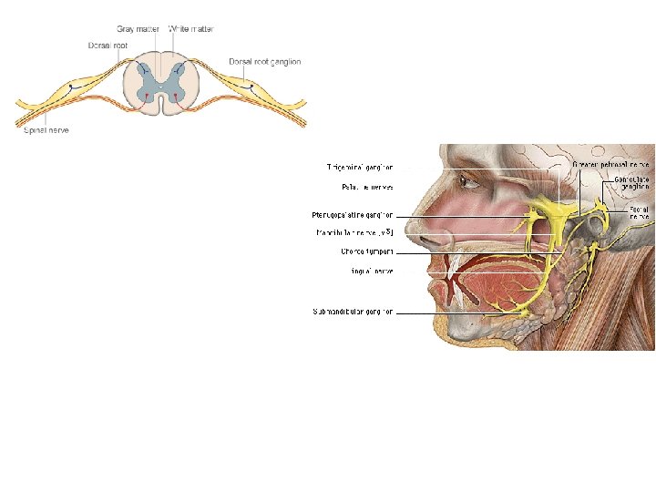
It gives rise to 31 pairs of spinal nerves (cervical, thoracic, lumbar, sacral and coccygeal). All are mixed nerves. Spinal nerves arise as rootlets then combine to form dorsal and ventral roots.
Dorsal and ventral roots merge laterally and form the spinal nerve. Dorsal root is related to the sensory information. Anterior (ventral) root is related to the motor information.
@ cervical region gives origin to the brachial plexus @ lower thoracic and lumbar regions gives origin to the lumbosacral plexus spinal cord is fusiformly enlarged; cervical and lumbar enlargements.
Inferiorly, the spinal cord tapers off into the conus medullaris, from the apex of which a prolongation of the pia mater, the filum terminale, descends to be attached to the posterior surface of the coccyx. The cord possesses a deep longitudinal fissure called the anterior median fissure in the midline anteriorly and a shallow furrow called the posterior median sulcus on the posterior surface.
Gray Matter On cross section, the gray matter is seen as an H-shaped pillar with anterior and posterior gray columns, or horns, united by a thin gray commissure containing the small central canal. A small lateral gray column or horn is present in the thoracic and upper lumbar segments of the cord. As in other regions of the central nervous system, the gray matter of the spinal cord consists of a mixture of nerve cells and their processes, neuroglia, and blood vessels. The nerve cells are multipolar, and the neuroglia forms an intricate network around the nerve cell bodies and their neurites.
White Matter divided into anterior, lateral, and posterior white columns or funiculi. The anterior column on each side lies between the midline and the point of emergence of the anterior nerve roots; the lateral column lies between the emergence of the anterior nerve roots and the entry of the posterior nerve roots; the posterior column lies between the entry of the posterior nerve roots and the midline.
Although some nerve tracts are concentrated in certain areas of the white matter, it is now generally accepted that considerable overlap is present. For purposes of description, the spinal tracts are divided into ascending, descending, and intersegmental tracts.
On entering the spinal cord, the sensory nerve fibers of different sizes and functions are sorted out and segregated into nerve bundles or tracts in the white matter. Some of the nerve fibers serve to link different segments of the spinal cord, while others ascend from the spinal cord to higher centers and thus connect the spinal cord with the brain. It is the bundles of the ascending fibers that are referred to as the ascending tracts.
The ascending tracts conduct afferent information, which may or may not reach consciousness. The information may be divided into two main groups: (1) exteroceptive information, which originates from outside the body, such as pain, temperature, and touch (2) proprioceptive information, which originates from inside the body, for example, from muscles and joints.
General information from the peripheral sensory endings is conducted through the nervous system by a series of neurons. Ascending pathway 3 neurons (simple one) First-order neuron: cell body @ dorsal (posterior) root gangliaon of spinal nerve Peripheral projection connects with sensory receptor ending. Central projection enters the spinal cord via posterior root & synapse on the second-order neuron.
The second-order neuron gives rise to an axon that decussates (crosses to the opposite side) and ascends to a higher level of the central nervous system, where it synapses with the third-order neuron. The third-order neuron is usually in the thalamus and gives rise to a projection fiber that passes to a sensory region of the cerebral cortex.
This three-neuron chain is the most common arrangement, but some afferent pathways use more or fewer neurons. Many of the neurons in the ascending pathways branch and give a major input into the reticular formation, which, in turn, activates the cerebral cortex, maintaining wakefulness. Other branches pass to motor neurons and participate in reflex muscular activity.