Otosclerosis Dr Vishal Sharma Definition Hereditary disease of











- Slides: 76
Otosclerosis Dr. Vishal Sharma
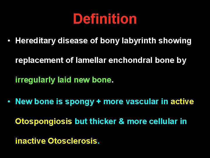
Definition • Hereditary disease of bony labyrinth showing replacement of lamellar enchondral bone by irregularly laid new bone. • New bone is spongy + more vascular in active Otospongiosis but thicker & more cellular in inactive Otosclerosis.
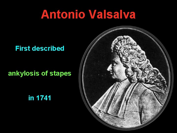
Antonio Valsalva First described ankylosis of stapes in 1741
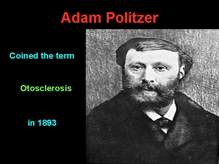
Adam Politzer Coined the term Otosclerosis in 1893
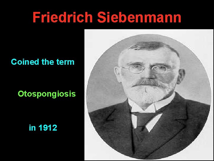
Friedrich Siebenmann Coined the term Otospongiosis in 1912
Epidemiology • Exact etiology is unknown (? Viral) • Autosomal dominant: variable penetrance • Race: common in white races & Indians • Female : Male = 2 : 1 • Age: Common in 20 - 40 years • Hormonal influence: es in pregnancy, menopause, stress (trauma, surgery)
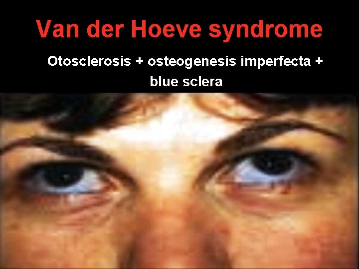
Van der Hoeve syndrome Otosclerosis + osteogenesis imperfecta + blue sclera
Types of Otosclerosis A. Stapedial B. Cochlear: otosclerotic focus is seen over Round window Promontory C. Stapedial + cochlear: mixed type D. Malignant: rapidly progressing cochlear lesion with severe sensori-neural deafness.
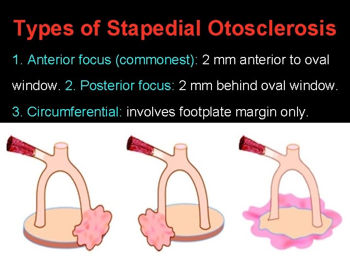
Types of Stapedial Otosclerosis 1. Anterior focus (commonest): 2 mm anterior to oval window. 2. Posterior focus: 2 mm behind oval window. 3. Circumferential: involves footplate margin only.
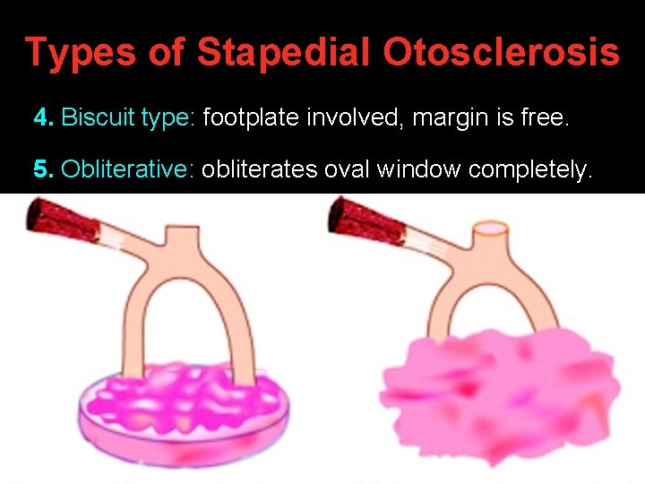
Types of Stapedial Otosclerosis 4. Biscuit type: footplate involved, margin is free. 5. Obliterative: obliterates oval window completely.
Symptoms of Otosclerosis 1. Deafness: Bilateral, slowly progressive Conductive: stapedial otosclerosis Sensori-neural: cochlear otosclerosis Mixed: stapedial + cochlear otosclerosis 2. Soft, modulated, monotonous voice 3. Tinnitus & vertigo: in cochlear lesion
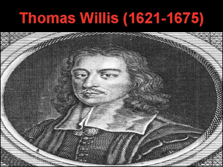
Symptoms of Otosclerosis 4. Paracusis Willisii: Pt has better hearing in noisy surroundings (people increase their voice intensity & pt’s speech discrimination becomes better).
Thomas Willis (1621 -1675)
Otoscopy Normal T. M. is seen in most cases. Pinkish colour over promontory seen in otospongiosis (2 - 10 % cases) Schwartze sign / Flamingo pink blush.
Tuning Fork Tests Rinne Weber A. B. C. Stapedial Negative (BC > AC) Lateralizes to Deaf ear Normal Cochlear Positive (AC > BC) Lateralizes to Better ear Decreased Mixed Negative (BC > AC) Lateralizes to Better ear Decreased
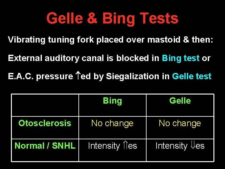
Gelle & Bing Tests Vibrating tuning fork placed over mastoid & then: External auditory canal is blocked in Bing test or E. A. C. pressure ed by Siegalization in Gelle test Bing Gelle Otosclerosis No change Normal / SNHL Intensity es
Pure Tone Audiometry • Low frequency conductive deafness • Carhart’s notch in bone conduction at 2 KHz
Carhart’s notch • Proposed theories 1. Stapes fixation disrupts normal ossicular resonance (2000 Hz) 2. Normal compression mode of bone conduction is disturbed because of relative perilymph immobility 3. Mechanical artefact • Carhart’s notch reverses with stapes surgery
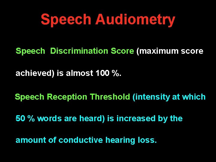
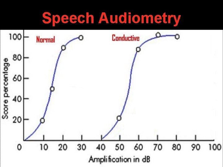
Speech Audiometry Speech Discrimination Score (maximum score achieved) is almost 100 %. Speech Reception Threshold (intensity at which 50 % words are heard) is increased by the amount of conductive hearing loss.
Speech Audiometry
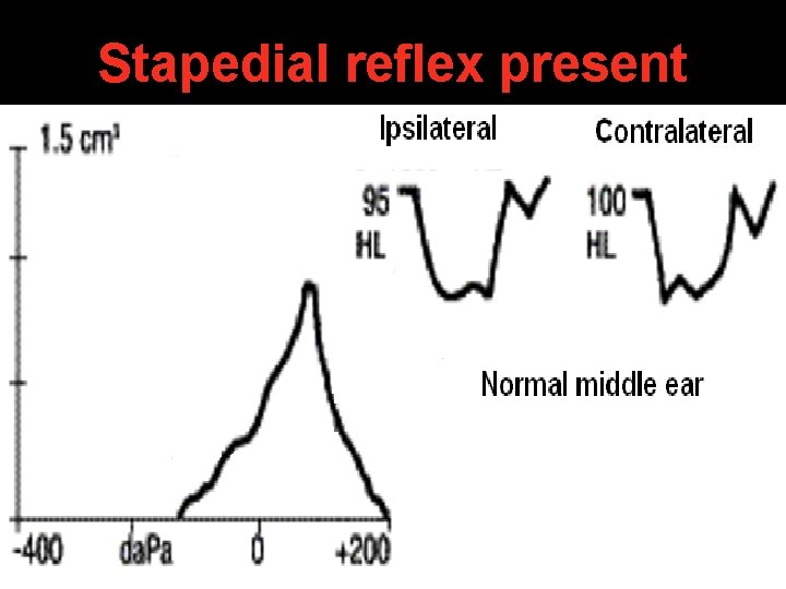
Impedance Audiometry • As curve seen in 40 % cases of otosclerosis. • Normal middle ear pressure + decreased middle ear compliance. • Others have A curve.
Stapedial reflex present
Stapedial reflex absent
C. T. scan temporal bone 200 coronal oblique cuts are taken
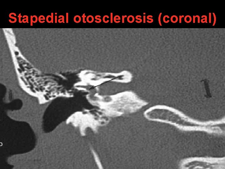
Stapedial otosclerosis (coronal)
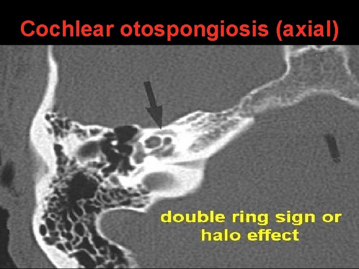
Cochlear otospongiosis (axial)
Differential Diagnosis • Otitis Media with Effusion: type B tympanogram • Adhesive Otitis Media: absence of T. M. mobility • Tympanosclerosis: white patch on T. M. • Ossicular discontinuity: type Ad tympanogram • Congenital ossicular chain fixation: tympanotomy • Malleus head fixation: tympanotomy
History of development of stapes surgery
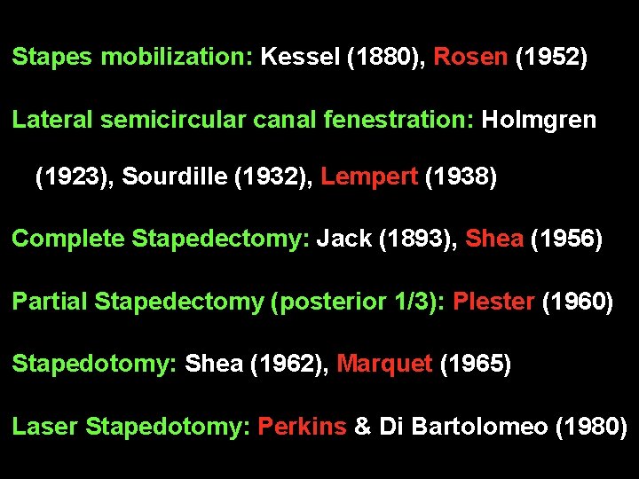
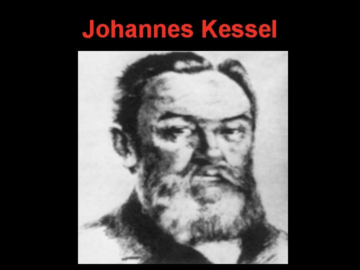
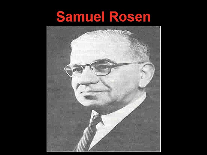
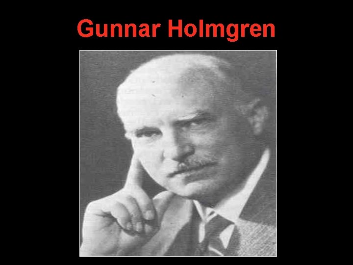
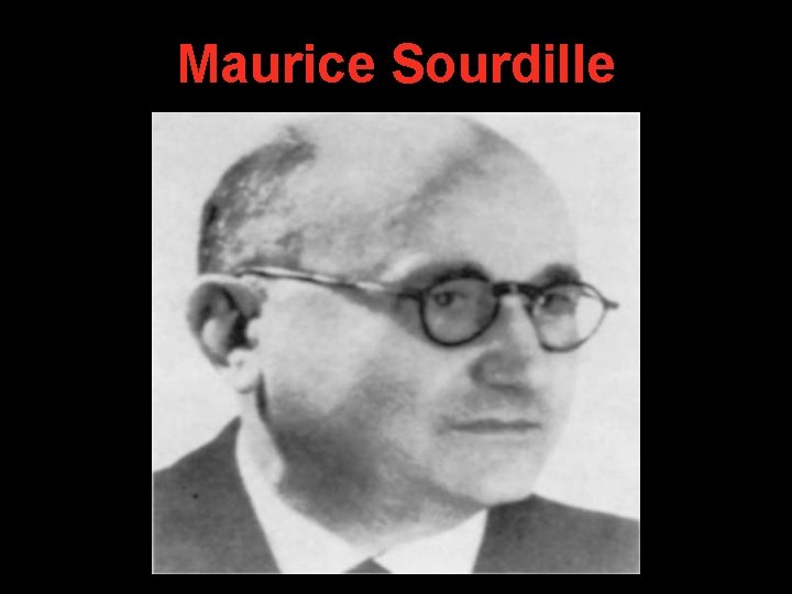
Stapes mobilization: Kessel (1880), Rosen (1952) Lateral semicircular canal fenestration: Holmgren (1923), Sourdille (1932), Lempert (1938) Complete Stapedectomy: Jack (1893), Shea (1956) Partial Stapedectomy (posterior 1/3): Plester (1960) Stapedotomy: Shea (1962), Marquet (1965) Laser Stapedotomy: Perkins & Di Bartolomeo (1980)
Johannes Kessel
Samuel Rosen
Gunnar Holmgren
Maurice Sourdille
Julius Lempert
John J. Shea Jr.
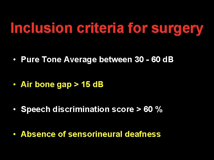
Inclusion criteria for surgery • Pure Tone Average between 30 - 60 d. B • Air bone gap > 15 d. B • Speech discrimination score > 60 % • Absence of sensorineural deafness
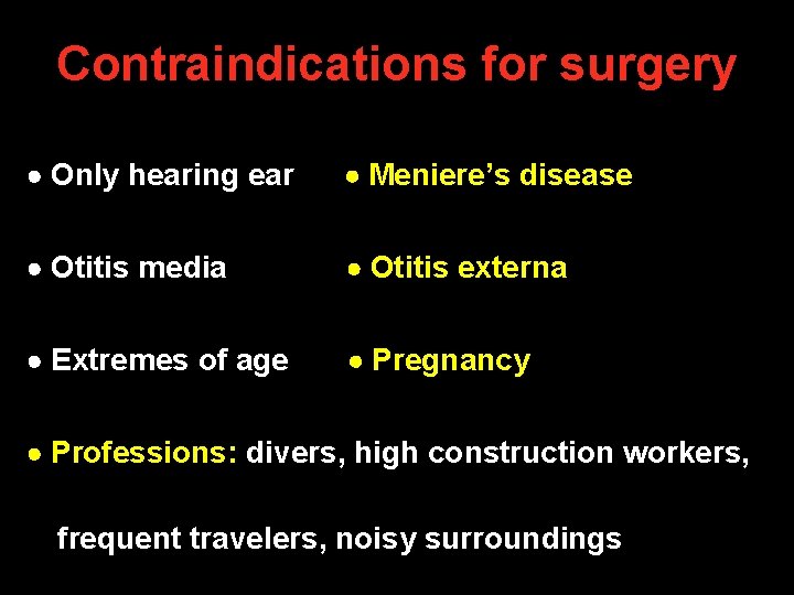
Contraindications for surgery Only hearing ear Meniere’s disease Otitis media Otitis externa Extremes of age Pregnancy Professions: divers, high construction workers, frequent travelers, noisy surroundings
Surgical steps for Stapedotomy
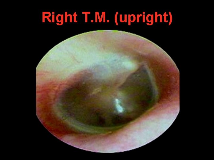
Right T. M. (upright)
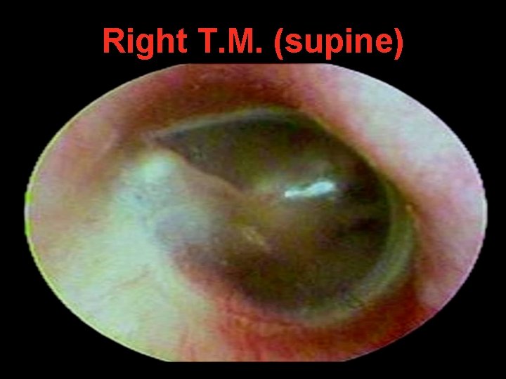
Right T. M. (supine)
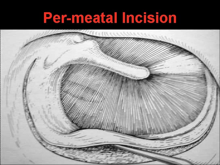
Per-meatal Incision
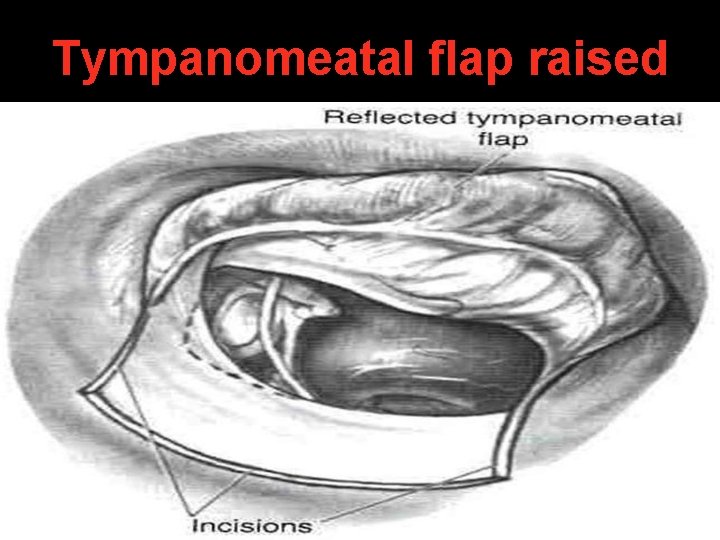
Tympanomeatal flap raised
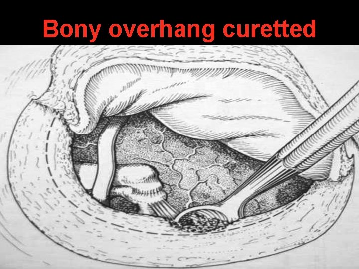
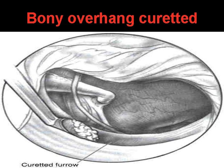
Bony overhang curetted
Bony overhang curetted
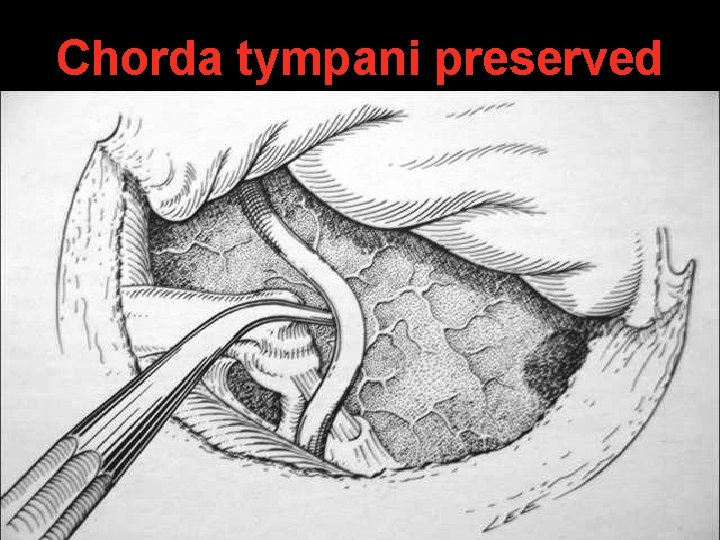
Chorda tympani preserved
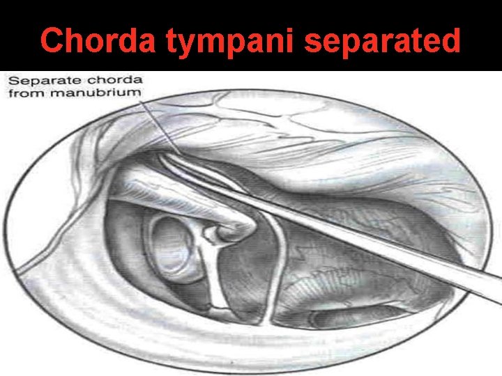
Chorda tympani separated
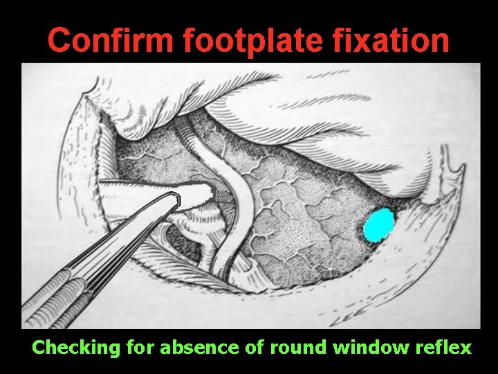
Confirm footplate fixation Checking for absence of round window reflex
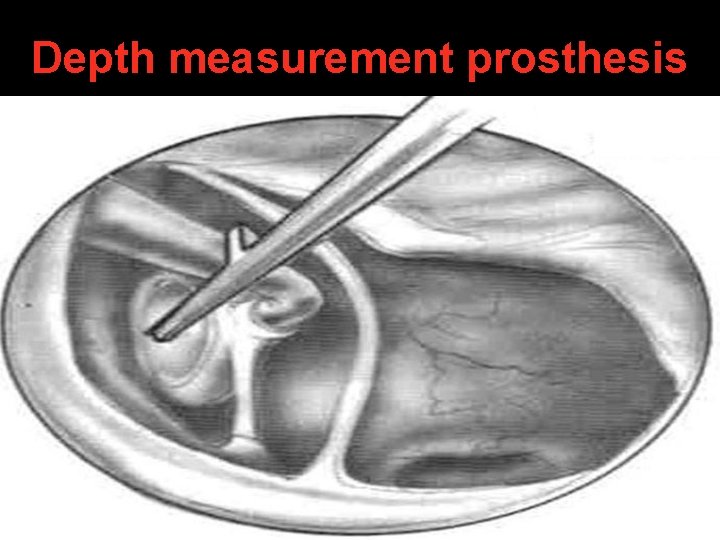
Depth measurement prosthesis
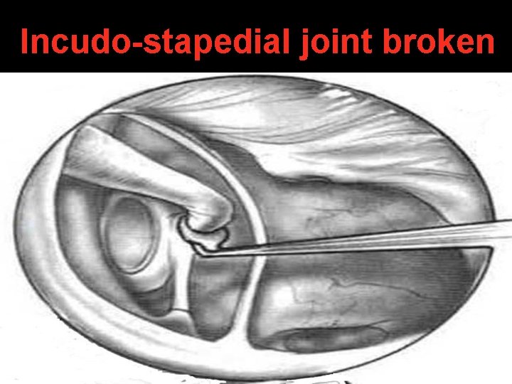
Incudo-stapedial joint broken
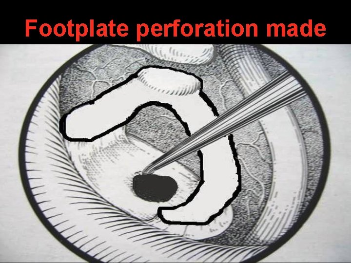
Footplate perforation made
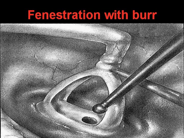
Fenestration with burr
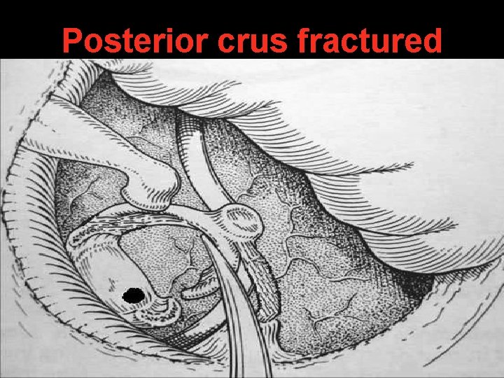
Posterior crus fractured
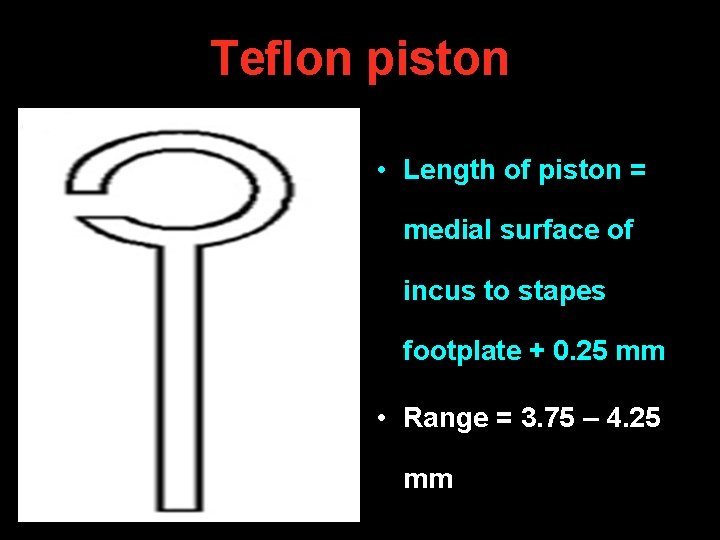
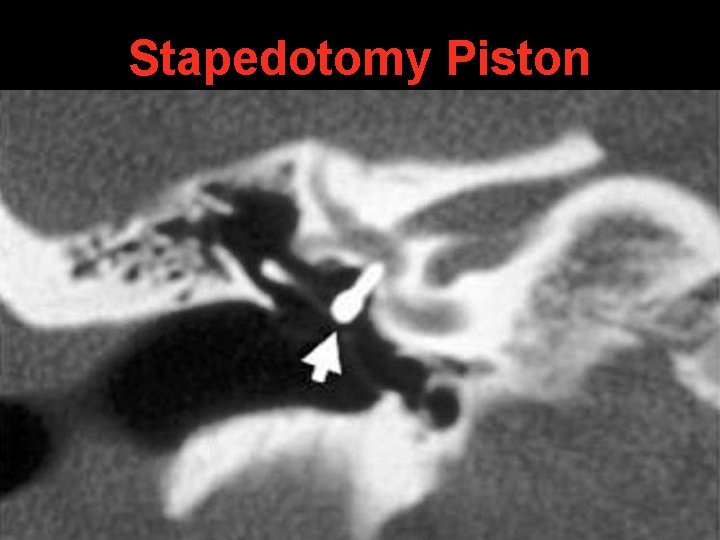
1 cm Teflon piston • Length of piston = medial surface of incus to stapes footplate + 0. 25 mm • Range = 3. 75 – 4. 25 mm
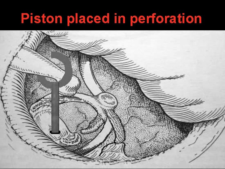
Piston placed in perforation
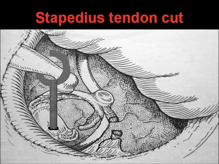
Stapedius tendon cut
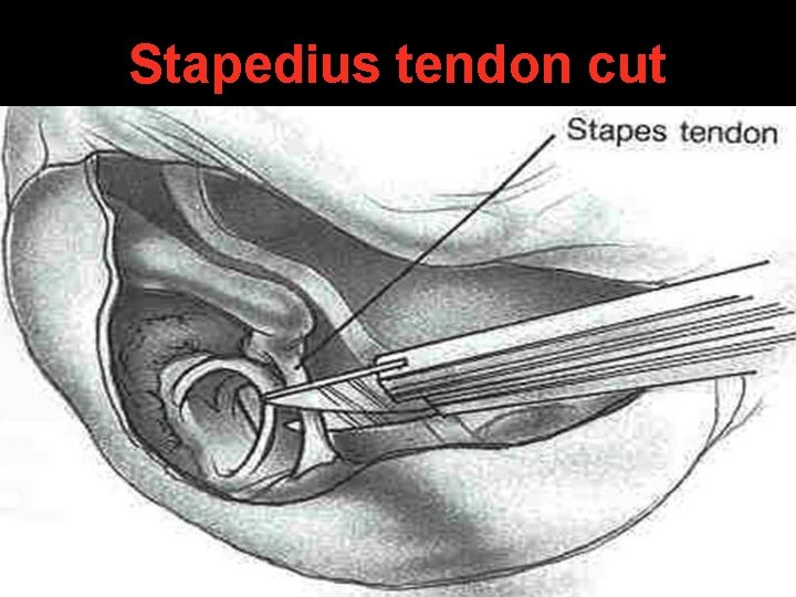
Stapedius tendon cut
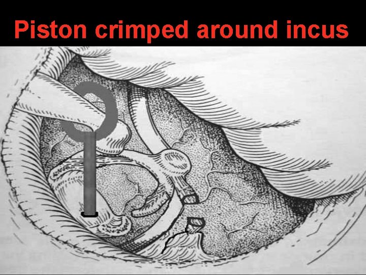
Piston crimped around incus
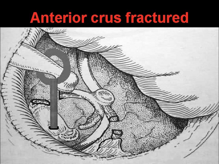
Anterior crus fractured
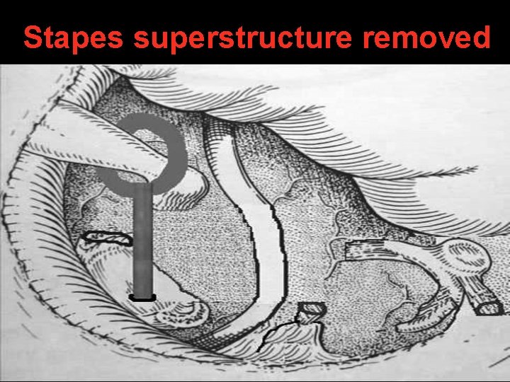
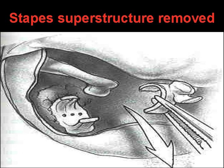
Stapes superstructure removed
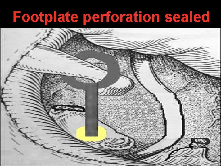
Footplate perforation sealed
Tympanomeatal flap put back
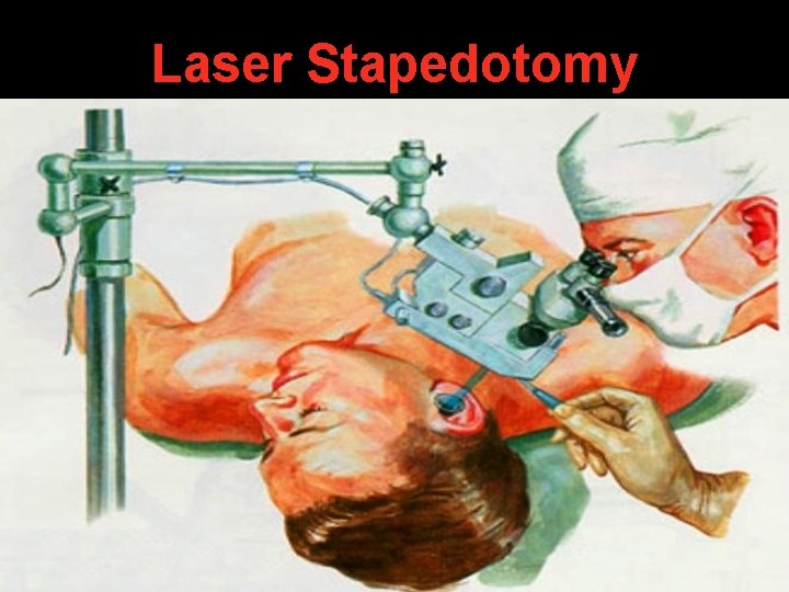
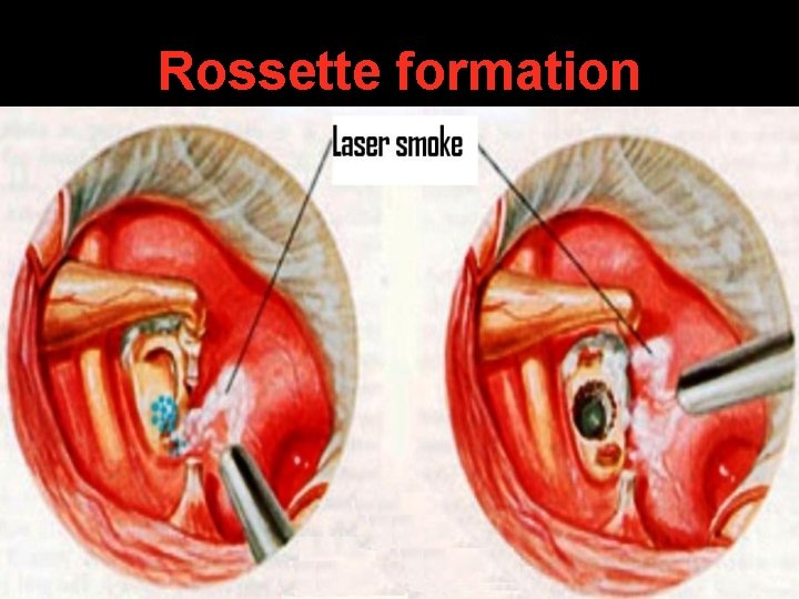
Laser Stapedotomy
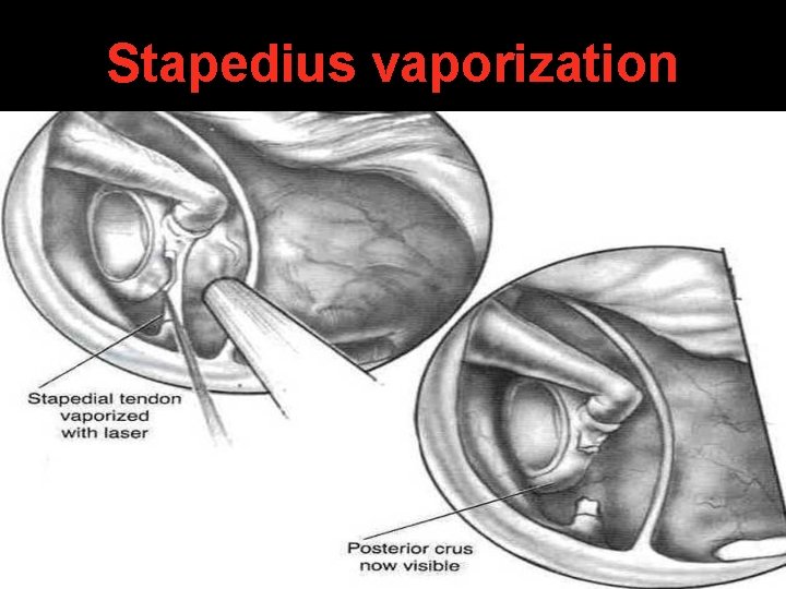
Stapedius vaporization
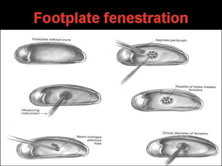
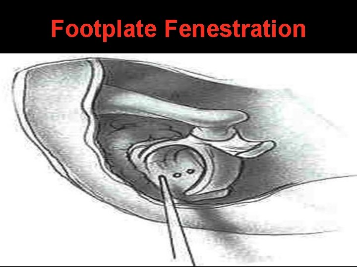
Footplate fenestration
Rossette formation
Stapedotomy Piston
Stapedectomy
Footplate Fenestration
Stapes superstructure removed
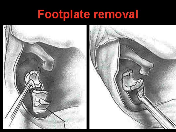
Footplate removal
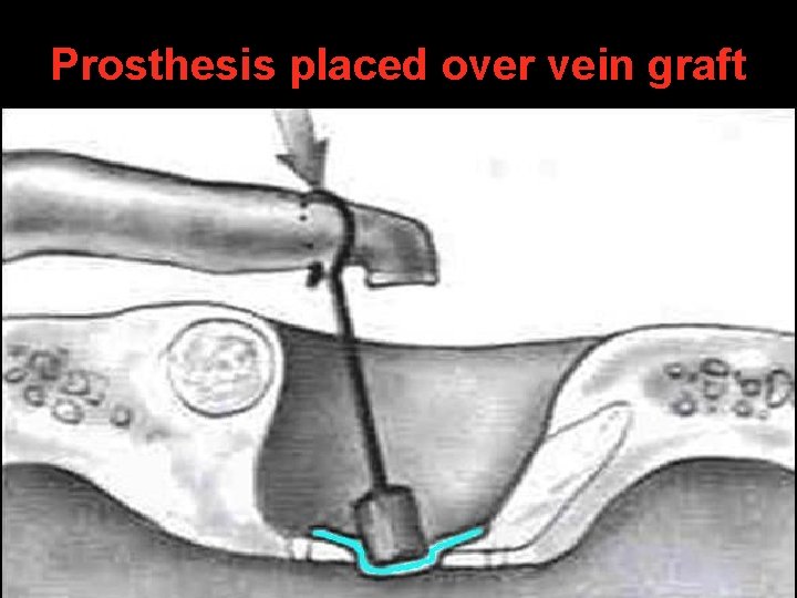
Prosthesis placed over vein graft
Complications of stapes surgery
Intra-operative Post-operative Floating footplate Otitis media Submerged footplate Oval window granuloma Dislocated incus Perilymph fistula Perforated TM Sensori-neural HL Damage to facial nerve Persistent air-bone gap or chorda tympani Vestibular dysfunction Persistent stapedial artery Delayed facial palsy in Perilymph flooding laser surgery
Sodium Fluoride • Given in cochlear otosclerosis & active focus of otospongiosis (Schwartze sign). • Acts by: a. Decreases bone resorption b. Increases bone formation c. Prevents enzymatic damage to cochlea • 20 mg orally, thrice daily for 3 - 6 months
Hearing Aid For patients who: • Are unfit for surgery • Refuse surgery
Thank You