Hereditary Hemolytic Anemia Youngshil Park M D Division
- Slides: 48
Hereditary Hemolytic Anemia Young-shil, Park, M. D. Division of Hematology-Oncology Department of Pediatrics Kyung Hee University Hospital at Gangdong
Introduction p Hemolysis n p the premature destruction of red blood cells (RBCs) Normal RBC n n survival time ; 110– 120 days (half-life, 55– 60 days) approximately 0. 85% of RBCs (the senescent ones) ; removed each day and replaced by the marrow to maintain the RBC count
p During hemolysis n n n RBC survival is shortened the RBC count falls erythropoietin is increased the stimulation of marrow activity results in heightened RBC production an increased percentage of reticulocytes in the blood
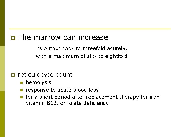
p The marrow can increase its output two- to threefold acutely, with a maximum of six- to eightfold p reticulocyte count n n n hemolysis response to acute blood loss for a short period after replacement therapy for iron, vitamin B 12, or folate deficiency
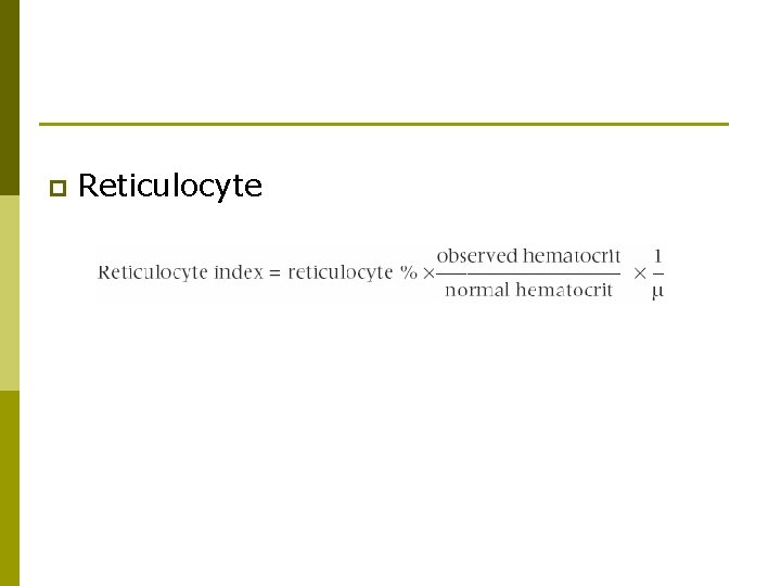
p Reticulocyte
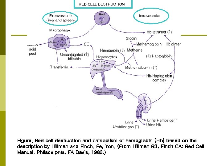
Figure. Red cell destruction and catabolism of hemoglobin (Hb) based on the description by Hillman and Finch. Fe, iron. (From Hillman RS, Finch CA: Red Cell Manual. Philadelphia, FA Davis, 1983. )
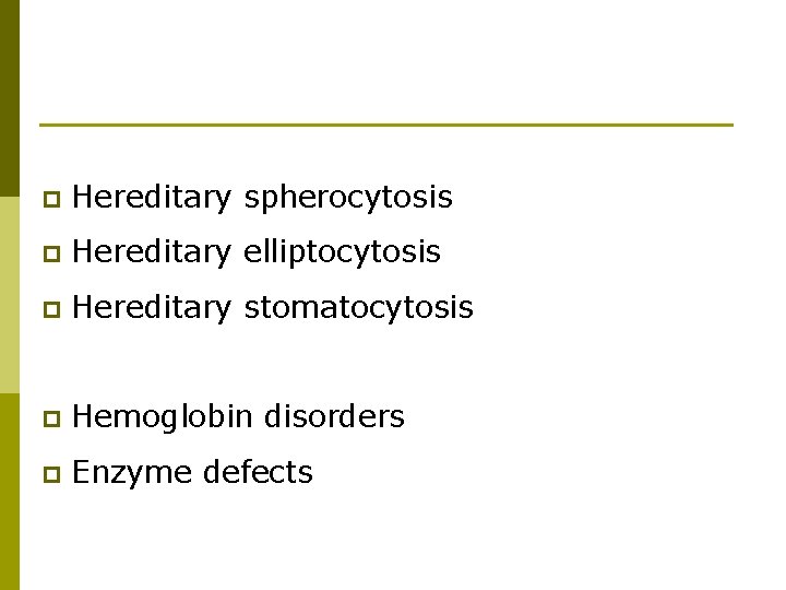
p Hereditary spherocytosis p Hereditary elliptocytosis p Hereditary stomatocytosis p Hemoglobin disorders p Enzyme defects
Hereditary spherocytosis
p p common cause of hemolysis and hemolytic anemia prevalence of approximately 1/5, 000 in people of Northern European descent most common inherited abnormality of the red blood cell (RBC) membrane. without anemia with minimal hemolysis or severe hemolytic anemia
Etiology p autosomal dominant less frequently, autosomal recessive p 25% of patients have no previous family history p
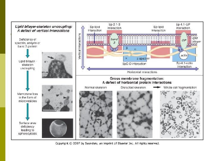
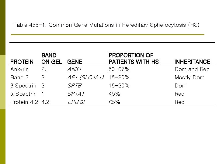
p most common molecular defects ; abnormalities of spectrin or ankyrin
Table 458 -1. Common Gene Mutations in Hereditary Spherocytosis (HS) BAND PROTEIN ON GEL Ankyrin 2. 1 Band 3 3 β Spectrin 2 α Spectrin 1 Protein 4. 2 PROPORTION OF GENE PATIENTS WITH HS ANK 1 50 -67% AE 1 (SLC 4 A 1) 15 -20% SPTB 15 -20% SPTA 1 <5% EPB 42 <5% INHERITANCE Dom and Rec Mostly Dom Rec
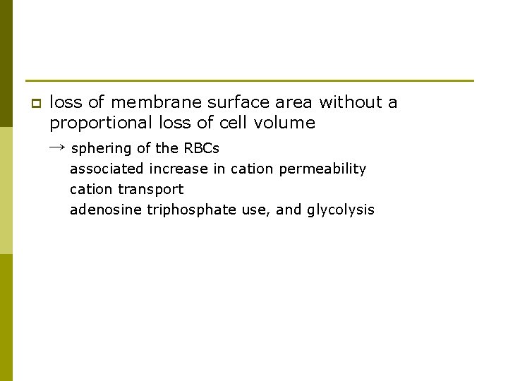
p loss of membrane surface area without a proportional loss of cell volume → sphering of the RBCs associated increase in cation permeability cation transport adenosine triphosphate use, and glycolysis
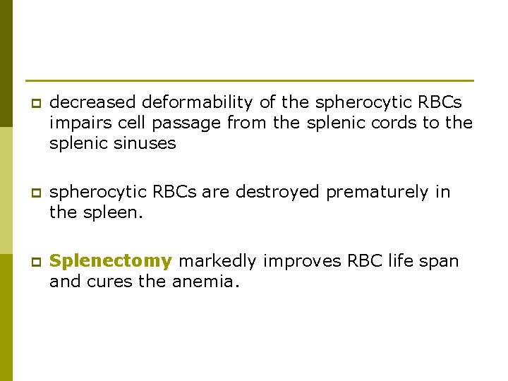
p decreased deformability of the spherocytic RBCs impairs cell passage from the splenic cords to the splenic sinuses p spherocytic RBCs are destroyed prematurely in the spleen. p Splenectomy markedly improves RBC life span and cures the anemia.
Clinical manifestations p in the newborn n p anemia and hyperbilirubinemia ⇒ phototherapy or exchange transfusions The severity in infants and children is variable
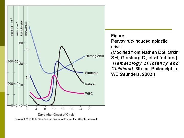
p p After infancy n splenomegaly n pigmentary (bilirubin) gallstones ; as early as age 4– 5 yr susceptible to aplastic crisis n n primarily as a result of parvovirus infection Such erythroid marrow failure ⇒ profound anemia (hematocrit <10%) high-output heart failure, hypoxia cardiovascular collapse, and death
Figure. Parvovirus-induced aplastic crisis. (Modified from Nathan DG, Orkin SH, Ginsburg D, et al [editors]: Hematology of Infancy and Childhood, 6 th ed. Philadelphia, WB Saunders, 2003. )
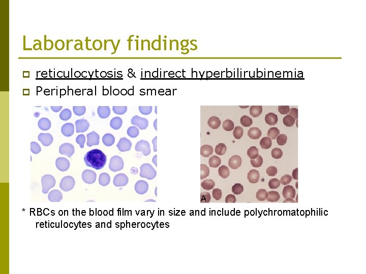
Laboratory findings p p reticulocytosis & indirect hyperbilirubinemia Peripheral blood smear * RBCs on the blood film vary in size and include polychromatophilic reticulocytes and spherocytes
p Erythroid hyperplasia is evident in the marrow aspirate or biopsy. p Marrow expansion ; on routine roentgenographic examination p decreased haptoglobin and the presence of gallstones on ultrasonography
p The diagnosis of hereditary spherocytosis usually is established n n n clinically from the blood film (many spherocytes and reticulocytes ) from the family history from splenomegaly
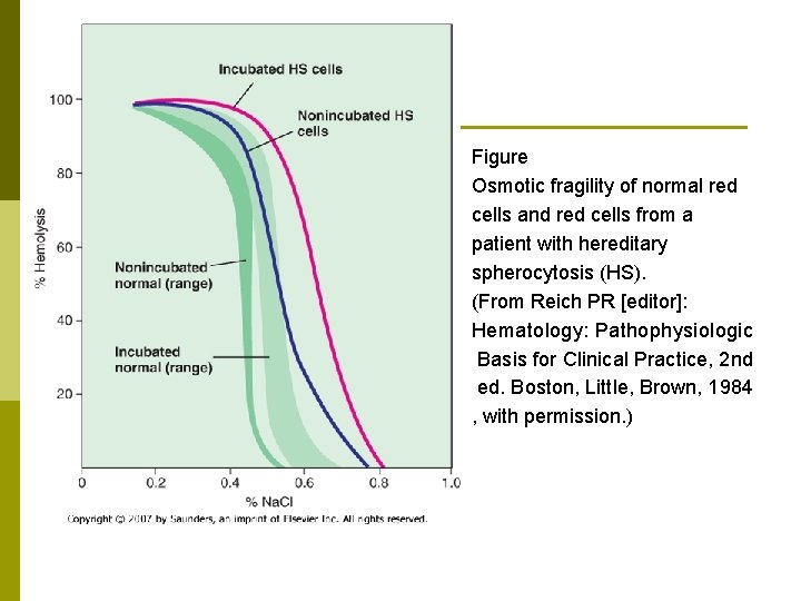
p confirmed with an osmotic fragility test p Other tests, such as the cryohemolysis test, osmotic gradient ektacytometry, and the eocin-5 -maleimide test p RBC membrane protein analysis using gel electrophoresis
Figure Osmotic fragility of normal red cells and red cells from a patient with hereditary spherocytosis (HS). (From Reich PR [editor]: Hematology: Pathophysiologic Basis for Clinical Practice, 2 nd ed. Boston, Little, Brown, 1984 , with permission. )
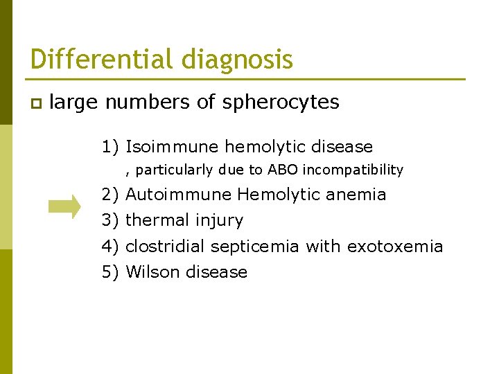
Differential diagnosis p large numbers of spherocytes 1) Isoimmune hemolytic disease , particularly due to ABO incompatibility 2) Autoimmune Hemolytic anemia 3) thermal injury 4) clostridial septicemia with exotoxemia 5) Wilson disease
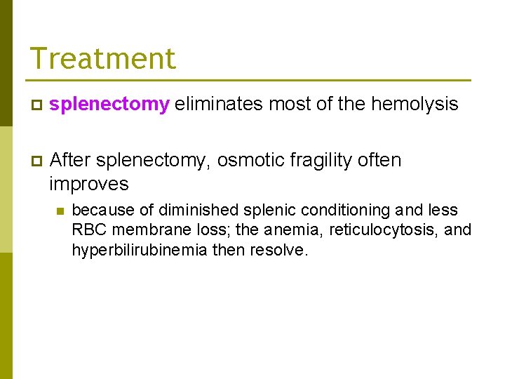
Treatment p splenectomy eliminates most of the hemolysis p After splenectomy, osmotic fragility often improves n because of diminished splenic conditioning and less RBC membrane loss; the anemia, reticulocytosis, and hyperbilirubinemia then resolve.
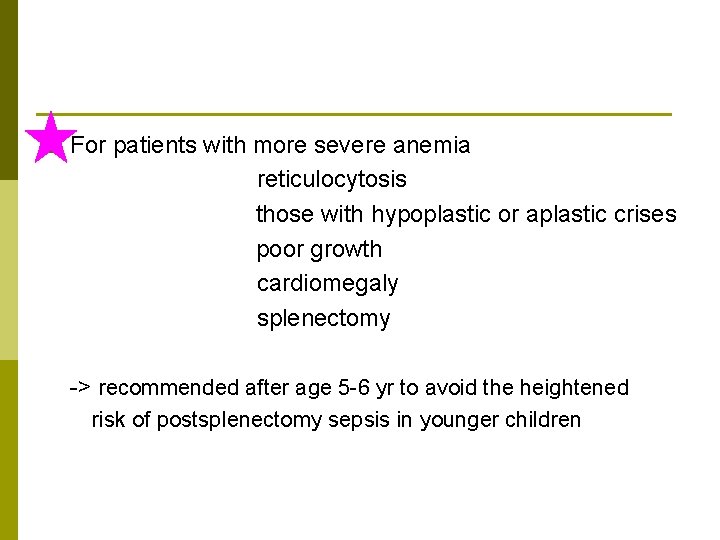
p For patients with more severe anemia reticulocytosis those with hypoplastic or aplastic crises poor growth cardiomegaly splenectomy -> recommended after age 5 -6 yr to avoid the heightened risk of postsplenectomy sepsis in younger children
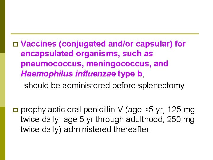
p Vaccines (conjugated and/or capsular) for encapsulated organisms, such as pneumococcus, meningococcus, and Haemophilus influenzae type b, should be administered before splenectomy p prophylactic oral penicillin V (age <5 yr, 125 mg twice daily; age 5 yr through adulthood, 250 mg twice daily) administered thereafter.
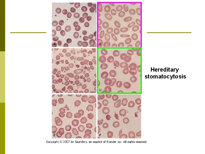
Hereditary Elliptocytosis p less common disorder than spherocytosis that also varies markedly in severity p The blood film is the most important test to establish hereditary elliptocytosis p Increased thermal instability p no treatment is necessary Splenectomy p
Hereditary stomatocytosis
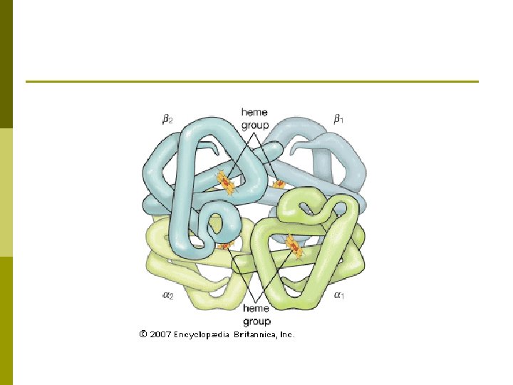
Hemoglobinopathies Hemoglobin ; tetramer consisting of 2 pairs of globin chains. p Abnormalities in these proteins are referred to as hemoglobinopathies. p
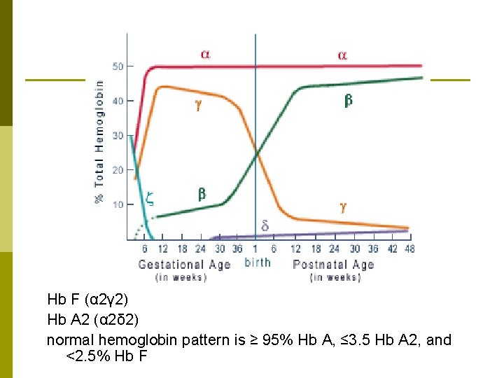
Hb F (α 2γ 2) Hb A 2 (α 2δ 2) normal hemoglobin pattern is ≥ 95% Hb A, ≤ 3. 5 Hb A 2, and <2. 5% Hb F
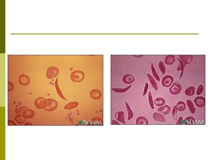
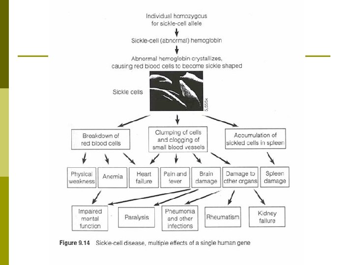
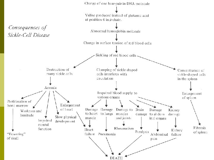
Sickle Cell Disease p Hemoglobin S (Hb S) n result of a single base pair change, thymine for adenine, at the 6 th codon of the β-globin gene In sickle cell anemia, Hb S ; commonly as high as 90% of the total hemoglobin. Hb SS p In sickle cell disease, Hb S is >50% of all hemoglobin. Hb AS p
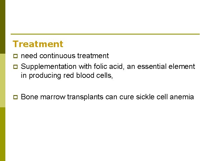
Treatment p need continuous treatment Supplementation with folic acid, an essential element in producing red blood cells, p Bone marrow transplants can cure sickle cell anemia p
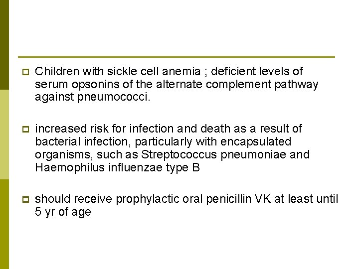
p Children with sickle cell anemia ; deficient levels of serum opsonins of the alternate complement pathway against pneumococci. p increased risk for infection and death as a result of bacterial infection, particularly with encapsulated organisms, such as Streptococcus pneumoniae and Haemophilus influenzae type B p should receive prophylactic oral penicillin VK at least until 5 yr of age
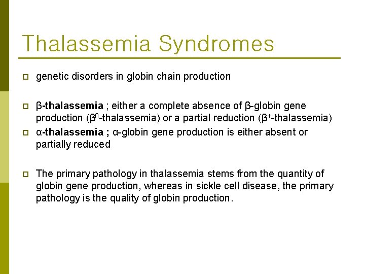
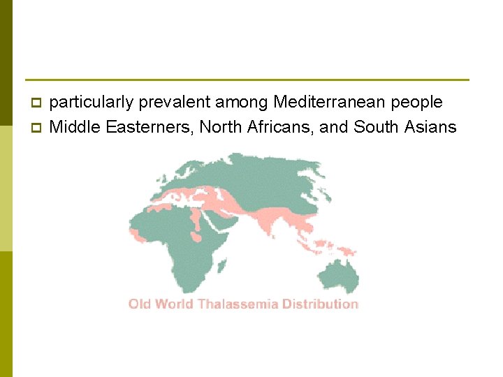
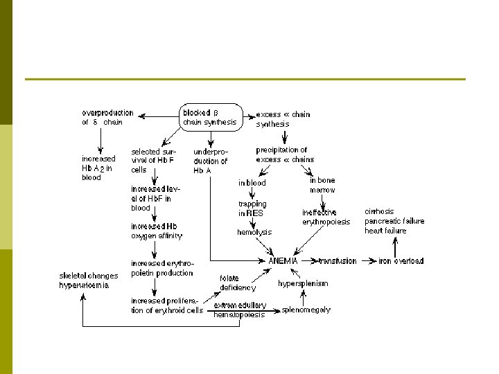
Thalassemia Syndromes p genetic disorders in globin chain production p β-thalassemia ; either a complete absence of β-globin gene production (β 0 -thalassemia) or a partial reduction (β+-thalassemia) α-thalassemia ; α-globin gene production is either absent or partially reduced p p The primary pathology in thalassemia stems from the quantity of globin gene production, whereas in sickle cell disease, the primary pathology is the quality of globin production.
p p particularly prevalent among Mediterranean people Middle Easterners, North Africans, and South Asians
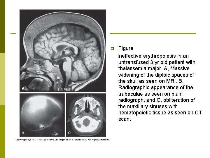
p Figure Ineffective erythropoiesis in an untransfused 3 yr old patient with thalassemia major. A, Massive widening of the diploic spaces of the skull as seen on MRI. B, Radiographic appearance of the trabeculae as seen on plain radiograph, and C, obliteration of the maxillary sinuses with hematopoietic tissue as seen on CT scan.
chronic blood transfusion therapy p iron chelation p Splenectomy p allogeneic hematopoietic transplantation p
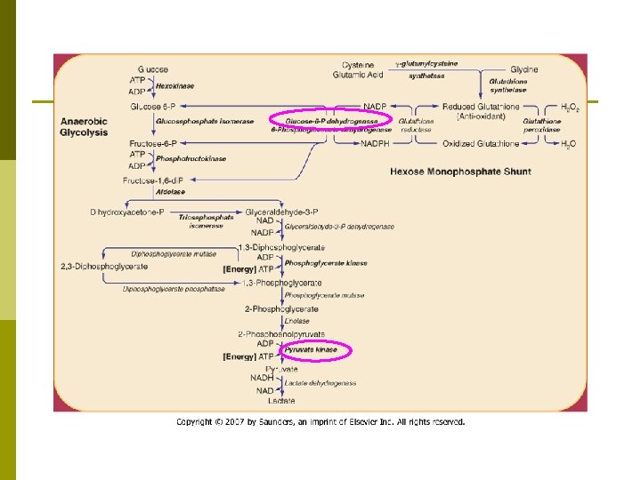
Enzyme defects p p Various red blood cell (RBC) enzymatic defects produce hemolytic anemias. generally not have a prominent morphologic alteration
1. Pyruvate Kinase Deficiency p The clinical manifestations vary from severe neonatal hemolytic anemia to mild, well-compensated hemolysis first noted in adulthood. p Severe jaundice and anemia in the neonatal period kernicterus p p The hemolysis in older children and adults varies in severity, with hemoglobin values ranging from 8 -12 g/d. L associated with some pallor, jaundice, and splenomegaly.
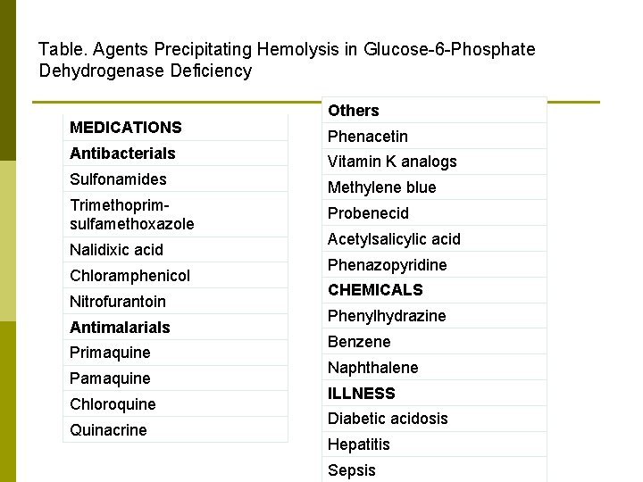
2. Glucose-6 -Phosphate Dehydrogenase Deficiencies p episodic hemolytic anemia induced by infections, certain drugs or, rarely, fava beans, and spontaneous chronic nonspherocytic hemolytic anemia p In the usual pattern of G 6 PD deficiency, symptoms develop 24 -48 hr after a patient has ingested a substance that has oxidant properties.
Table. Agents Precipitating Hemolysis in Glucose-6 -Phosphate Dehydrogenase Deficiency MEDICATIONS Antibacterials Sulfonamides Trimethoprimsulfamethoxazole Nalidixic acid Chloramphenicol Nitrofurantoin Antimalarials Primaquine Pamaquine Chloroquine Quinacrine Others Phenacetin Vitamin K analogs Methylene blue Probenecid Acetylsalicylic acid Phenazopyridine CHEMICALS Phenylhydrazine Benzene Naphthalene ILLNESS Diabetic acidosis Hepatitis Sepsis