HEREDITARY HAEMOLYTIC ANAEMIAS HEREDITARY SPHEROCYTOSIS BY DR FATMA

HEREDITARY HAEMOLYTIC ANAEMIAS HEREDITARY SPHEROCYTOSIS BY DR. FATMA ALQAHTANI CONSULTANT HAEMATOLOGIST

HEREDITARY HAEMOLYTIC ANAEMIA n MEMBRANE DEFECTS * Hereditary Spherocytosis * Hereditary Elliptocytosis * Hereditary Stomatocytosis etc….

HEREDITARY HAEMOLYTIC ANAEMIA n METABOLIC DEFECTS: Deficiency of: * Glucose-6 -phosphate dehydrogenase * Pyruvate kinase * Triose phosphate isomerase * Pyrimidine-5 -nucleotidase * Glutathione synthetase etc….

HEREDITARY HAEMOLYTIC ANAEMIA n HAEMOGLOBIN DEFECTS: * Defective synthesis e. g. Thalassaemia (Alpha or Beta) * Abnormal variants e. g. Hb S, Hb C, Unstable Hb

GENETIC ABNORMALITIES OF THE RED CELL MEMBRANE n n n Hereditary Spherocytosis Hereditary Elliptocytosis Hereditary Stomatocytosis Hydrocytosis (high MCV, low MCHC) Xerocytosis (low MCV, high MCHC shrunken RBCs due to K loss) Acanthocytosis – Abeta-lipoproteinaemia
 Electrophoretic Molecular weight Band*")
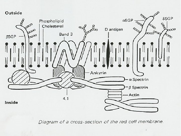
GENETIC ABNORMALITIES OF THE RED CELL MEMBRANE HEREDITARY SPHEROCYTOSIS (HS) Electrophoretic Molecular weight Band* (kd) NAME Number Location / function of molecules Chromosomal Gene Assignment cloned / RBC ( x 105 ) α Yes 1 2 240 220 2 2 5 42 5 Cs: forms protofilaments of 10 -13 monomers 7 pter – q 22 Yes ‘ Band ‘ 4. 1 78 2 Crosslinks spectrin heterodimers 1 p 32 – 1 pter Yes Ankyrin 2. 1 210 1 Links spectrin to band 3 ‘ Band ‘ 3 95 10 29 4 4 q 28 Yes Spectrin β Actin Glycophorin A PASI, 2 IMP = integral membrane protein CS: form heterodimers, tetramers 1 q 22 – 1 q 25 Yes IMP: anion transport: links to ankyrin CS = cytoskeleton IMP: sialoglycoprotein * as currently numbered on SDS – gels
 ? Spectrin genes Abnormal spectrin Decreased Synthesis of spectrin")
PATHOGENESIS OF HEREDITARY SPHEROCYTOSIS (HS) ? Spectrin genes Abnormal spectrin Decreased Synthesis of spectrin ? Gene for other membrane protein Spherocytes OF Decreased binding of spectrin Decreased spectrin in membrane Glucose requirement Decreased deformability
 n DEFINITION: A congenital disorder which is characterized by: * *")
HEREDITARY SPHEROCYTOSIS (HS) n DEFINITION: A congenital disorder which is characterized by: * * spherocytes increased osmotic fragility autosomal dominant inheritance ( ! recessive ) beneficial response to splenectomy
 The diagnosis of HS is not always easy since: * *")
HEREDITARY SPHEROCYTOSIS (HS) The diagnosis of HS is not always easy since: * * * The degree of spherocytosis is variable The changes in osmotic fragility are not always clear cut Sporadic cases can occur * Other haemolytic anaemias may respond to splenectomy
 n Role of spleen: * Results post splenectomy - Decrease in")
HEREDITARY SPHEROCYTOSIS (HS) n Role of spleen: * Results post splenectomy - Decrease in the rate of haemolysis (ameliorates the degree of anaemia) - Decrease in the number of spherocytes (but can not cure the red cell abnormality) * Spleen is the major site of red cell destruction * RBCs retained for long time in the splenic pulp as a result of decreased deformability + Unfavorable environmental conditions in the splenic pulp (acid p. H & decreased glucose) Failure of the cation pump Loss of water Loss of RBCs discoid shape Vicious circle
 Clinical Manifestations: • Most of the cases present in childhood or")
HEREDITARY SPHEROCYTOSIS (HS) Clinical Manifestations: • Most of the cases present in childhood or as teenagers • HS has been rarely diagnosed at: * Neonatal period (persistent jaundice) * The age of 60 (asymptomatic) • The disease has a wide spectrum of severity • The most consistent findings according to frequency are: * * * Jaundice Splenomegaly Anaemia
 Clinical Manifestations (cont…): • Haemolysis can be compensated for, with normal")
HEREDITARY SPHEROCYTOSIS (HS) Clinical Manifestations (cont…): • Haemolysis can be compensated for, with normal haemoglobin in about 1/3 of the patients • Patients may be more yellow than sick • Cholelithiasis is a complication of HS • HS as any other congenital haemolytic anaemia has a STEADY STATE and EPISODIC CHANGES
 Laboratory Tests and Findings: * Peripheral blood film > 1 -2%")
HEREDITARY SPHEROCYTOSIS (HS) Laboratory Tests and Findings: * Peripheral blood film > 1 -2% spherocytes in significant * MCHC is increased or in the upper limit of normal range (due to decreased water content) * Increased osmatic fragility (O. F) Shift to the right of the entire curve or only part of it Draw backs of this test: Laborious test Needs fresh defibrinated blood Not specific for HS (can be increased in AIHA) Insufficiently sensitive (10 -25%) of patients genetically proven to have HS have normal O. F) * Acidified glycerol lysis time. The rate of haemolysis (The time required for 50% lysis). Normal values > 1800 seconds) * Auto haemolysis (Screening test). 48 hours incubation under sterile conditions
 Spherocytes")
HEREDITARY SPHEROCYTOSIS (HS) Spherocytes
 Elliptocytes")
HEREDITARY ELLIPTOCYTOSIS (HE) Elliptocytes
 Stomatocytes")
HEREDITARY STOMATOCYTOSIS (HST) Stomatocytes

HEREDITARY SPHEROCYTOSIS
 Autohaemolysis Test Condition Lysis in a typical case (%) No addition")
HEREDITARY SPHEROCYTOSIS (HS) Autohaemolysis Test Condition Lysis in a typical case (%) No addition Normal + 27 mmol glucose 1. 7 0. 15 10. 1 1. 3 Pyruvate kinase deficiency 5. 5 6. 1 G 6 PD deficiency with CNSHA* 2. 9 1. 8 Hereditary spherocytosis * In the more common forms of G 6 PD deficiency without chronic non spherocytic haemolytic anaemia (CNSHA) the autohaemolysis test is normal
 Differential diagnosis n IF SPHEROCYTOSIS is prominent: Acquired haemolytic anaemia DAT")
HEREDITARY SPHEROCYTOSIS (HS) Differential diagnosis n IF SPHEROCYTOSIS is prominent: Acquired haemolytic anaemia DAT FAMILY DATA Red cell fragmentation poikilocytosis Fever n favours microangiopathic process may favor rare infectious cause for haemolysis (Clostridium welchii) IF SPHEROCYTOSIS is not prominent with chronic course: PNH ---- Ham test Enzymopatheis ---O. F. usually normal or decreased Autohaemolysis not corrected by glucose
 Complications n n n Leg ulcers Gall stones (Often asymptomatic) Aplastic")
HEREDITARY SPHEROCYTOSIS (HS) Complications n n n Leg ulcers Gall stones (Often asymptomatic) Aplastic crises
 Management n n n No cure The aim is to minimize")
HEREDITARY SPHEROCYTOSIS (HS) Management n n n No cure The aim is to minimize the consequences of the genetic abnormality Splenectomy Avoid below the age of 5 years unless haemolytic anaemia is very severe (rare is HS) Pneumococcal vaccination – (regular penicillin for at least 2 years) Treatment of complication’s as arises
 Genetic status Degree of")
Different forms of elliptocytosis Sub type of hereditary elliptocytosis (HE) Genetic status Degree of haemolysis Example of known molecular lesion Remarks Common Nonhaemolytic Heterozygote Absent Structurally abnormal α– spectrin One parent has similar picture Mild Heterozygote Minimal Glycoprotein C deficiency One parent has similar picture Intermediate Heterozygote Moderate One parent has similar picture Severe Heterozygote Severe One parent has HE: some times both Pyropoikilocyt osis Homozygote Severe Structurally abnormal α– spectrin Usuallybot parents asymptomatic. Or one has HE Spherocytic Heterozygote Homozygote Mild Severe Absence of protein 4. 1 Deletion revealed at DNA level (Conoboy et al. , 1986) Stomatocytic Not well defined Absent or mild Common in Melanesians ? Protects against malaria
- Slides: 24