Iron Cant live without enough of it Cant


 Fleming RE et al. N Engl J Med")


 ●")


")

 ●")
 Advantages Disadvantages May")


- Slides: 55
Iron: Can’t live without enough of it Can’t live with too much of it Dr. Lawrence Wolfe Associate Chief of Hematology Deputy Chief of Operations Cohen Children’s Medical Center Professor of Pediatrics Hofstra North. Shore LIJ School of Medicine
Transferrin
Ferritin
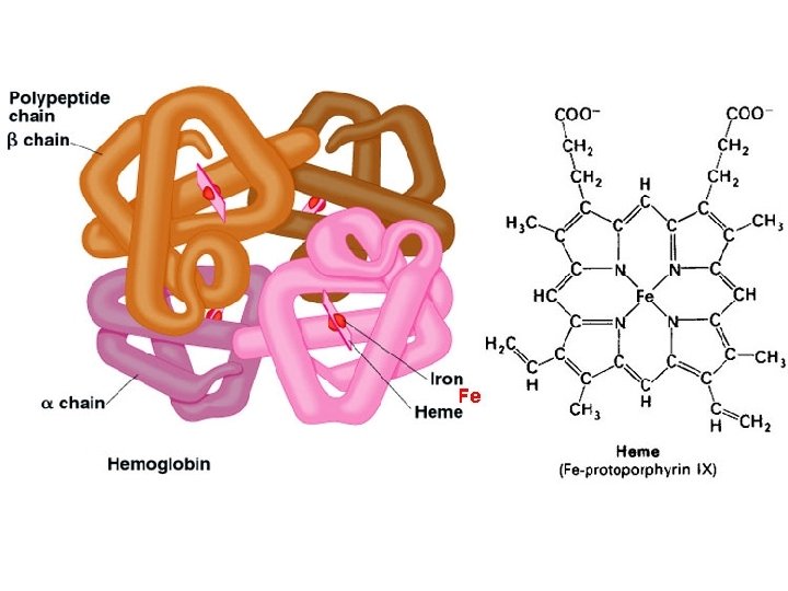
Iron plays an essential role in several metabolic processes Heme iron compounds Non-heme iron compounds Hemoglobin, myoglobin (oxygen transport) NAD dehydrogenase Cytochrome a, b, c (oxidative energy) Xanthine oxidase Cytochrome P 450 (drug metabolism) Ribonucleotide reductase Catalase, peroxidase (electron acceptor) Succinate dehydrogenase
Normal distribution and storage of body iron Utilization Duodenum (average, 1– 2 mg per day) Transferrin Muscle (myoglobin) (300 mg) Functional iron pool Utilization Transferrin Circulating erythrocytes (hemoglobin) (1800 mg) Storage iron Liver (1000 mg) Dietary iron Sloughed mucosal cells Desquamation/menstruation Other blood loss (average, 1– 2 mg per day) Iron loss Bone marrow (300 mg) Reticuloendothelial macrophages (600 mg) The human body has many mechanisms to absorb, transfer and store iron, but none to excrete it Andrews NC. N Engl J Med 1999; 341: 1986– 1995
Regulation of cellular iron release (1) Fleming RE et al. N Engl J Med 2005; 352: 1741– 1744
Specialized cells have mechanisms for exporting iron into plasma Spleen 1. 1% of the red cell mass is processed daily by spleen macrophages (10– 15 mg Fe/day) Macrophage Erythrocyte 2. Iron released from processed hemoglobin is either exported back into plasma (via ferroportin) or stored as ferritin Fe Fe 2+ Ferritin Ferroportin activity is regulated by hepcidin produced in the liver Courtesy of Professor IV Cabantchik idin c Hep 3. The released iron is absorbed by transferrin Ceruloplasmin Fe 2+ Fe 3+ Transferrin
Dietary iron absorption • 1– 2 mg of iron are absorbed from the diet every day. The same amount is lost through cell sloughing and bleeding • Dietary heme and non-heme iron are taken up by duodenal enterocytes Absorption of iron is finely controlled in response to body iron stores and iron needs
Dietary iron absorption • Iron is then released into the plasma and loaded onto transferrin • Release of iron into plasma is controlled by hepcidin Absorption of iron is finely controlled in response to body iron stores and iron needs
Version 6, 2010 11 The labile iron pool Iron is taken up by cells from circulating transferrin via transferrin receptors Tf. R 1 and Tf. R 2 Fe 3+ Transferrin Tf. R 2 ? ? ? DMT 1 Fe 2+ Iron is used mostly by mitochondria for heme and iron-sulfur cluster synthesis Heme synthesis LIP Fe-S cluster biogenesis Mitochondria Ferritin Courtesy of Professor IV Cabantchik ? DMT 1 Endosome Excess iron is stored or withdrawn into ferritin PLASMA Fe 2+ Tf. R 1 Following various steps iron is delivered into the cytosol LIP of Fe 2+ Dcyb Other iron utilization pathways Most cells have no iron-release mechanisms PLASMA
Version 6, 2010 12 Primary and secondary causes of iron overload Primary (hereditary) ● Resulting from a primary defect in the regulation of iron balance, eg, hereditary hemochromatosis Secondary (acquired) ● Caused by another condition or by its treatment – Ineffective erythropoiesis and anemias requiring repeated blood transfusion (eg DBA, thalassemia, sickle cell disease [SCD] and MDS) – Toxic ingestion Feder JN et al. Nat Genet 1996; 13: 399– 408; Porter JB. Br J Haematol 2001; 115: 239– 252
Version 6, 2010 13 Transfusion therapy results in iron overload ● 1 blood unit contains 200 mg iron ● A 60 kg patient with thalassemia receiving 45 units of blood annually has transfusional iron intake of 9 g iron/year – 0. 4 mg iron/kg body wt/day ● In addition, up to 4 mg/day may be absorbed from the gut – Up to 1. 5 g iron/year 200– 250 mg iron: Whole blood: 0. 47 mg iron/m. L ‘Pure’ red cells: 1. 16 mg iron/m. L Porter JB. Br J Haematol 2001; 115: 239– 252 ● Overload can occur after 10– 20 transfusions Iron overload is an inevitable consequence of multiple blood transfusions
Version 6, 2010 14 Normal distribution and turnover of body iron Erythron 2 g 20– 30 mg/day Parenchyma 0. 3 g Liver 1 g Transferrin 2– 3 mg/day 1– 2 mg/day Reticuloendothelial macrophages 0. 6 g 20– 30 mg/day Gut Iron balance is achieved in the normal state Hershko C et al. Ann NY Acad Sci 1998; 850: 191– 201
Version 6, 2010 15 Imbalance of distribution and turnover of body iron with transfusion therapy Erythron Transfusions 20– 40 mg/day NTBI Parenchyma Transferrin Reticuloendothelial macrophages Gut Iron balance is disturbed by blood transfusion because the body cannot remove the excess iron NTBI, non-transferrin-bound iron Hershko C et al. Ann NY Acad Sci 1998; 850: 191– 201
Version 6, 2010 16 Iron overload leads to formation of NTBI Transferrin saturation due to: • Frequent blood transfusions, or • Ineffective erythropoiesis leading to increased iron absorption Normal: no NTBI produced Subsequent formation of NTBI in plasma Pituitary Parathyroid Thyroid Iron overload Heart Transferrin saturation 100% 30% Uncontrolled iron loading of organs Fe Fe Fe Liver Pancreas Gonads Fe Fe
Version 6, 2010 17 Toxic radical formation increases with higher levels of ‘free’ iron in cells ● NTBI appears when transferrin iron-binding capacity is exceeded: – Redox-active component is labile plasma iron (LPI), which is chelatable and prevalent at >70% transferrin saturation ● LPI is potentially toxic: – Enters cells via unregulated pathways – Raises LCI, producing a labile iron pool ● LCI can catalyze formation of noxious HO· radicals to cause reactive oxygen species (ROS) – LCI (Fe 3+ and Fe 2+) reacts with reactive oxygen intermediates (O 2 and H 2 O 2) produced by respiration or by other incomplete reductions of O 2 and forms noxious HO· radicals (Haber-Weiss cycle) Fe 3+ + O 2 Fe 2+ + O 2 Fe 2+ + H 2 O 2 Fe 3+ + OH– + HO· (Fenton reaction) Sustained levels of LCI cause persistent oxidative stress
Version 6, 2010 18 Uncontrolled uptake of labile iron leads to cell and organ damage Non-transferrin iron Storage iron Uncontrolled uptake Transferrin iron Controlled uptake Labile Iron Free-radical generation Organelle damage Porter JB. Am J Hematol 2007; 82: 1136– 1139 Functional iron
Version 6, 2010 19 Iron overload negatively affects organ function Labile iron Free radical generation Lipid peroxidation Organelle damage Lysosomal fragility TGF-β 1 Collagen synthesis Enzyme leakage Cell death Fibrosis TGF, transforming growth factor Cohen AR and Porter JB. In Disorders of Hemoglobin: Genetics, Pathophysiology, and Clinical Management, Steinberg MH et al. (Eds); 2001: 979– 1027
Version 6, 2010 20 Excess iron is deposited in multiple organs, resulting in organ damage Iron overload Capacity of serum transferrin to bind iron is exceeded NTBI circulates in the plasma; some forms of NTBI (eg LPI) load tissues with excess iron Excess iron promotes the generation of free hydroxyl radicals, propagators of oxygenrelated tissue damage Cardiac failure Liver cirrhosis/ fibrosis/cancer Insoluble iron complexes are deposited in body tissues and end-organ toxicity occurs Diabetes mellitus Infertility Endocrine disturbances→ growth failure
Version 6, 2010 21 Organ systems susceptible to iron overload Clinical sequelae of iron overload Pituitary → impaired growth, infertility Thyroid → hypoparathyroidism Heart → cardiomyopathy, cardiac Liver → hepatic cirrhosis Pancreas → diabetes mellitus Gonads → hypogonadism Iron overload end-organ iron toxicities are inevitable in the absence of intervention therapy
Version 6, 2010 22 Post-mortem cardiac iron deposits correlate with blood transfusions in the pre-chelation era Patients with cardiac iron (%) 100 80 ● 131 transfused adult patients – 101 leukemias – 30 other anemias 60 40 20 0 0– 25 26– 50 51– 75 76– 100 101– 200 201– 300 Units of blood transfused Cardiac iron deposition increases with the number of blood transfusions in unchelated patients Buja LM & Roberts WC. Am J Med 1971; 51: 209– 221
Version 6, 2010 23 Repeated transfusions lead to iron overload in SCD Liver iron (mg Fe/g dw) 30 25 R=0. 795 20 15 10 5 0 0 20 40 60 80 100 120 140 160 Transfusion duration (months) There is a significant correlation between the duration of transfusions and liver iron in patients with SCD Harmatz P et al. Blood 2000; 96: 76– 79
24 Liver with iron overload Version 6, 2010 Normal liver
25 Pancreas with massive iron overload Version 6, 2010 Normal pancreas
26 Version 6, 2010 Dilated cardiomyopathy with iron overload Hypertrophic cardiomyopathy
Version 6, 2010 27 Methods used to assess iron overload ● Transferrin saturation ● Serum ferritin ● LIC ● Iron distribution (eg cardiac iron)
Version 6, 2010 28 Measuring LIC by liver biopsy Advantages Disadvantages Direct measurement of LIC Invasive; painful; potentially serious complications, eg bleeding Validated reference standard Risk of sampling error, especially in patients with cirrhosis Quantitative, specific and sensitive Inadequate standardization between laboratories Allows for measurement of nonheme storage iron Difficult to follow-up Provides information on liver histology/pathology Positive correlation with morbidity and mortality
Version 6, 2010 29 Iron burden is clinically significant across all transfusion-dependent anemias Mean baseline liver iron concentration (LIC) in deferasirox trials (measure of TOTAL body iron) LIC (mg Fe/g dw) 25 20 Increased mortality >15 Increased organ dysfunction 7– 15 5 Clinical concern >2– 7 0 Normal <1. 2 15 10 Thalassemia Other anemias SCD Patients were not achieving iron balance with current chelation regimens Cappellini MD et al. Blood 2006; 107: 3455– 3462; Porter J et al. Eur J Haematol 2008; 80: 168– 176; Vichinsky E et al. Br J Haematol 2007; 136: 501– 508 Studies 107, 108 and 109
Version 6, 2010 30 Ferritin and serum ferritin Serum ferritin >1000 ng/m. L is a marker of excess body iron ● Ferritin is primarily an intracellular protein that: – Stores iron in a form readily accessible to cells – Releases it in a controlled fashion ● The molecule is shaped like a hollow sphere that stores ferric (Fe 3+) iron in its central cavity – The storage capacity of ferritin is approximately 4500 Fe 3+ ions per molecule ● Ferritin is found in all tissues but primarily in the liver, spleen and bone marrow ● A small amount is also found in the blood as serum ferritin Harrison PM & Arosio P. Biochim Biophys Acta 1996; 1275: 161– 203
Version 6, 2010 31 Measuring and interpreting serum ferritin Advantages Disadvantages Easy to assess Indirect measurement of iron burden Inexpensive Fluctuates in response to inflammation, abnormal liver function, ascorbate deficiencies Repeat serial measures are useful for monitoring chelation therapy Individual measures may not provide reliable indication of iron levels and response to chelation therapy Positive correlation with morbidity and mortality Allows longitudinal follow-up of patients Serial measurement of serum ferritin is a simple, reliable, indirect measure of total body iron
Version 6, 2010 32 Correlation between serum ferritin and LIC in various underlying anemias MDS DBA Other anemias β-thalassemia Regression line Change in LIC (mg Fe/g dw per year) 15 10 5 0 − 5 − 10 − 15 − 2000 − 1500 − 1000 − 500 0 500 1000 1500 2000 Change in serum ferritin (ng/m. L per year) Porter J et al. Eur J Hematol 2008; 80: 168– 176 2500 3000
Version 6, 2010 33 Relationship between serum ferritin and myocardial iron Myocardial iron (µmol/g) 40 35 Patient group (n=14) 30 25 20 15 ● 11 MDS ● 1 Diamond-Blackfan anemia (DBA) ● 1 chronic hemolysis of unknown origin ● 1 acute myeloid leukemia (AML) in complete remission 10 5 0 1000 Serum ferritin (ng/m. L) 10000 Cardiac iron loading associated with serum ferritin levels >1800 ng/m. L Jensen PD et al. Blood 2003; 101: 4632– 4639
Version 6, 2010 34 Measuring LIC with MRI Advantages Disadvantages Non-invasive Indirect measurement of LIC Assesses iron content throughout the liver Requires magnetic resonance imager with dedicated imaging method Increasingly widely available worldwide Children under the age of 7 years require a general anesthetic Status of liver and heart can be assessed in parallel Validated relationship with LIC Allows longitudinal patient follow-up
Version 6, 2010 35 Relationship between R 2* MRI and liver biopsy (3) ● R 2* increases linearly with iron Patients Controls Fit 2000 1800 1600 ● Calibration independently validated in 23 biopsies R 2* (Hz) 1400 1200 1000 800 600 400 R=0. 97 200 0 0 10 20 30 40 50 Biopsy LIC (mg/g dry tissue) 60 Paired R 2* measurements demonstrated no mean difference, making MRI suitable for longitudinal examination Wood JC et al. Blood 2005; 106: 1460– 1465
Version 6, 2010 36 Measuring LIC with biomagnetic liver susceptometry (SQUID) Advantages Disadvantages May be repeated frequently Indirect measurement of LIC Allows follow-up Limited availability High cost Highly specialized equipment requiring dedicated technician Not validated for LIC assessment and may underestimate levels compared with biopsy* *During clinical development of deferasirox, LIC data by biopsy were shown to be related to data by SQUID by a factor of 0. 46 SQUID, Superconducting QUantum Interference Device Piga A et al. Presented at ASH 2005 [Blood 2005; 106(11): abst 2689]
Version 6, 2010 37 Measuring cardiac iron with MRI Advantages Rapidly assesses iron content in the septum of heart Disadvantages Indirect measurement of cardiac iron Relative iron burden can be estimated Requires magnetic resonance imager reproducibly with dedicated imaging method Functional parameters can be examined concurrently Cardiac methodologies remain to be standardized Iron status of liver and heart can be assessed in parallel Allows longitudinal follow-up MRI is a non-validated method to rapidly and effectively assess cardiac iron
Version 6, 2010 38 T 2* MRI: Emerging new standard for cardiac iron 90 80 70 LVEF (%) 60 50 40 Cardiac T 2* value of 37 ms in a normal heart 30 20 10 0 0 10 20 30 40 50 60 70 Heart T 2* (ms) 80 90 100 Myocardial T 2* values <20 ms are associated with progressive and significant decline in LVEF, left ventricular ejection fraction Anderson LJ et al. Eur Heart J 2001; 22: 2171– 2179 Cardiac T 2* value of 4 ms in a significantly iron-overloaded heart
Version 6, 2010 39 LVEF monitoring as a predictor of high risk Survival probability 1. 00 LVEF normal (n=47) 0. 75 LVEF falls (n=34) 0. 50 27/27 alive, if comply 7/7 dead, if poor intensification 0. 25 0 0 5 10 15 Follow-up (years) Davis BA et al. Blood 2004: 104: 263– 269 20 ● LVEF decrease of >10% or to below 45% results in significant increase in risk of: – Cardiac failure (P<0. 001) – Cardiac death (P=0. 001)
Version 6, 2010 40 Thresholds for parameters used to evaluate iron overload Iron-overloaded state Normal Mild Moderate Severe LIC, mg Fe/g dw <1. 2 3– 7 >7 >15 Serum ferritin, ng/m. L <300 Transferrin saturation, % 20– 50 Parameter >100 to <2500 >50 Myocardial T 2*, ms >20 8– 20 <8 Alanine aminotransferase (ALT), U/L <250 >250 LPI, μM 0– 0. 4 >0. 4 Increased risk of complications/cardiac disease
Version 6, 2010 41 Guidelines for starting treatment of iron overload in patients with β-thalassemia major Thalassaemia International Federation guidelines for the clinical management of thalassemia (2008)1 recommend that chelation therapy is considered when patients: Have received 10– 20 transfusion episodes OR Have a serum ferritin level of >1000 ng/m. L Italian Society of Hematology practice guidelines for the management of iron overload in patients with thalassemia (2008) 2 recommend that chelation therapy is considered when patients: Have received >10 transfusion episodes OR Have a serum ferritin level of >1000 ng/m. L 1 Thalassaemia 2 Angelucci International Federation. Guidelines for the clinical management of thalassemia, 2 nd Edition revised 2008; E et al. Haematologica 2008; 93: 741– 752
Version 6, 2010 42 What is chelation therapy? Toxic Chelator Metal Excretion
Version 6, 2010 43 Properties of an ideal chelator Efficacy ● Maintenance of iron balance or achievement of negative iron balance ● High and specific affinity for ferric iron (Fe 3+) ● Effective tissue and cell penetration ● High-chelating efficiency ● No iron redistribution ● Slow metabolism and elimination rate ● 24 -hour chelation coverage Convenience ● Oral bioavailability ● Half-life compatible with once-daily dosing ● Good compliance Tolerability ● Good adverse event (AE) profile
Version 6, 2010 44 Comparison of chelators Property DFO Deferiprone Deferasirox 25– 60 75– 100 20– 30 Sc, iv (8– 12 hours, 5 days/week) Oral 3 times daily Oral Once daily Half-life 20– 30 minutes 3– 4 hours 8– 16 hours Excretion Urinary, fecal Urinary Fecal Local reactions, ophthalmologic, auditory, growth retardation, allergic Gastrointestinal disturbances, agranulocytosis/ neutropenia, arthralgia, elevated liver enzymes Gastrointestinal disturbances, rash, renal impairment, hepatic impairment, ophthalmologic, auditory Licensed outside US/Canada Licensed Usual dose (mg/kg/day) Route Main adverse effects in prescribing information Status
Version 6, 2010 45 Chelating agents DFO ● Indicated for the treatment of chronic iron overload in patients with transfusion-dependent anemias 1 Deferiprone ● Treatment of iron overload in patients with thalassemia major for whom DFO therapy is contraindicated or inadequate. 2 Only limited data are available for pediatric patients aged 6– 10 years and no data for patients <6 years Deferasirox ● Treatment of iron overload due to blood transfusions in adults and children aged 2 years and older 3 (Please substitute with regional prescribing information where slide is being presented) 1 Desferal® Prescribing Information. Novartis 2007; 2 Ferriprox® [package insert]. Apotex Europe Ltd 2004; 3 EXJADE® (deferasirox) Basic Prescribing Information. Novartis Pharma AG
Version 6, 2010 46 Limitations of DFO therapy Poor oral bioavailability and a short plasma half-life Slow subcutaneous infusion 3– 7 times weekly Inconvenient administration Injection-site reactions and pain 1 Gabutti Poor compliance reported to lead to increased mortality 1 V & Piga A. Acta Haematol 1996; 95: 26– 36 Equipment not widely available in many countries
Version 6, 2010 47 Possible chelation regimens Deferasirox monotherapy MON TUE WED THU FRI SAT SUN 100% chelation coverage Continuous chelation coverage with once-daily oral deferasirox
Version 6, 2010 48 Protection from LPI appearance with standard chelation regimens Deferiprone x 3 LPI (µmol/l) 1. 2 1. 0 0. 8 0. 6 0. 4 0. 2 0 1. 2 LPI (µmol/l) 1. 2 0 2 4 6 8 10 12 24 --DFO-- Deferiprone x 3 0 1. 0 0. 8 0. 6 0. 4 0. 2 0 2 4 6 8 10 12 24 Deferiprone (75 mg/kg/day orally in three daily doses) 0 1. 2 1. 0 0 --DFO-- 0 Treatment (h) 2 4 6 8 10 12 24 DFO (40 mg/kg/day overnight) Deferasirox (30 mg/kg/day once daily) Deferasirox The grey shaded area denotes the normal range of LPI 0 2 4 6 8 10 12 24 Treatment (h) LPI values were determined in blood samples taken every 2 hours, starting at 8 am (0 hours)until 8– 10 pm and ending at 8 am the following day (24 hours) Zanninelli G et al. Br J Haematol 2009; 147: 744– 751
Version 6, 2010 49 LIC and liver prognosis ● The liver is the primary site of iron storage in the body Threshold LIC levels (mg Fe/g dw) 1. 2 Normal 15 7 Mild iron overload Moderate iron overload Increased risk of liver fibrosis Jensen PD et al. Blood 2003; 101: 4632– 4639; Angelucci E et al. Blood 2002; 100: 17– 21 Severe iron overload Progression of liver fibrosis Deranged ALT
Version 6, 2010 50 Iron in the liver and chelation therapy Levels >7 mg Fe/g dw associated with increased risk of hepatic fibrosis and Normal iron diabetesliver mellitus levels are <1. 2 mg Levels >15 mg Fe/g dw The liver is the primary site of iron storage in the body associated with greatly increased risk of cardiac disease and death in unchelated patients Iron overload can be monitored by measuring LIC correlates significantly with total body iron stores Optimal maintenance level Chelation therapy for chelation effectively therapy in/ maintains patients liver with iron thal reduces major is <7 mg levels Fe/g dw
Version 6, 2010 51 When does cardiac iron loading begin? Sickle cell 70 ● >13 years of transfusions Thalassemia 60 ● >35 grams T 2* 50 ● Data only for patients with thalassemia major receiving chelation 40 30 Abnormal T 2* 20 10 0 0 5 10 15 20 Transfusion (years) Wood JC et al. Blood 2004; 103: 1934– 1936 25 ● Prior chelation therapy
Version 6, 2010 52 Continuous, 24 -hour iv DFO rescue for cardiac disease 70 LVEF (%) 60 50 Mean=49% P=0. 002 40 30 20 Mean=36% Pre-DFO Post-DFO (6– 12 months) Continuous chelation coverage by iv DFO infusion can reverse iron induced cardiac dysfunction Davis BA & Porter JB. Blood 2000; 95: 1229– 1236
Version 6, 2010 53 Continuous, 24 -hour DFO therapy improves cardiac T 2* and cardiac function ● Six subjects showed improved cardiac T 2* Baseline 120 108 P=0. 03 106 110 P=0. 03 g m. L % LVEF 7 6 100 80 40 8 102 90 50 P=0. 003 104 100 60 9 P=0. 02 70 98 60 96 P=0. 02 ms 70 12 months 5 4 3 50 94 40 92 2 30 90 1 20 88 0 LVEDVI = left ventricular end-diastolic volume index; LVESVI = left ventricular end-systolic volume index Anderson LJ et al. Br J Haematol 2004; 127: 348– 355 LVESVI LV mass index Cardiac T 2*
Version 6, 2010 54 Prospective improvement in myocardial T 2* with DFO and deferiprone monotherapy DFO 43 mg/kg/day x 5. 7 sc Deferiprone 92 mg/kg/day orally Myocardial T 2* (geometric mean ± SEM) 18 17 Deferiprone (change 3. 5 ms; n=29; P<0. 001) 16 DFO (change 1. 7 ms; n=32; P<0. 001) 15 14 13 12 Baseline 6 months 12 months Significant improvement in myocardial T 2* with both DFO and deferiprone SEM, standard error of the mean Pennell DJ et al. Blood 2006; 107: 3738– 3744
Version 6, 2010 55 All available chelators remove cardiac iron ● Liver iron and heart iron do not correlate after chelation is started Effect of chelation therapy on cardiac iron DFO 1 Deferiprone 1 Deferasirox 2, 3 Improvement in T 2* Increase in LVEF (for patients with T 2* >20 ms) Reversal of cardiac dysfunction (symptomatic or asymptomatic) No data yet 1 Pennell DJ et al. Blood 2006; 107: 3738– 3744; 2 Pennell DJ et al. Blood 2010; 115: 2364– 2371; 3 Pennell DJ et al. Blood 2009; 114(22): abst 4062