
Unit 1 OTOSCLEROSIS PRESENTED BY REVIEWED BY Ms
Unit 1 OTOSCLEROSIS PRESENTED BY: REVIEWED BY: Ms. Pavithra K Mr. Jobin Lecturer, MSN dept Asst. Professor, MSN dept
LEARNING OBJECTIVES At the end of the class students will be able to • define Otosclerosis • Enlist the causes of Otosclerosis • describe the patho-physiology of Otosclerosis • list down the clinical features of Otosclerosis • enumerate the management of Otosclerosis
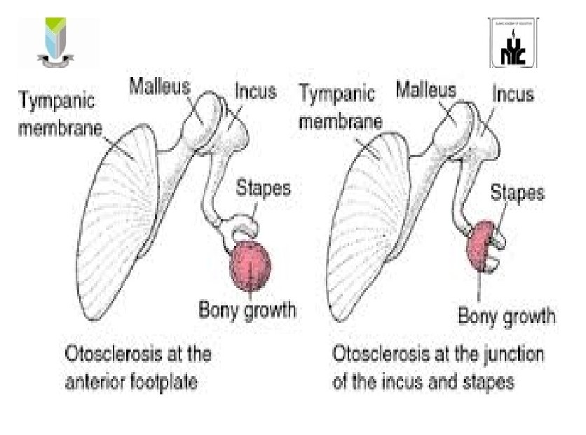
• Otospongiosis • Otosclerosis is a primary disease of the ossicles and bony otic capsule characterized by abnormal removal of mature bone by osteoclasts and replacement with woven bone of greater thickness, cellularity and vascularity.
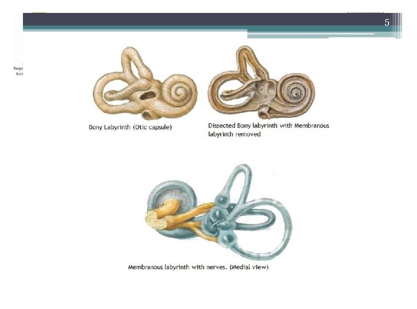
• a primary disease of the ossicles and bony otic capsule in which irregular spongy bone replaces the normal layer which is characterized by – footplate of the stapes and cause conductive hearing loss – Cochlea and cause sensory neural hearing loss
Incidence • • 0. 5 -1% of total population Female: Male (2: 1) 20 -30 years of age Usually bilateral (85%)
Etiology • • Exact cause is unknown Remnants of embryonic cartilage Genetic / heriditary Hormonal – Pregnancy and menopause Osteogenesis Imperfecta Pagets Disease Imbalance in trypsin / Antitrypsin in the inner ear
• Anatomical anomalies in the temporal bone • Viral infection – measles • Autoimmune disorders
Types • • • Stapedial otosclerosis Cochlear otosclerosis Malignant otosclerosis Combined otosclerosis Histological otosclerosis
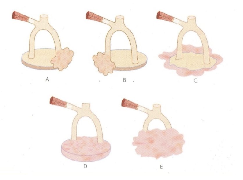
Stapedial otosclerosis • Stapes is involved • Sites: – Fissula ante fenestrum – Fissula post fenestrum – Circumferential – Biscuit type – Obliterative
• Cochlear otosclerosis – Round window and labyrinth is involved – Stapes fixation • Malignant otosclerosis – Severe type of cochlear otosclerosis – Starts in the early life
• Combined otosclerosis – Both stapes and cochlear • Histological otosclerosis – 9 -12% – Foot plate of stapes – Measeles virus
Phases • Early spongiotic phase • Late or sclerotic phase
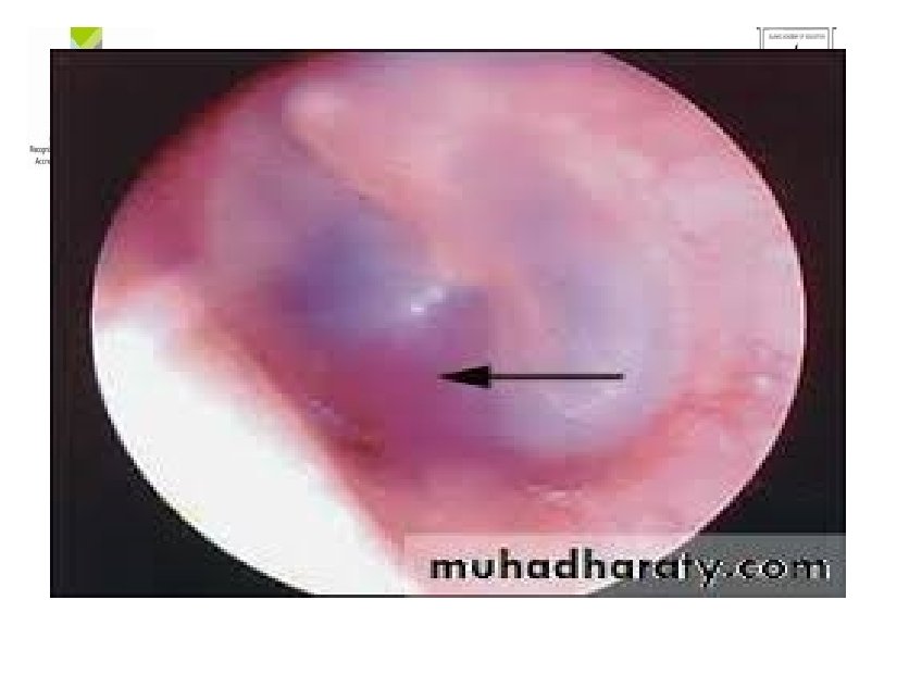
Early spongiotic phase • • • Otospongiosis osteoclasts and histiocytes Active reabsorption of bone Dialated blood vessels Schwartze’s sign – red blush seen over the ear drum
Late Sclerotic phase • • Formation of new bone in resorption areas New bone – dense and sclerotic Initially starts in the stapes area Then to membranous labyrinth
Pathophysiology • • Due to etiological Destruction of the normal bone Abnormal enzymatic activity Osteoclastic activity Osteoblastic -----Calcification Fixation of stapes Alters the conduction of sound Conductive hearing loss
• Otitic capsule • Bony labyrinth and nerve • Sensori neural hearing loss
Clinical manifestations • slow, progressive , conductive or sensori – neural hearing loss • Tinnitus • Otalgia • Vertigo • Dizziness • Balance problems
• Paracusis of willis – Ability to hear a conversation better in a noisy environment than in a quiet one • Schwartze sign • Rinne test - BC>AC • Weber test – Affected ear
Diagnostic Evaluation • • History collection Physical examination Otoscopy -Schwartze sign Rinne test - BC>AC Weber test – Affected ear Audiometry CT scan
Management Goals • Reduction of the osteoblastic activity • Inhibit proteolytic enzymes action • Slows the progression of sensori neural hearing loss
Medical Management • Tab sodium fluoride with vit D and calcium carbonate • Dose- 50 -75 mg/day • 3 months – 2 years Uses: • Arrest the further progression of cochlear loss • Anti-enzymatic action on proteolytic enzymes
• Bisphonates – Alendronate, Ibandronate – Reduced the bone resorption • Analgesics • Antibiotics • Hearing aids
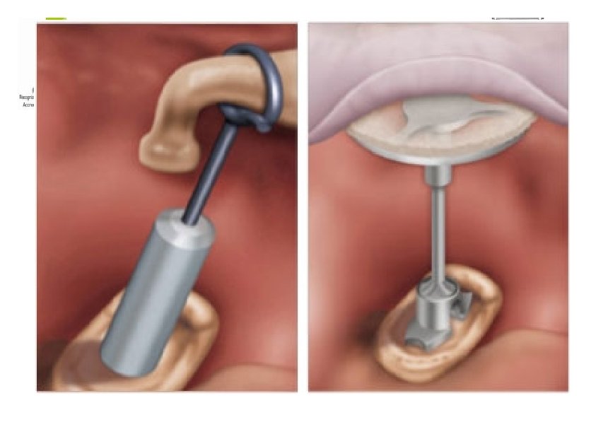
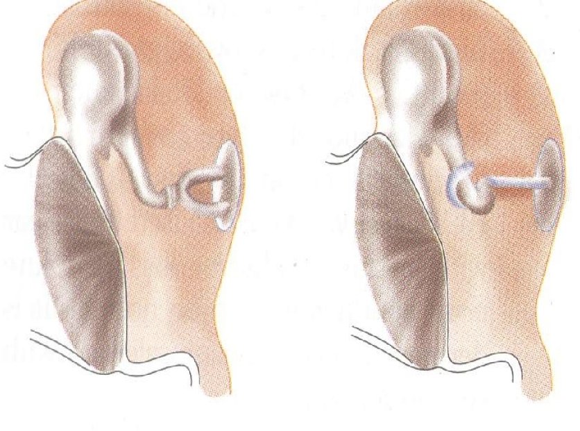
Surgical Management • Stapedectomy – partial removal of the stapes • Stapedotomy – opening in the stapes footplate followed by prosthesis insertion
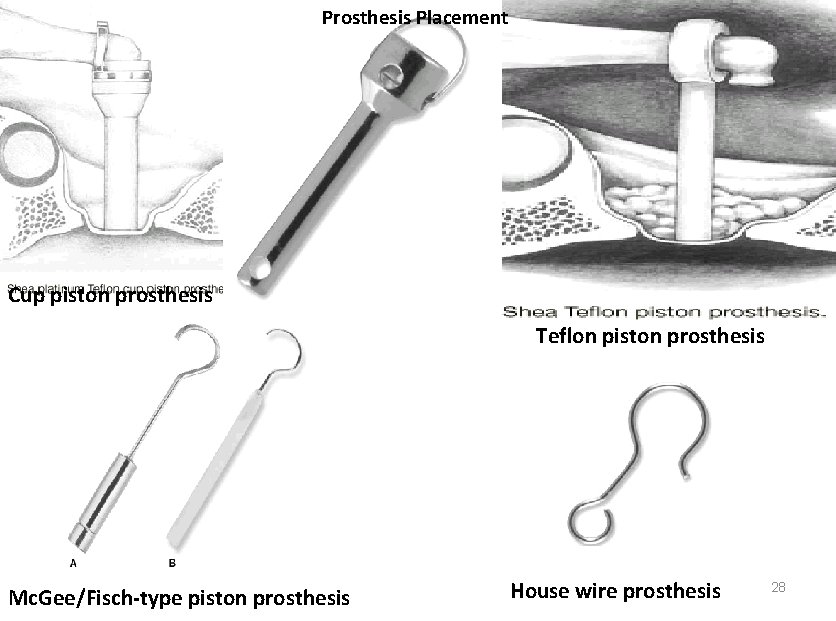
Prosthesis Placement Cup piston prosthesis Teflon piston prosthesis Mc. Gee/Fisch-type piston prosthesis House wire prosthesis 28
Nursing Management • Do not blow the nose for three weeks following surgery. • Keep the mouth open during sneezing or coughing • Avoid any heavy lifting straining or bending for three weeks following surgery • Keep the head elevated on 2 -3 pillows if possible. • place a cotton wool ball coated in Vaseline in the car canal to prevent getting wet during head bath.
• Replace the cotton wool ball daily for one week • Take oral antibiotic as prescribed. • If there is a separate incision a small amount of drainage may occur. If the drainage is profuse or develops a foul odour it should be reported. • Popping sounds, a plugged sensation, ringing or fluctuating hearing may be occur during healing
• Avoid travel by air for three weeks following surgery. • notice any swelling, redness or excessive pain • Some dizziness may occur after surgery. If severe or is associated with nausea or vomiting should be reported. • Teach the use of hearing aids
Nursing diagnosis • Disturbed Sensory Perception Auditory related to decreased sensory reception as evidenced by decreased hearing • Impaired Verbal Communication related to hearing loss • Acute Pain related to the abnormal bone growth or surgical procedure • Self-esteem disturbance related to changes in body function.
• Risk for Injury related to the vertigo. • Activity intolerance related to the vertigo. • Deficient knowledge related to disease condition or therapeutic regimen
EVALUATION • • • What is Otosclerosis. What are the causes of Otosclerosis. Eneumerate the pathophysiology of Otosclerosis. Enlist the clinical features of Otosclerosis. Explain the management of Otosclerosis.
REFERENCE • Smeltzer – Brunner & Suddharth Textbook of Medical Surgical Nursing, Wolters kluwer publishers, 12 th edition 2009. • Black – Medical Surgical Nursing, Elsevier publishers, 8 th edition 2009. • Nettina – Lippincott manual of Nursing Practice, Wolters kluwer publishers, 7 th edition 2014. • Lewis – Medical Surgical Nursing, Elsevier publishers, 10 th edition, 2017
- Slides: 37