HLA system MHC glycoproteins MHC glycoproteins class I
")
 § MHCgp. I present peptide fragments from")


 process foreign antigens and present them complexed with")


 § Allospecific serums (obtained from multiple natal to 6 weeks after")
")
 PCR-SSP (Polymerase chain reaction with sequential specific primers)")
 PCR-SSO § PCR reaction with sequence-specific oligonucleotides §")
 PCR-SBT § Sequencing based typing § We get")


, need")

,")
 is heterodimer consisting of a and b (g,")


+Ag peptid (APC) 2. Co-stimulating")


 and CD")


- Slides: 57
HLA system (MHC glycoproteins)
MHC glycoproteins class I (Major histocompatibility complex) § MHCgp. I present peptide fragments from itracellular proteins (which are produced by cell, including viral peptides if are present) on the cell surface for cytotoxic T lymphocytes ( CD 8+) § Expressed on all nucleated cells § 3 isotypes of classical MHC gp. (HLA - A, -B, -C) § 3 isotypes non-classical MHC gp. (HLA - E, -F, -G; molecule CD 1)
MHC gp I structure § MHC gp class I consists of transmembrane chain a and associated b 2 microglobulin § a 1, a 2 - binding site for peptides § Peptide binding is necessary for a stable conformation of MHC gp
MHC gp. I peptide binding § MHC gp I binds peptides long 8 - 10 amino acids § Certain MHC gp molecule binds peptides sharing identical structural features binding motif § The binding of endogenous peptides occurs in the endoplasmic reticulum during biosynthesis of MHC gp I § These peptides are produced from intracellular proteins that are cleaved by the proteasomes
MHC gp. I peptide binding
Non-classical MHC gp I § HLA - E, -F, -G; CD 1 molecules § Structurally similar to classical MHC gp § Less polymorphic § Expressed only on some cells § They specialize in binding of specific ligands
Non-classical MHC gp I § HLA-E and HLA-G - expressed on the trophoblast cells § Complexes of HLA-E and HLA-G with peptides are recognized by NK cells inhibitory receptors and contribute to the tolerance of the fetus in utero
MHC glycoproteins class II § MHC gp. II present peptide fragments from extracellular proteins on the cell surface for helper T lymphocytes (CD 4+) § Expressed on the APC (dendritic cells, monocytes, macrophages, B lymphocytes) § 3 isotypes of MHC gp. II (DR, DQ, DP)
MHC gp II structure § MHC gp II consist of 2 associated transmembrane chains a and b § a 1, b 1 - binding site for peptide § Peptide binding is necessary for a stable coformation of MHC gp and ensure its long presentation on the cell surface
MHC gp II peptide binding § MHC gp II binds peptides long 15 - 35 amino acids § Certain MHC gp molecule binds peptides sharing identical structural features –binding motif § Invariant chain blocks the binding site for the peptide § Exogenous peptides binds to MHC gp II in the endosome § Peptide fragments from endocytosed extracellular proteins
MHC gp II peptide binding
Antigen prezentation An antigen-presenting cell (APC) process foreign antigens and present them complexed with MHC‘s on their surfaces to T cells.
MHC glycoproteins polymorphism § HLA complex is located on chromosome 6 § For MHC gp is typical high polymorphism (hundreds of different alelic forms of isotypes) § Codominant inheritance of alelic forms
MHC glycoproteins polymorphism § Increases resistance to disease § Causes complications in the organ transplantation § Association of certain alleles with autoimmune diseases and increased susceptibility to infections
HLA typing = determmination of HLA antigens on the surface of lymphocytes § Carry out during the testing before transplantation and in determination of paternity § serotyping § genotyping
Serotyping (microlymfocytotoxic test) § Allospecific serums (obtained from multiple natal to 6 weeks after birth, or commercially prepared sets of typing serums (monoclonal antibodies)) § Principle - the incubation of lymphocytes with typing serums in the presence of rabbit complement, then is added the vital dye which stained dead cells - cells carrying specific HLA are killed by cytotoxic Ab against the Ag, the percentage of dead cells is a measure of serum toxicity (forces and antileukocyte antibody titre) § In positive reaction is more than 10% dead cells (serological typing can be done also by flow cytometry)
Serotyping (microlymfocytotoxic test)
Molecular genetic methods - genotyping a) PCR-SSP (Polymerase chain reaction with sequential specific primers) § Extracted DNA is used as a substrate in a set of PCR reactions § Each PCR reaction contains primers pair specific for a certain allele (or group of alleles) § Positive and negative reactions are evaluated by electrophoresis
Molecular genetic methods - genotyping b) PCR-SSO § PCR reaction with sequence-specific oligonucleotides § Hybridization with enzyme or radiolabeled oligonucleotides probes specific for individual alleles
Molecular genetic methods - genotyping c) PCR-SBT § Sequencing based typing § We get the exact sequence of nucleotides, which compares with a database of known sequences of HLA alleles
T cells
T cells § Cellular component of antigen-specific mechanisms § Several subsets of T lymphocytes (TH 1, TH 2, Treg, TC…) § Regulation of immune processes and destruction of virus-infected cells or tumor cells § TCR recognize peptide-MHC complex § T cell are activated by APC
T cell development § T cells originate in bone marrow and then migrate to the thymus where they mature (ab. T lymphocytes), the final differentiation is after activation by antigen processed and presented by APC § gd. T cells can develop outside thymus (the minority population) § T cells are after activation stimulated to proliferation and differentiation into effector cells and memory cells
T cell development
T cell development Pluripotent hematopoietic stem cells Pro-thymocytes – double negative T cells - are coming from the bone marrow to the thymus, where they begin to rearrange TCRb genes, express on their surface pre-TCR (Composed of b chain, pre-TCRa and CD 3 complex), then begin TCRa genes rearrangement Cortical thymocytes – double positive T cells - express on their surface TCR (composed of chains a, b and CD 3) and CD 4 and CD 8 co-receptor (double positive T lymphocyte), selection of the cells with dysfunctional TCR and autoreactive cells Medullary thymocytes (mature T cell) - retain the expression of CD 4 or CD 8, then migrate to secondary lymphoid organs
T cell selection § Positive selection - the elimination of cells with dysfunctional TCR, thymocytes that recognize MHC gp with low affinity are positively selected, then maintain the expression of CD 4 or CD 8 (depending which class of MHC gp binds to the TCR). § Negative selection - the elimination of autoreactive cells, which strongly bind MHCgp with normal peptides (autoantigens) which are expressed on the surface of thymic cells § 98% of pro-thymocytes in the thymus during its development dies § Mature T cells (Medullary thymocytes) leave thymus and migrate to secondary lymphoid organs
T cell selection
T cell surface markers § TCR - recognizes Ag peptide bound to MHC molecule § CD 3 - TCR component, participates in signal transduction § CD 4 or CD 8 - co-receptors, bind to MHC gp § CD 28 - costimulatory receptor, binds to CD 80, CD 86 on APC § CTLA-4 (CD 152) - inhibitory receptor, binds to CD 80, CD 86
Interaction between APC and T cell
T cell subpopulations § ab-T lymphocytes - have TCRab, major type (95 -98%), need thymus for development, recognize peptide antigens in the complex with MHC gp § gd-T lymphocytes - (2 -5%) may develop outside thymus, some are able to recognize native Ag, apply in defense of the skin and mucous membranes
CD 4+ T cells Express the CD 4 coreceptor (co-receptor for MHC class II gp), TCRab, precursors of helper T cells (TH), TH differentiate to several subtypes, which secret different cytokines TH 0 - produce a mixture of cytokines such as TH 1 and TH 2 TH 1 - IL-2, IFNg (activates macrophages ) TH 2 - IL-4, IL-5, IL-6, IL-10 (B lymphocytes assistance) TH 3 – TGFb Treg - regulatory T cells arise in the thymus from a part of autoreactive lymphocytes, suppress the activity of autoreactive T cell clones (IL-10, TGFb)
CD 8+ T cells Expressing the CD 8 co-receptor (co-receptor for MHC gp I), TCRab, precursors of cytotoxic T cells (TC) TC – recognize and destroy virus –infected cells or the cells infected with other intracellular parasites and some cancer cells
TCR § TCR (T cell receptor) is heterodimer consisting of a and b (g, d) chains § associated with CD 3 complex, which is necessary for signal transduction § N-terminal parts of a and b (g, d) chains form the binding site for Ag
T cell activation § T cell are activated by APC (DC, monocyte, macrophage, B cell) § TCR recognize peptide-MHC complex § TCR cooperate with coreceptors CD 4 (binds to MHC gp II) or CD 8 (binds to MHC gp I)
T cell activation § For full activation are necessary 2 signals § The first signal : TCR binding to peptide-MHC complex § The second signal comes from T cell co-stimulatory receptor CD 28 which binds to CD 80, CD 86 on APC § Without costimulation, the T cell becomes anergic (prevention of inappropriate responses to self-peptides)
T cell activation 1. Signal: TCR – MHC gp I(II)+Ag peptid (APC) 2. Co-stimulating signal: CD 28 (T lymphocyte) – CD 80, CD 86 (APC)
TCR cooperation with co-receptors CD 4, CD 8
Antigen-specific mechanisms
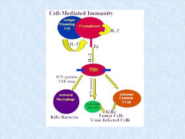
TH 1 based immune response
TH 1 based immune response - inflammatory reaction § TH 1 cells cooperate with macrophages and activate them (NO production - destroy intracellular parasites) § Activated macrophages secrete some cytokines (IL-1, TNF, . . . ) that help to stimulate T cells and stimulate local inflammation, which helps suppress infection § Interaction between TH 1 cells and macrophages is a fundamental mechanism of delayed-type immunopathological reactions (DTH Delayed-type hypersensitivity)
TH 1 based immune response § The infected macrophage produces protein fragments derived from intracellular parasites, some of which are presented on the surface in the complex with MHC gp class II § Macrophages and dendritic cells stimulated by certain microorganisms produce IL-12 § TH precursor, which detects the infected macrophage and receives signals via the TCR, CD 28 and receptor for IL-12 proliferates and differentiates into effector TH 1 cells that produce IFNg and IL-2. § IFNg activates macrophage NO synthase IL-2 is growth factor for T cells
Interaction between APC and TH precursor
TH 2 based immune response
TH 2 based immune response § TH 2 cells cooperate with B lymphocytes (which were stimulated by Ag) by cytokine production (IL-4, IL-5, IL-6, IL-10) and direct intercellular contact (CD 40 L) § For stimulation of B lymphocytes is usually necessary cooperation between APC → TH 2 cell → B lymphocyte § In minimal model, where the B cell becomes a good APC (CD 80, CD 86) is sufficient cooperation between TH 2 cell → B lymphocyte
TH 2 based immune response • TH precursor, which detects the infected macrophage and receives signals through the TCR, CD 28 , IL-4 receptor and IL-2 receptor proliferates and differentiates in the effector TH 2, which provide B lymphocytes auxiliary signals via secreted cytokines IL-4, IL-5, IL-6, IL-10 and molecule CD 40 L, which bind to the costimulatory receptor on B lymphocytes CD 40
TH 2 based immune response § Interaction between CD 40 (B lymphocytes) and CD 40 L (TH 2 cells) is essential for the initiation of somatic mutations, izotype switching and formation of memory cells § IL-4, IL-5, IL-6, IL-10: stimulation of B lymphocytes
Function of TH 2 cells
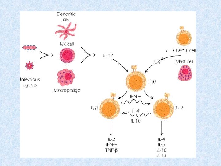
Mutual regulation of activities TH 1 versus TH 2 § Whether the TH precursor cell will develop into TH 1 or TH 2 decides cytokine ratio of IL-12 and IL-4 § IL-12 is produced by macrophages and dendritic cells stimulated by certain microorganisms § IL-4 is produced by activated basophils, mast cells and TH 2 cells § TH 1 cytokines (mainly IFNg) inhibit the development of TH 2 and stimulate the development of TH 1 (IL-2 stimulates also TH 2) § Cytokines produced by TH 2 (IL-4, IL-10) inhibit the development of TH 1 and stimulate the development of TH 2
TC based immune response
Cytotoxic T lymphocytes stimulation § TC recognize cells infected with viruses or other intracellular parasites, and some tumor cells § Precursor of TC, which recognizes a peptide-MHC gp. I complex on the surface of APC via TCR and receives signals via CD 28 proliferates and differentiates to clone mature effector cytotoxic cells (CTL) § For full TC activation is necessary IL-12 § CTL are spread by bloodstream into tissues; for activation of cytotoxic mechanisms is sufficient signal through the TCR (signal through a costimulatory receptor CD 28 is no longer necessary)
§ Professional APC are dendritic cells or macrophages that are infected with virus, or swallowed antigens from dead infected, tumor or stressed cells § In order APC could activate the TC precursor, APC must be stimulated by contact with TH 1 cell via CD 40, then the dendritic cell begins to express CD 80, CD 86 and secrete cytokines (IL-1, IL-12) = change of resting APC in activated
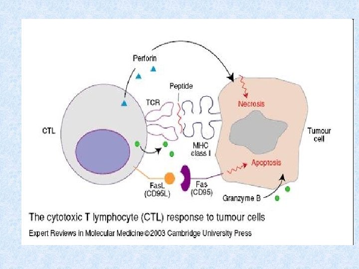
Tc effector functions § Cytotoxic granules containing perforin, granzymes and granulysin § Fas ligand (Fas. L) - which binds to the apoptotic receptor Fas (CD 95) presented on the surface of many different cells (also on the surface of TC) § TNFb § Activation of effector mechanismus leads to apoptotic death of the target cell.
Thank you for your attention
• T cell development http: //www. youtube. com/watch? v=od. LLr 6 mja. UQ • TLR receptors http: //www. youtube. com/watch? v=i. VMIZy-Y 3 f 8 • MHC II prezentation http: //www. youtube. com/watch? v=_8 JMVq 7 HF 2 Y • MHC I prezentation http: //www. youtube. com/watch? v=vr. FMWy. Jw. Gxw