HLA in more detail 1 What is HLA


- Slides: 28

HLA - in more detail 1

What is HLA? • HLA = Human Leukocyte Antigen – a system of cell surface proteins, originally defined by their antigenicity. – HLA antigens are also referred to as (major) histocompatibility antigens or major histocompatibility complex (MHC) antigens. 2

What is histocompatibility? • The word from histo- (tissue) and compatible. • Originally defined through tissue grafting – tissues from unrelated individuals are rejected. • The genes controlling rejection map to histocompatibility loci both MAJOR and minor – major because they control rapid rejection, minor much slower. 3

What is histocompatibility? • The major loci are very closely linked in one region of the genome: the major histocompatibility complex - MHC; – many individual minor loci are scattered about the genome. 4

What is histocompatibility? • The MHC loci are highly polymorphic – i. e. multiple alleles so that different individuals encode different MHC antigens. • This polymorphism defines the immunological identity of individuals. 5

The nature of MHC antigens – This discussion will be confined to the human HLA system • There are two classes of HLA antigen, I & II, which differ in structure, cellular distribution, and function. • There are 3 class I antigens A, B & C; • There are 3 class II antigens DR, DB & DQ. 6

The nature of HLA antigens • Nomenclature: initially HLA types were defined antigenically i. e. through their reaction with specific antibodies & assigned a type number – so HLA-A 1, -A 2, A 3 etc. • There are 20 HLA-A, 42 HLA-B, 8 HLA-C, 18 HLA-DR, and 6 HLA-DQ types defined in this way – and for most purposes this is enough. 7

The nature of HLA antigens • Nomenclature: BUT when sequences of HLA proteins were determine, it was found that these serotypes represent large families of molecular allelic types. • These are defined by a 4 to 8 digit number: – the first 2 digits are the old serotype hence HLA-A 2 becomes HLA-A*02001 etc. Go to http: //www. anthonynolan. org. uk/HIG/nomen/reports/nomen_r eport s. html for more detail 8

The nature of HLA antigens • You do not need to know about MHC genetics, except that – the HLA genes are polymorphic; – there are separate loci for A, B & C (class I) and pairs of loci for DR, DP & DQ (class II) (remember, 2 variable chains & for class II); – the loci on both chromosomes are expressed so that there are potentially 6 different HLA antigens on a cell surface. 9

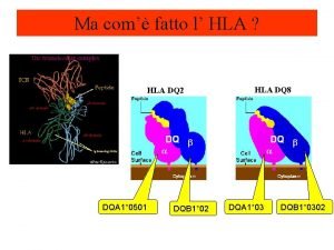
The nature of HLA antigens • Structure: All are heterodimeric cell surface proteins. – Class I are composed of a three-domain heavy chain which is polymorphic and a single domain light chain ( 2 -microglobulin) which is constant. – Class II are two equal chains, & , each with 2 domains: both chains are polymorphic (except DR ). 10

The nature of HLA antigens • Structure: Although class I and II differ in detailed structure they all have overall the same gross form: a 4 -domain structure with a distinct “cleft” in the membrane distal region. 11

Ribbon diagram of HLA A Note the domain structure: the heavy chain with three domains 1 2 3 plus 2 microglobulin. It is 2 microglobulin that anchors the molecule to the cell surface so that the cleft is distal to the cell. Note that the cleft has a floor of -pleated sheet and sides of -helix. 12

The expression of HLA antigens • Class I antigens A, B & C are variably expressed by all nucleated cells except hepatocytes and CNS cells – their expression is controlled at the level of transcription by cytokines, which can increase expression in cells already expressing or induce in those not expressing. 13

The expression of HLA antigens • Class II antigens DR, DP & DQ are expressed by limited cell types – constitutively only by B cells and dendritic cells; – inducibly by macrophages and some other cell types e. g. keratinocytes. – Again, their expression is controlled at the level of transcription by cytokines. 14

The interaction of HLA antigens and peptide • Remember that the role of HLA antigens is to “present” peptides to T cells. • X ray pictures show short peptides in the cleft with residues at the ends attached to the “floor” of the cleft. 15

The interaction of HLA antigens and peptide • Different allelic forms of the same HLA antigen associate with different sets of peptides – that is, one single HLA protein binds many different peptides (this is referred to as “promiscuous” binding); – but these peptides will have some relatedness i. e. common amino acids, particularly those called “anchor residues” that bind to the cleft. 16

The interaction of HLA antigens and peptide • The “polymorphic residues” i. e. those that differ between different alleles are concentrated in the -pleated region of the floor • this accounts for the binding of different peptide sets by different HLA antigens. 17

Ribbon diagram of HLA A Note the domain structure: the heavy chain with three domains 1 2 3 plus 2 microglobulin. It is 2 microglobulin that anchors the molecule to the cell surface so that the cleft is distal to the cell. Note that the cleft has a floor of -pleated sheet and sides of -helix. 18

The interaction of HLA antigens and peptide • A reasonable guess at why HLA antigens are polymorphic is to ensure that across a population there is sufficient diversity to ensure that all antigens are catered for – the start of a new story; – and it is not that HLA is there to frustrate transplant surgeons. 19

“Processing” • This is the means by which protein antigens are degraded to peptides which associate with HLA – it is different for class I and class II. 20

Class I Processing • This deals with intracellular, cytoplasmic “endogenous” proteins. • All proteins within the cell are sampled: proteasomes degrade cellular proteins to peptides which are transferred to the endoplasmic reticulum (ER) via the Transporter important in Antigen Presentation (TAP). 21

Class I Processing • Within the ER peptides complex with class I antigens and are then transported to the cell surface where they are presented to CTL – this amounts to intracellular surveillance: all cell proteins are sampled, but will only activate CTL if derived from a foreign protein. 22

Class I Processing - for intracellular surveillance CD 8+CTL 5. where it is presented for inspection by a passing CTL Interior of cell 1. Cytoplasmic protein “processed” to peptides thru’ proteasome 2. which enter ER via TAP 4. the HLA/peptide complex is transported to the cell surface 3. and associate with HLA class I Endoplasmic reticulum (ER) 23

Class II Processing • This deals with extracellular, “exogenous”, proteins taken up by the antigen presenting cell into some sort of phagosome. • Proteins are degraded to peptides within the phagosome. • The phagosome fuses with an exocytic vesicle containing class II antigens: the peptide binds to the class II antigen. 24

Class II Processing • The vesicle containing the peptide/class II complex travels to the cell surface where the complex is presented to helper T cells – this amounts to extracellular surveillance: all proteins in the cell’s environment are sampled, but will only activate helper cells with TCR for a foreign protein. 25

Class II Processing - for extracellular surveillance 1. Extracellular protein taken up into endo/ phagocytic vesicle 2. where it is degraded to peptides. CD 4+ Th 5. where it is presented for inspection by a passing helper T cell 4. & moves to the cell surface wh the complex inserts into the mem 3. Exo & endocytic vesicles fuse & class II/peptide complex forms Interior of APC Exocytic vesicle containing MHC class II 26

Conclusion • The next slide summarises everything • Except it doesn’t show CD 4 or CD 8: their role is to interact with class II and class I respectively in order to stabilise the interaction and they are also involved in signalling – so CD 4 cells recognise class II antigen plus peptide – and CD 8 ditto class I. 27

The TCR- peptide. HLA complex TCR Peptide (brown) hunched up with anchor residues P 1, P 8 interacting with HLA: TCR sees both peptide and HLA 28
 More more more i want more more more more we praise you
 More more more i want more more more more we praise you
 Major details vs minor details
 What are major details
 Pygalgie traitement
 Hla b35 artrite psoriasica
 Hla typizace
 Fcm 원리
 Color 05042008
 Palm sunday hla
 Beurteilung lehrplan 21 kanton bern
 Mhc and hla
 Hla b27 pozitivita
 Hla b44
 Ulcerative colitis hla
 Hla typizace
 Mhc and hla
 More love to thee o lord
 Aspire not to
 More choices more chances
 Example of newtons first law
 Human history becomes more and more a race
 5 apples in a basket riddle
 Knowing more remembering more
 The more you study the more you learn
 Skimming reading images
 Xoserve-detail-page-one (xoserveservices.com)
 Vary preposition
 Perimeter fire containment systems
