
PBS 803 Class 5 Antigen Capture and Presentation
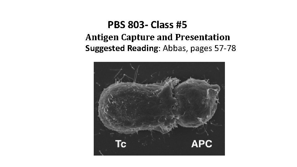
PBS 803 - Class #5 Antigen Capture and Presentation Suggested Reading: Abbas, pages 57 -78
Learning Objectives for Class #5 part I · List several examples of APC’s (e. g. dendritic cells, macrophages, B cells). · Describe the function of MHC molecules both in the initial antigen presentation and in subsequent cell-cell interactions of the immune system. · List the major structural features of the MHC gene products (e. g. , Class I molecules are 2 chains, a heavy chain and a beta-2 microglobulin chain). · Compare the tissue distribution of class I and class II MHC molecules. · Explain MHC polymorphism and the selective advantage of polymorphic MHC
Learning Objectives for Class #5 part 2 · Describe the concept of MHC-restricted antigen recognition and diagram this recognition process for both helper and CTLs · Define: haplotypes, genotypes, phenotypes, alleles · Compare and contrast the presentation of exogenous and endogenous antigens to T lymphocytes · Describe the conversion of polypeptides to peptide antigens via antigen processing (e. g. the roles of compartments, ubiquitin, proteases, TAP, CLIP etc. ) · Compare and contrast the presentation of exogenous and endogenous antigens to T lymphocytes. · Describe and explain the importance of cross-presentation
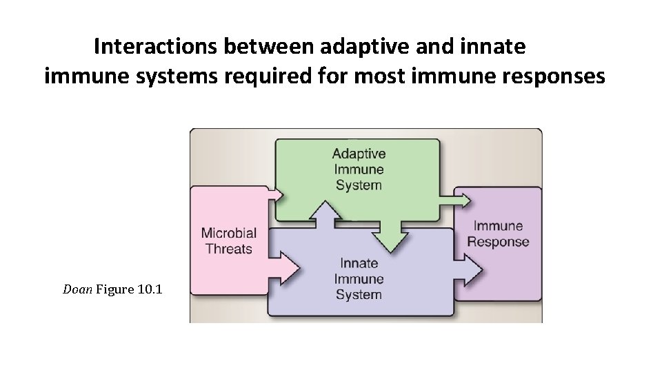
Interactions between adaptive and innate immune systems required for most immune responses Doan Figure 10. 1
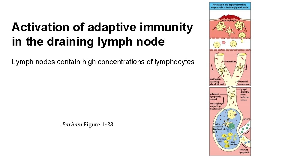
Activation of adaptive immunity in the draining lymph node Lymph nodes contain high concentrations of lymphocytes Parham Figure 1 -23
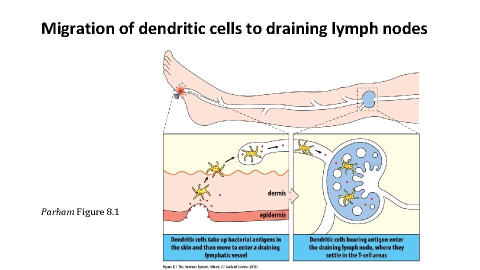
Migration of dendritic cells to draining lymph nodes Parham Figure 8. 1
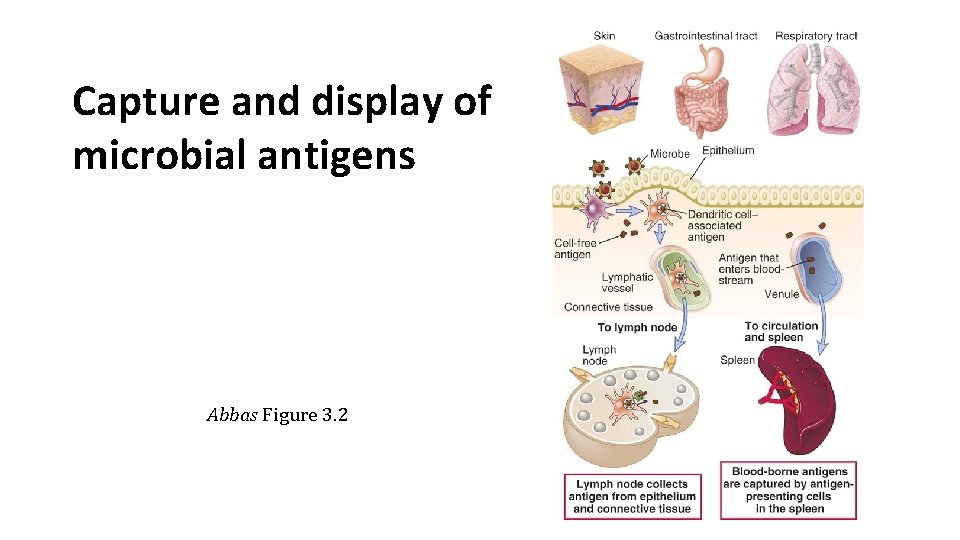
Capture and display of microbial antigens Abbas Figure 3. 2
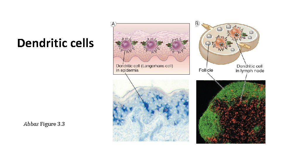
Dendritic cells Abbas Figure 3. 3
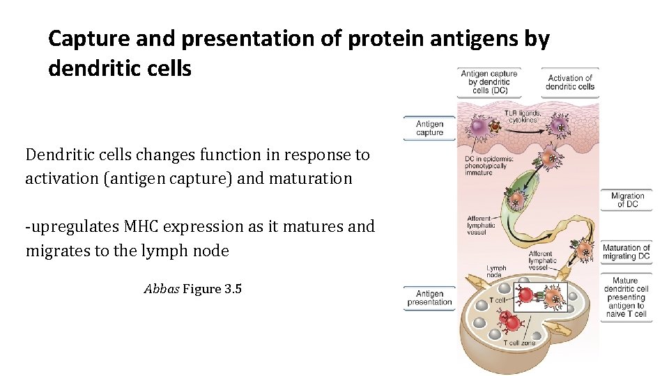
Capture and presentation of protein antigens by dendritic cells Dendritic cells changes function in response to activation (antigen capture) and maturation -upregulates MHC expression as it matures and migrates to the lymph node Abbas Figure 3. 5
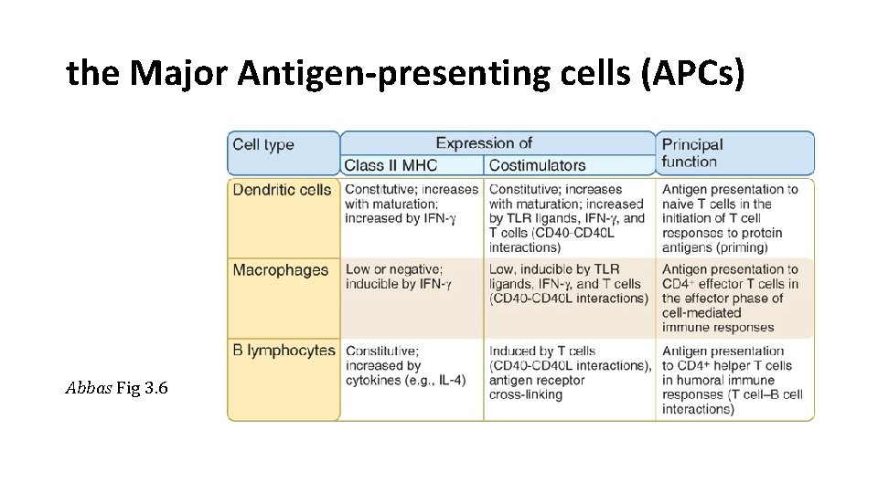
the Major Antigen-presenting cells (APCs) Abbas Fig 3. 6
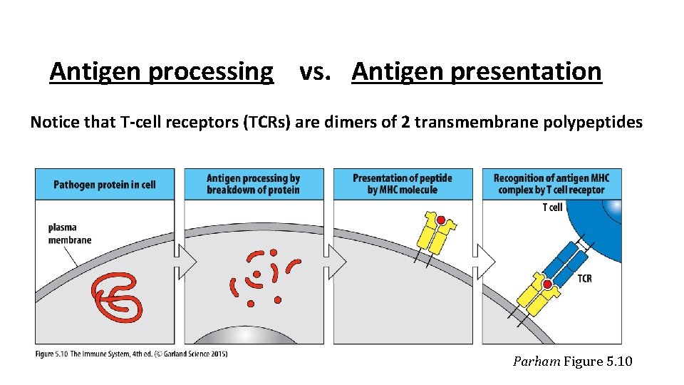
Antigen processing vs. Antigen presentation Notice that T-cell receptors (TCRs) are dimers of 2 transmembrane polypeptides Parham Figure 5. 10
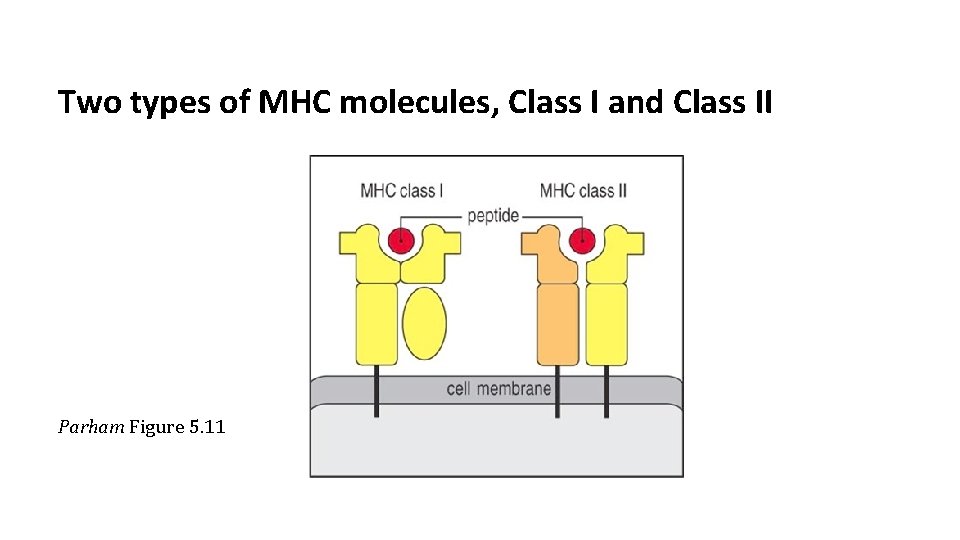
Two types of MHC molecules, Class I and Class II Parham Figure 5. 11
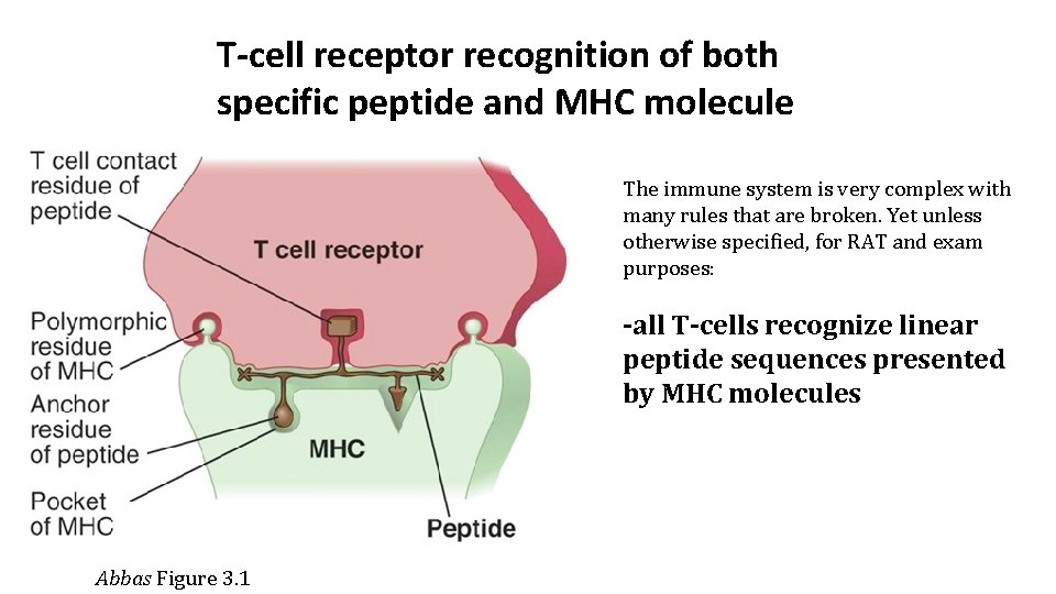
T-cell receptor recognition of both specific peptide and MHC molecule The immune system is very complex with many rules that are broken. Yet unless otherwise specified, for RAT and exam purposes: -all T-cells recognize linear peptide sequences presented by MHC molecules Abbas Figure 3. 1
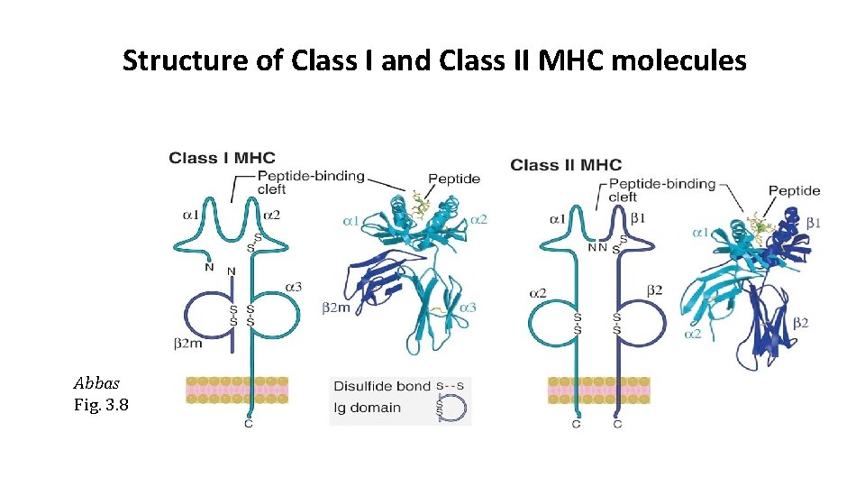
Structure of Class I and Class II MHC molecules Abbas Fig. 3. 8

Genetic organization of MHC Class I and Class II loci each person inherits 2 sets of haplotypes, one from each parent, increases diversity Haplotype –set of closely linked genes inherited as a unit from one parent Allele- one of multiple forms of a gene that can exist at the same locus within the genome Polymorphism – multiple alleles for a gene present within individual or population Doan Figure 6. 7
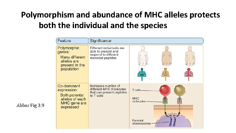
Polymorphism and abundance of MHC alleles protects both the individual and the species Abbas Fig 3. 9
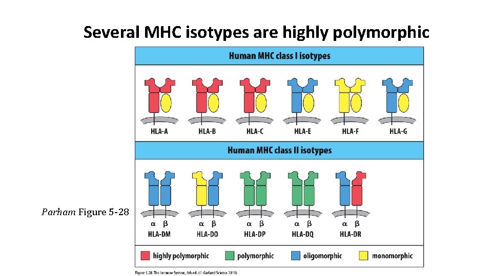
Several MHC isotypes are highly polymorphic Parham Figure 5 -28
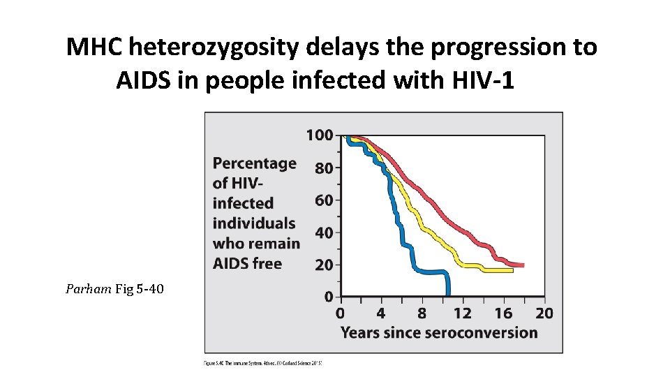
MHC heterozygosity delays the progression to AIDS in people infected with HIV-1 Parham Fig 5 -40
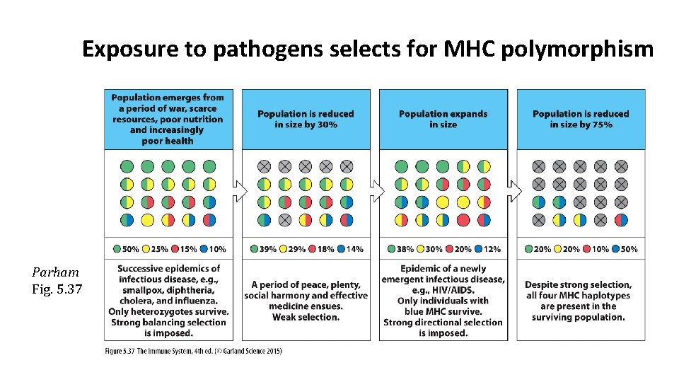
Exposure to pathogens selects for MHC polymorphism Parham Fig. 5. 37
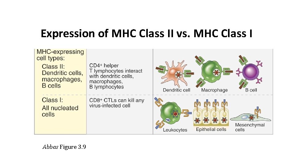
Expression of MHC Class II vs. MHC Class I Abbas Figure 3. 9
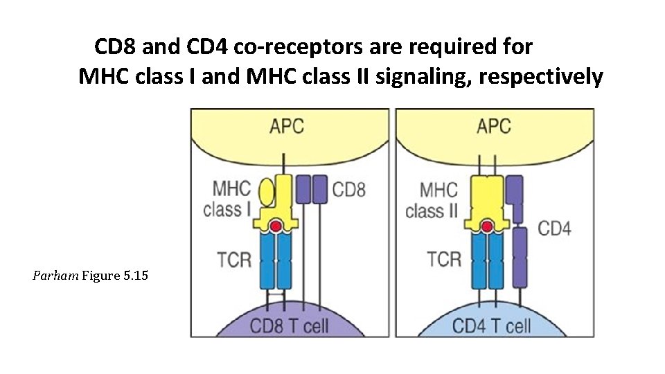
CD 8 and CD 4 co-receptors are required for MHC class I and MHC class II signaling, respectively Parham Figure 5. 15
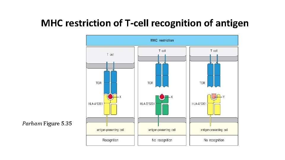
MHC restriction of T-cell recognition of antigen Parham Figure 5. 35
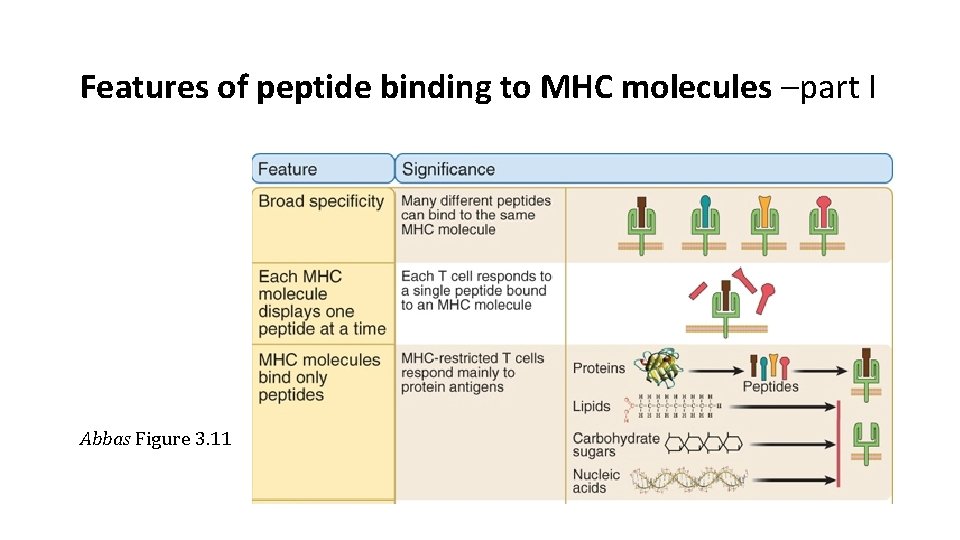
Features of peptide binding to MHC molecules –part I Abbas Figure 3. 11
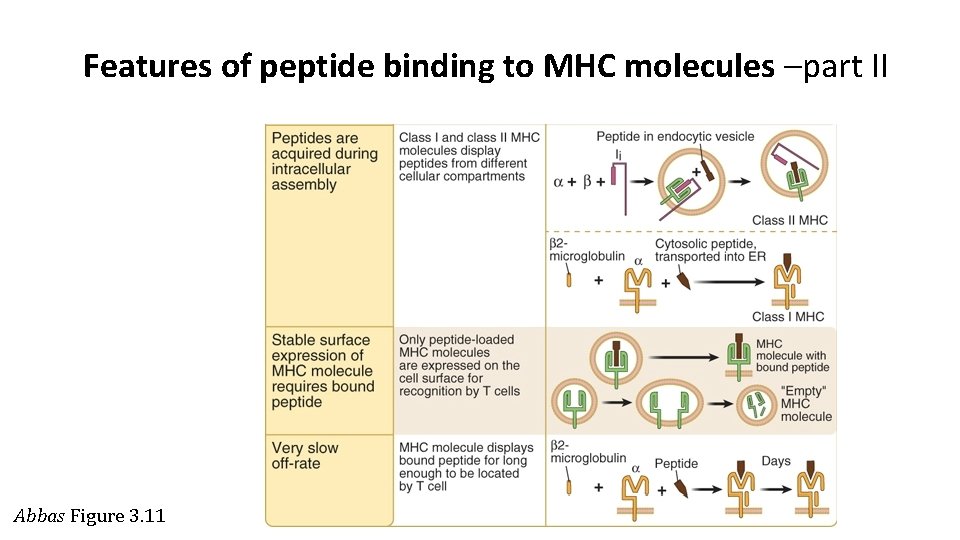
Features of peptide binding to MHC molecules –part II Abbas Figure 3. 11
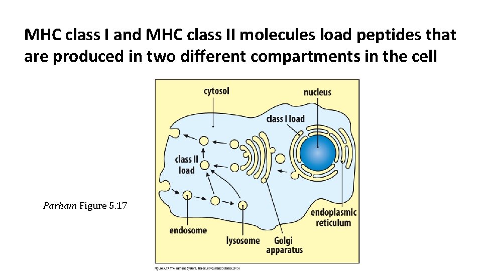
MHC class I and MHC class II molecules load peptides that are produced in two different compartments in the cell Parham Figure 5. 17
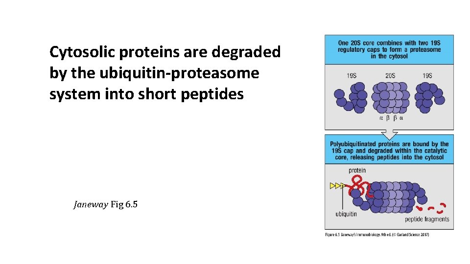
Cytosolic proteins are degraded by the ubiquitin-proteasome system into short peptides Janeway Fig 6. 5
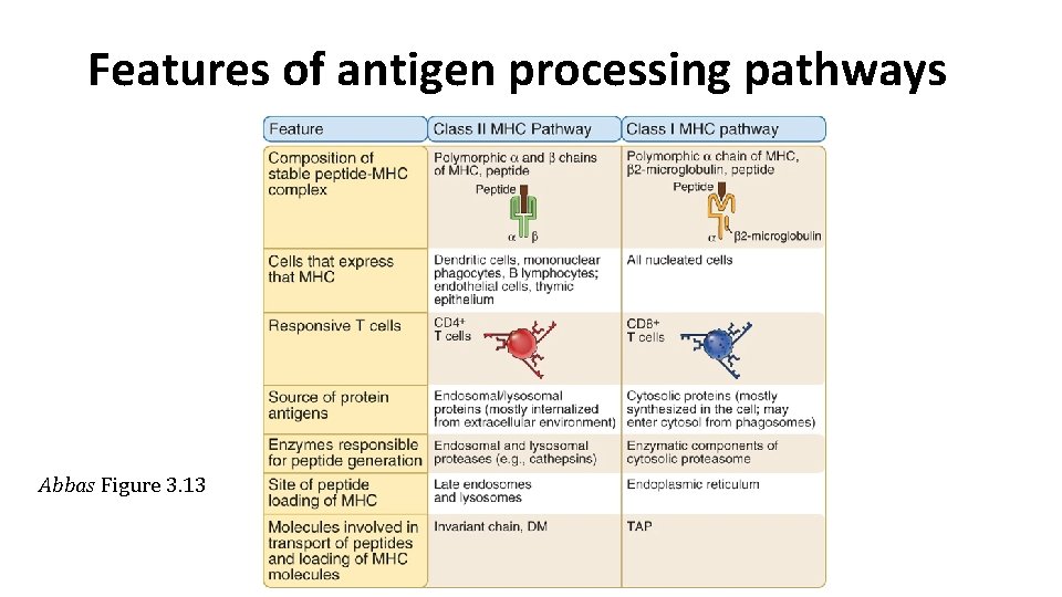
Features of antigen processing pathways Abbas Figure 3. 13
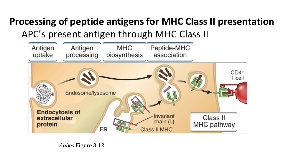
Processing of peptide antigens for MHC Class II presentation APC’s present antigen through MHC Class II Abbas Figure 3. 12
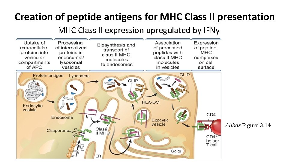
Creation of peptide antigens for MHC Class II presentation MHC Class II expression upregulated by IFNγ Abbas Figure 3. 14
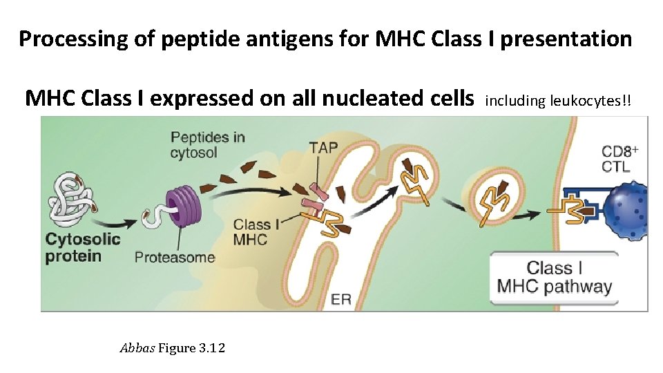
Processing of peptide antigens for MHC Class I presentation MHC Class I expressed on all nucleated cells including leukocytes!! Abbas Figure 3. 12
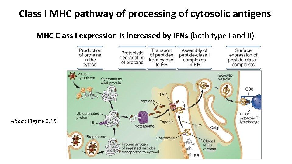
Class I MHC pathway of processing of cytosolic antigens MHC Class I expression is increased by IFNs (both type I and II) Abbas Figure 3. 15
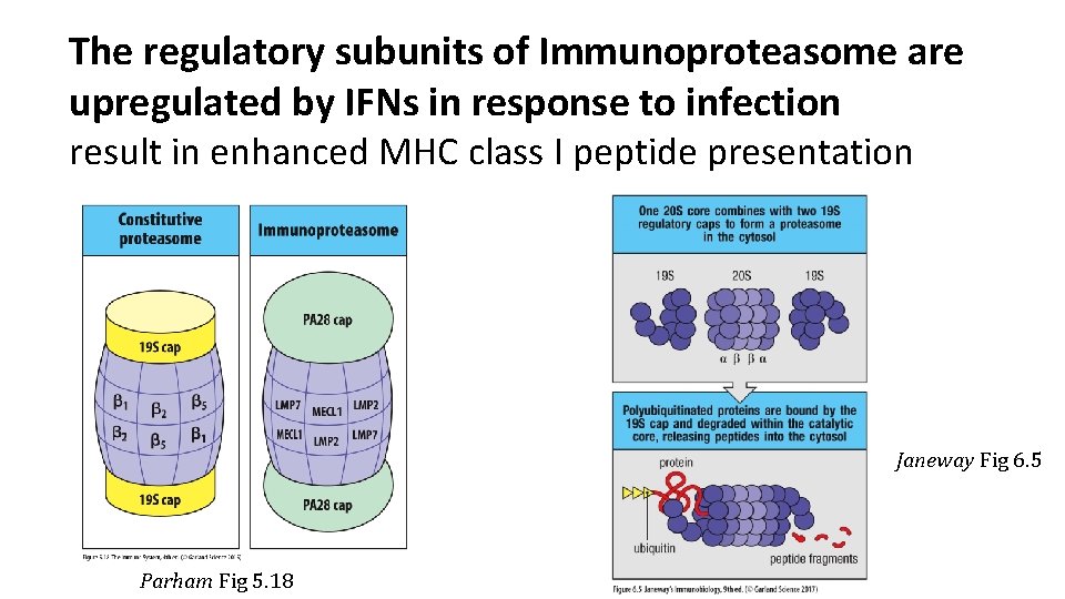
The regulatory subunits of Immunoproteasome are upregulated by IFNs in response to infection result in enhanced MHC class I peptide presentation Janeway Fig 6. 5 Parham Fig 5. 18
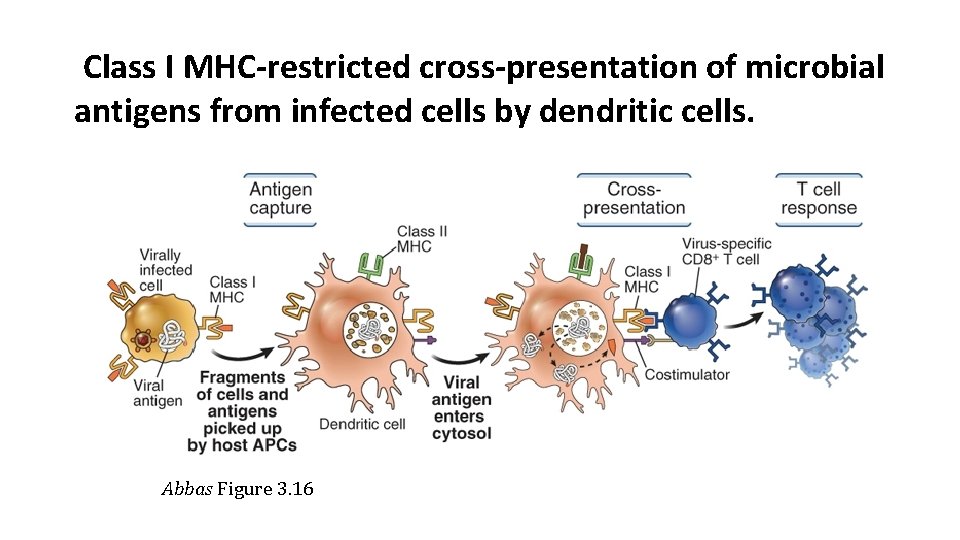
Class I MHC-restricted cross-presentation of microbial antigens from infected cells by dendritic cells. Abbas Figure 3. 16
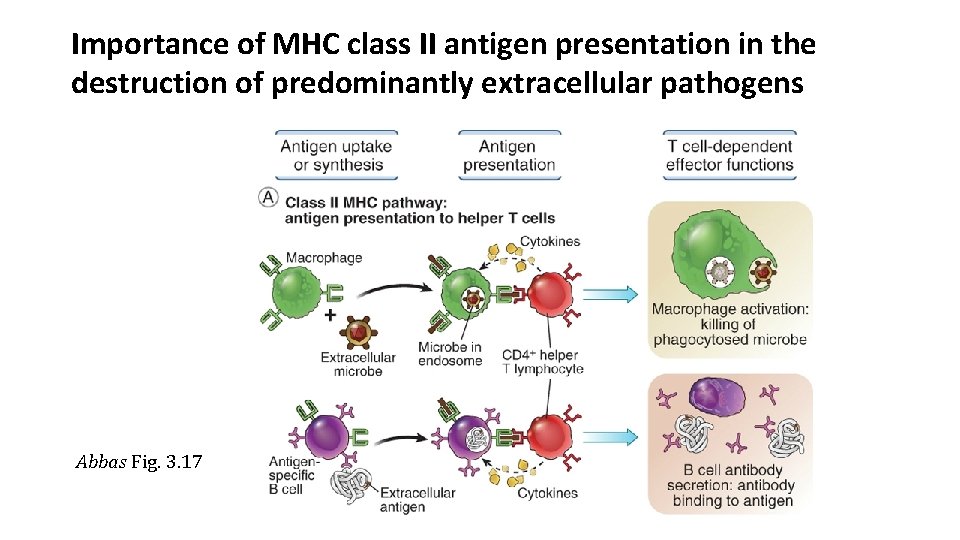
Importance of MHC class II antigen presentation in the destruction of predominantly extracellular pathogens Abbas Fig. 3. 17
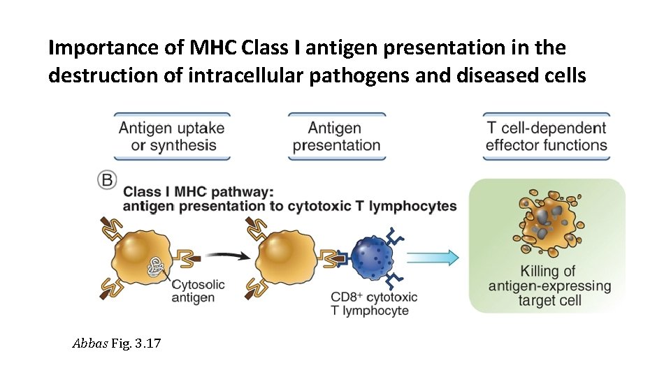
Importance of MHC Class I antigen presentation in the destruction of intracellular pathogens and diseased cells Abbas Fig. 3. 17
- Slides: 35