Hemolytic Anemia Dr Ali Khazaal Jumaa F I
 F.")


 2 - Alloimmune")
 2 - Membrane defect (")


 2 - rheumatic disorders (RA,")


 Free autoantibody may be detected by the indirect antiglobulin test")

 - Rituximab -")


 - Acquired erythrocyte fragmentation occurs when RBC are forced at")
")
 Thrombotic thrombocytopenic purpura (TTP) Hemolytic uremic syndrome (HUS) HELLP")

 DIC is characterized by systemic activation of blood coagulation, which")


 - Prolonged partial thromboplastin")


 is a hematological emergency characterized by clotting in small blood")


 - Congenital : This hereditary form of TTP")


 - Rituximab - chemotherapy")

 Hemolytic-uremic syndrome (HUS) is a clinical syndrome characterized by progressive")

![Typical (Stx–associated) HUS (t. HUS) - Typical HUS (Shiga-like toxin–associated HUS [Stx-HUS]) is the](https://slidetodoc.com/presentation_image_h2/6c8b138ab2ae33ddb3b52d53269f5cac/image-65.jpg "Typical (Stx–associated) HUS (t. HUS) - Typical HUS (Shiga-like toxin–associated HUS [Stx-HUS]) is the")
 HUS (a. HUS) Non–Stx-HUS, or atypical HUS, is less common and accounts")


 - Fever (56%), bloody diarrhea")
 Edema, fluid")


 Response Sepsis Malignancy DIC")
 plt Coagulopat hy (PT, PTT, d-dimer) Compleme ADAMTS 13 CNS Renal")
- Slides: 77
Hemolytic Anemia Dr. Ali Khazaal Jumaa F. I. B. M. S (Internal Medicine) F. I. B. M. S (Clinical Hematology)
Objectives: Definition of Hemolytic anemia Classification Clinical presentation Approach consideration Immune – mediated hemolytic anemia
Definition Hemolysis is the premature destruction of erythrocytes. A hemolytic anemia will develop if bone marrow activity cannot compensate for the erythrocyte loss. The severity of the anemia depends on whether the onset of hemolysis is gradual or abrupt and on the extent of erythrocyte destruction.
Classification Pathophysiological Congenital or acquired Site of hemolysis: Intramedullary Intravascular Extravascular
Extracorpuscular Immune - mediated 1 - Autoimmune (warm ab, cold ab) 2 - Alloimmune (incompatible b. transfusion) 3 - Drug - induced Mechanical injury 1 - Microangiopathic HA (DIC, TTP, HUS) 2 - Trauma (prosthetic valve, thermal, exercise) 3 - Hypersplenism
Intracorpuscular Congenital 1 - Hemoglobinopathy (SCD, thalassemia, unstable Hb) 2 - Membrane defect ( her. spherocytosis, her. elliptocytosis, her. ovalocytosis) 3 - Enzymopathy (G 6 PD def, pyruvate kinase def) Acquired 1 - Paroxysmal nocturnal hemoglobinurea (PNH) 2 -Infection (malaria)
History Signs and symptoms of hemolytic anemia are diverse and are due to anemia, the extent of compensation, previous treatment, and the underlying disorder. Patients with minimal or long-standing hemolytic anemia may be asymptomatic, and hemolysis is often found incidentally during routine laboratory testing. Clinical manifestations may include the following: - In intravascular hemolysis, iron deficiency due to chronic hemoglobinuria can exacerbate anemia and weakness. - Tachycardia, dyspnea, angina, and weakness occur in patients with severe anemia, as cardiac function is sensitive to anoxia.
- Persistent hemolysis may result in the development of bilirubin gallstones; these patients may present with abdominal pain. - Bronze skin color and diabetes occur in hematosiderosis; iron overload may occur in patients who have received multiple transfusions or those who have been administered iron therapy erroneously. - Dark urine may be due to hemoglobinuria or increased urobilinogen. - In addition to hemolysis, patients with thrombotic thrombocytopenic purpura (TTP) may experience fever, neurologic signs, renal failure, and thrombocytopenia.
- Leg ulcers may develop in patients with sickle cell anemia and other hemolytic disorders, as a result of decreased red blood cell (RBC) deformability and endothelial changes. - Venous thromboembolism occurs in % of adults with warm autoimmune hemolytic anemia. Children with hereditary spherocytosis (HS) are also at increased risk for thrombosis especially in those who are experiencing a hemolytic crisis. - Patients with PNH may present with pancytopenia and/or thrombotic events
Physical examination Anemia Jaundice Splenomegaly Disease - related
Approach Considerations Standard blood studies for the workup of suspected hemolytic anemia include the following: Complete blood cell count Reticulocyte count Peripheral blood smear Serum lactate dehydrogenase (LDH) Serum haptoglobin Indirect bilirubin
Haptoglobin It is a protein produced mostly by liver, functions to bind the free plasma hemoglobin that has been released by damaged red blood cells, which allows degradative enzymes to gain access to the hemoglobin while at the same time preventing loss of iron through the kidneys and protecting the kidneys from damage by free hemoglobin. A low serum haptoglobin level is a criterion for moderate-to-severe hemolysis. A decrease in serum haptoglobin is more likely in intravascular hemolysis than in extravascular hemolysis. However, it is an acute phase reactant. Therefore, haptoglobin levels can be normal or elevated despite significant hemolysis in patients with infections and in other reactive states.
Autoimmune Hemolytic Anemia
Hemolytic Anemia Resulting from Warm-Reacting Antibodies It is the result of host antibodies that react with autologous RBC. AIHA may be classified by as secondary or primary (idiopathic). AIHA may also be classified by the nature of the antibody. “Warm-reacting” antibodies are usually of the Ig. G type, have optimal activity at 37°C, and bind complement. “Cold-reacting” antibodies show affinity at lower temperatures. Warm antibody AIHA is the most common type.
Secondary AIHA 1 - lymphoproliferative disorders ( CLL, NHL) 2 - rheumatic disorders (RA, SLE) 3 - infections (Mycoplasma pneumoniae) 4 - solid tumors (CA ovary, CA stomach) 5 - chronic inflammatory disorders ( ulcerative colitis) 6 - drugs ( methyl dopa, fludarabine)
- Antibody-coated RBCs are trapped by macrophages primarily in the spleen, where they are ingested and destroyed and a spherocyte with a lower surface area to- volume ratio is released. . - Large quantities of Ig. G will increase trapping of RBCs by macrophages in the liver and spleen. - RBC may be destroyed by monocytes or lymphocytes by direct cytotoxic activity, without phagocytosis. - Antibodies may also attach to late erythroid precursors and suppress erythropoiesis.
Laboratory features - Anemia can range from mild to life-threatening. - Blood film reveals polychromasia (indicating reticulocytosis) and spherocytes
Platelet count Evans syndrome is a condition in which both autoimmune-mediated RBC and platelet destruction occur. Reticulocyte % A low neutrophil count, as a result of immune neutropenia, may also be present. Marrow examination usually reveals erythroid hyperplasia Unconjugated hyperbilirubinemia Haptoglobin LDH Urinary urobilinogen
Serologic Features The diagnosis of AIHA requires demonstration of immunoglobulin and/or complement bound to the RBCs. This is achieved by the direct antiglobulin test (DAT), Coomb’s test, in which rabbit antiserum to human Ig. G or complement is added to suspensions of washed patient’s RBCs. Agglutination of the RBCs signifies the presence of surface Ig. G or complement. RBC may be coated with: — Ig. G alone — Ig. G and complement — Complement only Rarely, anti-Ig. A and anti-Ig. M reactions are encountered.
Direct Coomb’s Test
Indirect Coomb’s test (IAT) Free autoantibody may be detected by the indirect antiglobulin test (IAT), in which the patient’s serum is incubated with normal donor RBCs, which are then tested for agglutination by the addition of antiglobulin serum.
- A positive IAT with a negative DAT probably does not indicate autoimmune disease but an alloantibody generated by a prior transfusion or pregnancy. - Occasional patients exhibit all the features of AIHA but have a negative DAT. The amount of their RBC-bound autoantibody is too low for detection by DAT but can often be demonstrated by more sensitive methods, such as enzymelinked immunoassay or radioimmunoassay.
Treatment - Observation : for asymptomatic pt - Corticosteroid (prednisolone, dexamethasone) - Rituximab - Immunosuppressant : cyclophosphamide, azathioprine, mycophenolate mofetil - Splenectomy - Rx of the underlying cause
Prognosis - Idiopathic warm-antibody AHA runs an unpredictable course characterized by remissions and relapses. - Survival at 10 years is approximately 70%. - In addition to anemia, deep venous thrombosis, pulmonary emboli, splenic infarcts, and other cardiovascular events occur during active hemolytic disease. - In secondary warm-antibody AHA, prognosis is related to the underlying disease. - AIHA related to infection is self-limited and responds well to glucocorticoids.
Cold Agglutinin-Mediated AIHA - This type of anemia is caused by autoantibodies that bind red cells best at temperatures below 37°C, usually below 31°C. - The complement system plays a major role in red cell destruction. - It is classified as either primary (chronic cold agglutinin disease) or secondary (generally as a result of Mycoplasma pneumoniae infection or Epstein-Barr virus, or lymphoproliferative disorders, like Waldenstrom macroglobulinemia). - Peak incidence for the primary (chronic) syndrome is in persons older than 50 years. - This disorder characteristically has monoclonal Ig. M cold agglutinins
Clinical Features - Cold-agglutinin–mediated hemolysis accounts for 10% to 20% of all cases of autoimmune hemolytic anemia. - Women are affected more commonly than men. - Acrocyanosis is frequently observed, but skin ulceration and necrosis are uncommon. - Splenomegaly may occasionally be seen in the idiopathic form. - The hemolysis caused by mycoplasma infection develops as the patient recovers from the infection and is self-limited, lasting 1 to 3 weeks.
Laboratory Features - Anemia is usually mild to moderate. On the blood film the red cells show autoagglutination, polychromasia, and spherocytosis. - High serum titers of cold agglutinins (generally Ig. M) - Positive DAT
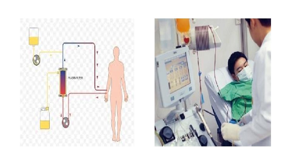
Treatment - Treatment of the underlying cause - Keeping the patient warm is important and may be the only treatment needed for mild conditions. - Rituximab - Fludarabine, Chlorambucil or cyclophosphamide have been used for more severe chronic cases. - Splenectomy and glucocorticoids generally are not helpful, although very high dose glucocorticoids may be useful in severely ill patients. - In critically ill patients, plasmapheresis may provide temporary relief.
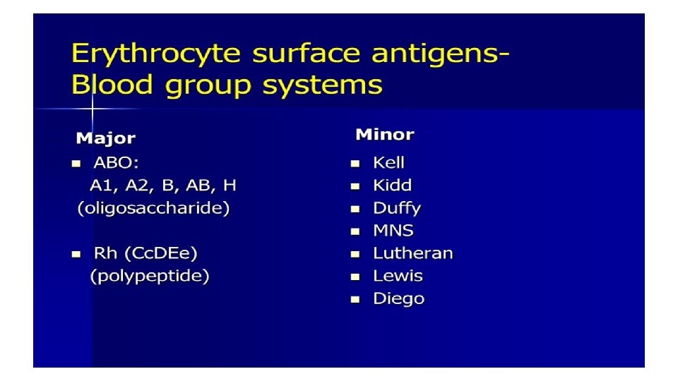
Transfusion – Induced Alloimmunization Allogeneic blood transfusion is a form of temporary transplantation. This procedure introduces a multitude of foreign antigens and living cells into the recipient that persist for a variable time. A recipient who is immunocompetent may mount an immune response to the donor antigens (ie, alloimmunization), resulting in various clinical consequences, depending on the blood cells and specific antigens involved. The antigens most commonly involved can be classified into the following categories: (1) human leukocyte antigens (HLAs), class I shared by platelets and leukocytes and class II present on some leukocytes; (2) granulocyte-specific antigens; (3) platelet-specific antigens (human platelet antigens [HPAs]); and (4) RBC-specific antigens.
Alloimmunization against RBCs Acute intravascular hemolytic transfusion reactions (rarely a consequence of alloimmunization and almost always caused by ABO antibodies). Delayed hemolytic transfusion reactions (DHTRs) (hemolysis caused by RBC alloantibodies typically presenting clinically 7– 14 days after transfusion). Hemolytic disease of the fetus and newborn (mother's alloimmunization against red blood cell fetal antigens, most often resulting from previous pregnancies).
Acute hemolytic transfusion reaction AHTR is a life-threatening reaction to receiving a blood transfusion. AHTRs occur within 24 hours of the transfusion and can be triggered by a few milliliters of blood. The reaction is triggered by naturally – occurring or pre-formed host antibodies destroying donor red blood cells. AHTR typically occurs when there is an ABO blood group incompatibility.
Clinical presentation Early acute hemolytic transfusion reactions are typically characterized by fever, which may be accompanied by rigors (chills). Mild cases are also typically characterized by abdominal, back, flank, or chest pain. More severe cases may be characterized by shortness of breath, low blood pressure, hemoglobinuria, and may progress to shock and disseminated intravascular coagulation. In anesthetized or unconscious patients, hematuria may be the first sign. Other symptoms include nausea, vomiting, and wheezing. DAT +ve
Management - Discontinuation of the transfusion - Fluid replacement - Close monitoring of vital and supportive care, which may include diuretics, blood pressure support - Treatment of disseminated intravascular coagulation (with fresh frozen plasma, cryoprecipitate, and platelet transfusion - Dopamine is used for blood pressure support because it causes vasodilation (dilation of blood vessels) in the kidneys as well as increasing the cardiac output.
Microangiopathic Hemolytic Anemia (MAHA) - Acquired erythrocyte fragmentation occurs when RBC are forced at high shear stress through partial vascular occlusions or over abnormal vascular surfaces. - In addition to signs of hemolysis such as anemia, reticulocytosis, decreased haptoglobin, elevated indirect bilirubin, and sometimes elevated serum lactic dehydrogenase, fragmented red cells (schistocytes) are evident in the blood film.
Schistocyte (Fragmented RBC)
Causes Disseminated intravascular coagulation (DIC) Thrombotic thrombocytopenic purpura (TTP) Hemolytic uremic syndrome (HUS) HELLP syndrome (hemolysis, elevated liver enzymes, low platelet) Cancer Malignant hypertension Scleroderma renal crisis Malfunctioning cardiac valves (called the "Waring Blender syndrome") Kasabach–Merritt syndrome Drugs (chemotherapy) Others diseases: renal allograft rejection, paroxysmal nocturnal hemoglobinuria, and vasculitides such as polyarteritis nodosa and granulomatosis with polyangiitis, antiphospholipid syndrome
Pathophysiology The endothelial layer of small vessels is damaged with resulting fibrin deposition and platelet aggregation. As red blood cells travel through these damaged vessels, they are fragmented resulting in intravascular hemolysis. The resulting schistocytes (red cell fragments) are also increasingly targeted for destruction by the reticuloendothelial system in the spleen.
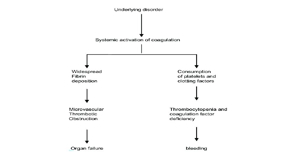
Disseminated intravascular coagulation (DIC) DIC is characterized by systemic activation of blood coagulation, which results in generation and deposition of fibrin, leading to microvascular thrombi in various organs and contributing to multiple organ. Consumption of clotting factors and platelets in DIC can result in lifethreatening hemorrhage. Derangement of the fibrinolytic system further contributes to intravascular clot formation, but in some cases, accelerated fibrinolysis may cause severe bleeding. Hence, a patient with DIC can present with a simultaneously occurring thrombotic and bleeding problem.
Causes of DIC Obstetric causes: Non- obstetric causes: - Amniotic fluid embolism - Abruptio placenta - Placenta previa - HELLP and eclampsia - Intrauterine death - Septic abortion - Acute fatty liver of pregnancy - Sepsis - Major trauma - Leukemia - Snake bite - Acute liver failure - ABO incompatibility - Transplant rejection - Massive transfusion
Acute versus chronic DIC exists in both acute and chronic forms. Acute DIC develops when sudden exposure of blood to procoagulants (eg, tissue factor [TF]) generates intravascular coagulation. Compensatory hemostatic mechanisms are quickly overwhelmed, and, as a consequence, a severe consumptive coagulopathy leading to hemorrhage develops. Abnormalities of blood coagulation parameters are readily identified, and organ failure frequently results. In contrast, chronic DIC reflects a compensated state that develops when blood is continuously or intermittently exposed to small amounts of TF. Compensatory mechanisms in the liver and bone marrow are not overwhelmed, and there may be little obvious clinical or laboratory indication of the presence of DIC. Chronic DIC is more frequently observed in patients with solid tumors and in those with large aortic aneurysms.
Epidemiology DIC may occur in 30 -50% of patients with sepsis, and it develops in an estimated 1% of all hospitalized patients. DIC occurs at all ages and in all races, and no particular sex predisposition has been noted.
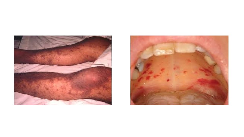
Physical Examination With acute DIC, the physical findings are usually those of the underlying condition; however, patients with the acute disease have petechiae on the soft palate, trunk, and extremities from thrombocytopenia and ecchymosis at venipuncture sites. These patients also manifest ecchymosis in traumatized areas. In patients with so-called chronic or subacute DIC, of which the primary manifestation is thrombosis from excess thrombin formation, the signs of venous thromboembolism may be present.
Laboratory tests - Low platelet - Prolonged prothrombin time (PT) - Prolonged partial thromboplastin time (PTT) - high levels of fibrin degradation products (FDPs) and D-dimer - Schistocytes in blood film
The massive fibrin deposition in DIC suggests that fibrinogen levels would be decreased. Accordingly, measurement of fibrinogen has been widely advocated as a useful tool for the diagnosis of DIC; however, it is in fact not very helpful. Fibrinogen, as a positive acute-phase reactant, is increased in inflammation, and whereas values may decrease as the illness progresses, they are rarely low. One study demonstrated that in up to 57% of DIC patients, the levels of fibrinogen may in fact remain within normal limits. Accordingly, testing for D-dimer or FDPs may be helpful for differentiating DIC from other conditions that may be associated with a low platelet count and prolonged clotting times, such as chronic liver disease.
Treatment - Treatment should primarily focus on addressing the underlying disorder. - Management of the DIC itself has the following basic features: Monitor vital signs Assess and document the extent of hemorrhage and thrombosis Correct hypovolemia Administer basic hemostatic procedures when indicated (plasma, platelet, cryoprecipitate, Packed RBC) - Heparin should be provided to those patients who demonstrate extensive fibrin deposition without evidence of substantial hemorrhage; it is usually reserved for cases of chronic DIC.
Thrombotic thrombocytopenic purpura (TTP) is a hematological emergency characterized by clotting in small blood vessels (thromboses), low platelet count and end organ damage. In its full-blown form, the disease consists of the following pentad: Microangiopathic hemolytic anemia (fragmented RBC) Thrombocytopenic purpura Neurologic abnormalities Fever Renal disease
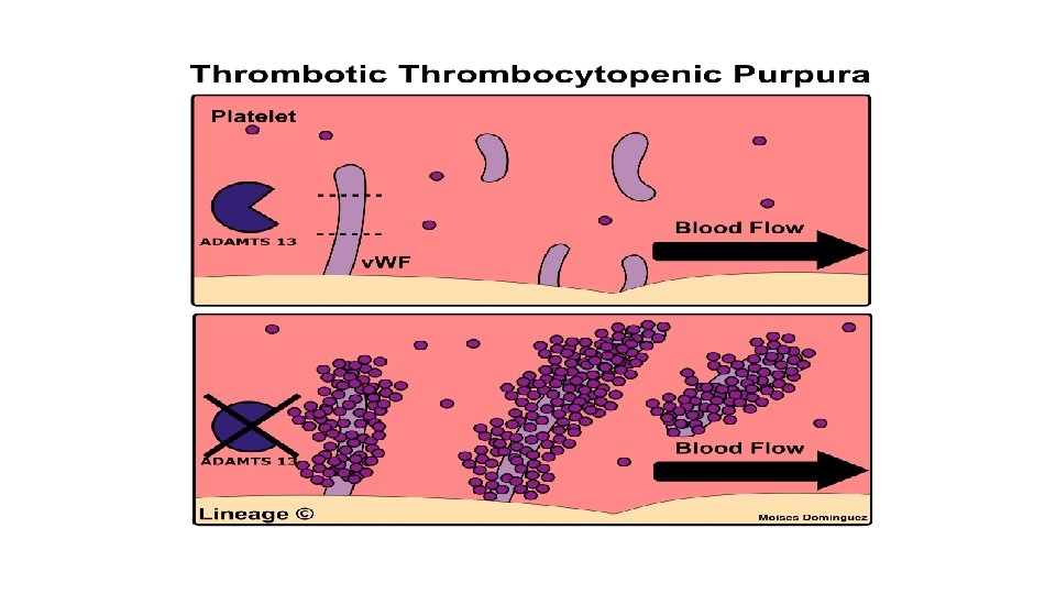
Pathophysiology TTP can affect any organ system, but involvement of the peripheral blood, the central nervous system, and the kidneys causes the clinical manifestations. The classic histologic lesion is one of bland thrombi in the microvasculature of affected organs. Patients with TTP have unusually large multimers of von Willebrand factor (v. WF) in their plasma, and they lack a plasma protease that is responsible for the breakdown of these ultralarge v. WF multimers. In the congenital form of TTP, mutations in the gene encoding this protease have been described. In the more common sporadic form, an antibody inhibitor can be isolated in most patients. This protease has been isolated and cloned and is designated ADAMTS 13 (A Disintegrinlike And Metalloprotease with Thrombo. Spondin type 1 motif 13).
Von Willebrand Factor VWF is a large multimeric glycoprotein present in blood plasma and produced constitutively as ultra-large VWF in endothelium, megakaryocytes, and subendothelial connective tissue. Functions: - Factor VIII is bound to VWF while inactive in circulation; factor VIII degrades rapidly when not bound to VWF. Factor VIII is released from VWF by the action of thrombin. In the absence of VWF, factor VIII has a half-life of 1 -2 hours; when carried by intact VWF, factor VIII has a half-life of 8 -12 hours. - VWF binds to collagen, e. g. , when collagen is exposed beneath endothelial cells due to damage occurring to the blood vessel. - VWF binds to platelet gp. Ib when it forms a complex; this binding occurs under all circumstances, but is most efficient under high shear stress (i. e. , rapid blood flow in narrow blood vessels).
Role in disease - Hereditary or acquired defects of VWF lead to von Willebrand disease (v. WD), a bleeding diathesis of the skin and mucous membranes. - In thrombotic thrombocytopenic purpura (TTP), ADAMTS 13 either is deficient or has been inhibited by antibodies directed at the enzyme. This leads to decreased breakdown of the ultra -large multimers of VWF and microangiopathic hemolytic anemia with deposition of fibrin and platelets in small vessels, and capillary necrosis.
Epidemiology - Incidence : 1 / 50 000 admissions - Mortality : Untreated, TTP has a mortality rate of as high as 90%. With plasma exchange, the mortality rate is reduced to 10 -20%. - Median age : 40 yr - Female : Male ratio = 2 : 1 - Prognosis : In general, survivors have no long-term sequelae, with the exception of residual neurologic deficits in a minority of patients. However, relapses are not uncommon, occurring in up to 33% of patients.
Clinical presentation History Patients with TTP typically report an acute or subacute onset of the following symptoms : - Neurologic manifestations include alteration in mental status, seizures, hemiplegia, paresthesias, visual disturbance, and aphasia - Fatigue may accompany the anemia - Severe bleeding from thrombocytopenia is unusual, although petechiae are common Clinical manifestations may also include the following: - Fever occurs in approximately 50% of patients - Patients may notice dark urine from hemoglobinuria.
Physical Patients with TTP or HUS have no characteristic physical findings. Findings upon examination depend on the severity of involvement of the target organ systems. Hemolytic anemia and thrombocytopenia cause pallor, jaundice, and petechiae. Abnormal findings on neurologic examination consist of mental status changes and/or focal neurologic deficits. Organomegaly is not typical.
Causes of TTP - Autoimmune (Idiopathic) - Congenital : This hereditary form of TTP is called the Upshaw–Schulman syndrome - Secondary : Predisposing factors are: Cancer Bone marrow transplantation Pregnancy Medication use: Antiviral drugs (acyclovir), Certain chemotherapy, Quinine, Platelet aggregation inhibitors (ticlopidine, clopidogrel, and prasugrel), Hormone altering drugs (estrogens, contraceptives, hormone replacement therapy). HIV-1 infection The mechanism of secondary TTP is poorly understood, as ADAMTS 13 activity is generally not as depressed as in idiopathic TTP, and inhibitors cannot be detected. Probable etiology may involve endothelial damage.
Investigations: - CBC, ret. count and B. film - Renal function test and s. electrolytes - Bilirubin - LDH - Haptoglobin - Brain imaging - PT, PTT - d- dimer, fibrinogen, FDP - ADAMTS 13 activity, anti-ADAMTS 13 antibody assay
Tretament - TTP is a medical emergeny that necessitate prompt initiation of plasma exchange - Only (20 -30%) of patients present with the classic pentad - The presence of MAHA (schistocytes, elevated LDH level, and indirect hyperbilirubinemia) and thrombocytopenia in the absence of other obvious causes (eg, DIC, malignant hypertension) is justification to begin total plasma exchange— preferably within 4– 8 hours
Other lines of Rx : - Corticosteroid: (prednisolone 1 mg/kg) - Rituximab - chemotherapy : vincristine - immunosuppressant : azathioprine - capalcizumab: (recently approved), It is an antibody fragment that targets the A 1 -domain of von Willebrand factor (v. WF), and inhibits the interaction between v. WF and platelets, thereby reducing both v. WFmediated platelet adhesion and platelet consumption. - Platelet transfusion : ? ?
Complete response criteria differ depending on the investigator, but they generally include the following: - Resolution of neurologic symptoms - Normalization of platelet count, LDH, and bilirubin levels - Normalization of RFT
Hemolytic Uremic Syndrome (HUS) Hemolytic-uremic syndrome (HUS) is a clinical syndrome characterized by progressive renal failure that is associated with MAHA (nonimmune, Coombs-negative) and thrombocytopenia. HUS is the most common cause of acute kidney injury in children and is increasingly recognized in adults.
Pathophysiology Damage to endothelial cells is the primary event in the pathogenesis of hemolytic-uremic syndrome (HUS). The cardinal lesion is composed of arteriolar and capillary microthrombi (thrombotic microangiopathy) and red blood cell (RBC) fragmentation. HUS is classified into two main categories, depending on whether it is associated with Shiga-like toxin (Stx) or not, typical (t. HUS) and atypical (a. HUS). Shiga-like toxin is so called because it was initially identified in studies of Shigella dysenteriae, but this toxin is also elaborated by Escherichia coli.
Typical (Stx–associated) HUS (t. HUS) - Typical HUS (Shiga-like toxin–associated HUS [Stx-HUS]) is the classic, primary or epidemic, form of HUS. - Stx-HUS is largely a disease of children and often results in diarrhea. One fourth of patients present without diarrhea. - Acute kidney injury occurs in 55 -70% of patients, but they have a favorable prognosis, and as many as 70 -85% of patients recover renal function.
Atypical (non–Stx-associated) HUS (a. HUS) Non–Stx-HUS, or atypical HUS, is less common and accounts for 5 -10% of all cases. As the name implies, non–Stx-HUS does not result from infection by Stx-producing bacteria. The term atypical HUS : complement-mediated thrombotic microangiopathy It may occur at all ages, but a. HUS is most frequent in adults and occurs without prodromal diarrhea. Patients have an unfavorable prognosis. a. HUS can occur in sporadic cases or in families. Overall, patients with a. HUS have a poor outcome, and as many as 50% may progress to end-stage renal disease (ESRD) or irreversible brain damage. Up to 25% of patients die during the acute phase.
Various triggers for sporadic non-Stx–HUS have been identified, including the following: Nonenteric infections Viruses Drugs (quinine, chemotherapy, clopidogril) Malignancies Transplantation Pregnancy Antiphospholipid syndrome, systemic lupus erythematosus
Epidemiology - Incidence : 2 -6 cases / 100 000 population/ yr - peaking in the summer and fall - Hemolytic-uremic syndrome (HUS) occurs infrequently in blacks - Both sexes are affected equally with HUS - HUS occurs mainly in young children; however, adolescents and adults are not exempt. In young children, spontaneous recovery is common. In adults, the probability of recovery is low when HUS is associated with severe hypertension.
Mortality/Morbidity For t. HUS, acute renal failure occurs in 55 -70% of patients; up to 70 -85% recover renal function. For a. HUS, patients have poor outcomes, with up to 50% progressing to ESRD or irreversible brain damage. As many as 25% die during the acute phase. Complications of HUS may include the following: Renal failure Stroke Coma Seizures Bleeding
History findings may include the following: Prodromal gastroenteritis (83%) - Fever (56%), bloody diarrhea (50%) for 2 -7 days before the onset of renal failure Irritability, lethargy Seizures (20%) Acute renal failure (97%) Anuria (55%)
Physical Examination Physical findings may include the following: Confusion Tachypnea Hypertension (47%) Edema, fluid overload (69%) Pallor, often severe Jaundice
Investigations: - Urinalysis: mild proteinuria is frequently present; RBCs and RBC casts may be present -Stool culture: Obtain a sample for stool culture. Evaluate especially for E coli and Shigella bacteria. - Low s. C 3 level - CBC, ret. count and B. film - Renal function test and s. electrolytes - Bilirubin, LDH, Haptoglobin - Renal U/S, Brain imaging - PT, PTT, d- dimer, fibrinogen, FDP - ADAMTS 13 activity, anti-ADAMTS 13 antibody assay
Management of t. HUS: - No specific Rx - Supportive therapy is still the mainstay during the acute phase - There is no clear consensus on the use of antibiotics. The evidence is to avoid antibiotics unless patient is septic. Some studies demonstrated that certain antibiotics that target DNA synthesis, including ciprofloxacin and trimethoprim-sulfamethoxazole, might increase shiga toxin production. - Renal transplantation is safe and effective for children who progress to end -stage renal disease (ESRD). The recurrence rate in patients who undergo renal transplantation for HUS is 0 -10%. - Other treatments, including plasma exchange and use of intravenously infused immunoglobulin (Ig. G), fibrinolytic agents, antiplatelet agents, corticosteroids, and antioxidants have proved ineffective in controlled clinical trials.
Management of a. HUS: - Plasma exchange is the initial treatment of choice in all adult patients with atypical HUS and should be considered as early as possible in the disease course. - Two monoclonal antibodies were approved for the treatment of atypical HUS: eculizumab and ravulizumab. These monoclonal antibodies inhibit complement-mediated thrombotic microangiopathy. Both of these agents carry black box warnings regarding meningococcal infection, which include a recommendation to immunize patients with meningococcal vaccines at least 2 weeks before starting treatment.
Supportive therapy is as follows: - Maintain fluid and electrolyte balance - Adequate blood-pressure control and adequate renin-angiotensin blockade is helpful for patients who have chronic kidney disease after an episode of HUS - For seizure control, consider prophylactic phenytoin in patients with neurologic symptoms (20 -40% of patients have seizures) - Control azotemia - Optimize nutrition
Other explanation Clinical findings of MAHA PEX (2 -3 days) Response Sepsis Malignancy DIC Medications Continue until normal plt and LDH No response ADAMTS 13 activity < 10% ADAMTS 13 activity ≥ 10% Intensify PEX Add IS Consider a. HUS other explanation Eculizumab
Disease MAHA (schistcyte) plt Coagulopat hy (PT, PTT, d-dimer) Compleme ADAMTS 13 CNS Renal manifestati impairmen nt on (C 3) t DIC + ↓ ↑↑ ↔ ↔ +/- TTP + ↓ ↔↑ ↔ ↓ ++ + t. HUS + ↓ ↔↑ ↔ ↔ +/- ++ a. HUS + ↓ ↔↑ ↓ ↔ +/- ++