Hematology Morphology of RCB in health and disease

Hematology Morphology of RCB in health and disease 1 -Discuss aspects of red cell morphology related to size 2 -Discuss aspects of red cell morphology related to shape 3 - Discuss aspects of red cell morphology related to color

Normal RBC -Size: Normocyte -Colour: Normochromic

RED BLOOD CELL MORPHOLOGY Abnormal erythrocyte morphology is found in pathological states that may be : - abnormalities in size (anisocytosis). - In shape (poikilocytosis). -In hemoglobin content or the presence of inclusion bodies in erythrocyte. .

Variation in size Red blood cells can vary in size from smaller than normal, microcytes, to larger than normal, macrocytes. When red cells of normal size, microcytes and macrocytes are present in the same field, the term anisocytosis is used.

Anisocytosis: Variation in Size Microcytes Macrocytes

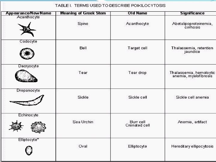
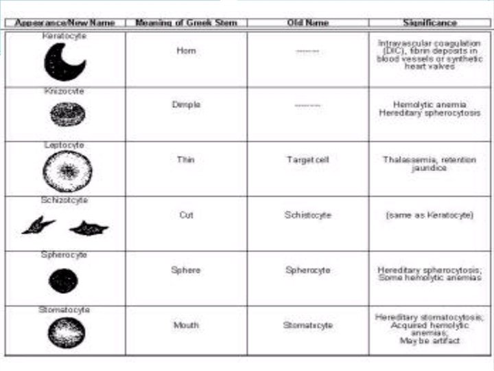
Variation in shape Poikilocytosis

Poikilocytosis: Abnormal Shape Target Cells Hypochromic
 1 -Microcytosis: Morphology: - Decrease in the red")
1 -Variation in erythrocyte size (anisocytosis) 1 -Microcytosis: Morphology: - Decrease in the red cell size. Red cells are smaller than ± 7µm in diameter. The nucleus of a small lymphocyte (± 8, µm) , is a useful guide to the size of a red blood cell. Smaller than a nucleus of the lymphocyte, central pallor is greater than 1/3 of the cell. Found in: - Iron deficiency anemia. - Thalassaemia. - Sideroblastic anemia. - Lead poisoning. - Anemia of chronic disease.
 Comment: Most erythrocytes presented in the picture are")
1 -Variation in erythrocyte size (anisocytosis) Comment: Most erythrocytes presented in the picture are microcytes (compare with the small lymphocyte). The degree of hemoglobinization is sufficient. Normal platelets and single ovalocytes are present. Staining: MGG Magnification: x 1000 1. microcyte 2. normocyte
 2 -Macrocytosis: Morphology: Increase in the size of")
1 -Variation in erythrocyte size (anisocytosis) 2 -Macrocytosis: Morphology: Increase in the size of a red cell. Red cells are larger than 9µm in diameter(diameter of 9 -14 microns , 1. 5 - 2 times larger than normal red cells). May be round or oval in shape, the diagnostic significance being different. Found in: - Folate and B 12 deficiencies (oval) - Ethanol (round) - Liver disease (round) - Reticulocytosis (round)

3 -Megalocytes are the result of decreased DNA synthesis, frequently due to vitamin B 12 and/or folic acid deficiencies. -Decreased DNA synthesis causes the nucleus in the developing red cells to mature at a slower than normal rate. -Since hemoglobin production is not affected, the mature red cell is larger than normal


4 -Pseudomacrocytes -appears larger than the lymphocyte but in contrast to megalocytes has an area of central pallor. -size is the result of exaggerated flattening and thus the presence of the central pallor. -in patients with cirrhosis of the liver, obstructive jaundice, post splenectomy. two types of macrocytes: True macrocytes (megalocytes). Increased MCV, MCH Pseudomacrocytes. Normal MCV, MCH
 RBCs may have different shapes: 1 -")
II- Variation of red cells shape (Poikilocytosis) RBCs may have different shapes: 1 - Spherocytosis: Morphology: Red cells are more spherical. Lack the central area of pallor on a stained blood film. cells which have a decreased surface-to-volume ratio. cell is thicker in diameter than normal red cells they appear to be round, darkly-stained cells without central pallor. Found in: - Hereditary spherocytosis - Immune haemolytic anemia - Zieve's syndrome - Microangiopathic haemolytic anemia
 2 -Target Cells: (Codocytes) Morphology: Red cells")
II- Variation of red cells shape (Poikilocytosis) 2 -Target Cells: (Codocytes) Morphology: Red cells have an area of increased staining which appears in the area of central pallor. -Codocytes appear in conditions which cause the surface of the red cell to increase disproportionately to its volume. -This may result from a decrease in hemoglobin, as in iron deficiency anemia, or an increase in cell membrane. Found in: -Obstructive liver disease - Severe iron deficiency - Thalassaemia - Haemoglobinopathies (S and C) - Post splenectomy
 3 - Ovalocytes: Morphology: oval shape red")
II- Variation of red cells shape (Poikilocytosis) 3 - Ovalocytes: Morphology: oval shape red blood cell Found in: - Thalassaemia major. - Hereditary ovalocytosis. - Sickle cell anemia
 4 - Elliptocytosis: Morphology: The red cells")
II- Variation of red cells shape (Poikilocytosis) 4 - Elliptocytosis: Morphology: The red cells are oval or elliptical in shape. Long axis is twice the short axis. Less than 1% of red cells in normal blood are oval. Found in: - Hereditary elliptocytosis - Megaloblastic anemia - Iron deficiency - Thalassaemia - Myelofibrosis
 5 - Tear Drop Cells(Dacrocyte): Morphology: Red")
II- Variation of red cells shape (Poikilocytosis) 5 - Tear Drop Cells(Dacrocyte): Morphology: Red cells shaped like a tear drop or pear Found in: - Bone marrow fibrosis - Megaloblastic anemia - Iron deficiency - Thalassaemia
 6 - Blister cell: Morphology: Have accentric")
II- Variation of red cells shape (Poikilocytosis) 6 - Blister cell: Morphology: Have accentric hallow area. Found in: Microangiopathic hemolytic anemia
 7 - Schistocytosis: Morphology: Fragmentation of the")
II- Variation of red cells shape (Poikilocytosis) 7 - Schistocytosis: Morphology: Fragmentation of the red cells. red cell fragments which are formed when fibrin strands come in contact with circulating red cells. The strands cut a small piece from the original cell. Found in: - DIC - Micro angiopathic haemolytic anemia - Mechanical haemolytic anemia

II- Variation of red cells shape (Poikilocytosis 8 - Stomatocytosis: Morphology: Red cells with a central linear slit or stoma. Seen as mouth-shaped form in peripheral smear. Found in: - Alcohol excess - Alcoholic liver disease - Hereditary stomatocytosis - Hereditary spherocytosis
")
II- Variation of red cells shape (Poikilocytosis 9 - Echinocyte , Burr (crenation ) cell: Morphology: Red cell with uniformly spaced, pointed projections. Red cell with 30 or more, short blunt projections which are regularly distributed on their surface. Found in: - hemolytic anemia - Uremia. - Megaloblastic anemia
: Morphology: Part")
II- Variation of red cells shape (Poikilocytosis 10 - Keratocytes (horn cell): Morphology: Part of the cell fuses back leaving two or three horn-like projections. The keratocyte is a fragile cell and remains in circulation for only a few hours. Found in: - Uraemia - Severe burns - EDTA artifact - Liver disease

II- Variation of red cells shape (Poikilocytosis 11 - Acanthocytosis: Morphology: are red blood cells with irregularly spaced projections, these projections very in width but usually contain a rounded end. -Smaller than normal and have little or no central pallor. -Acanthocytes have an excess of cholesterol -Large numbers of these cells on a smear can be of diagnostic significance. Found in: - Hereditary acanthocytosis, 50 -100% of blood cells. -Liver disease - Post splenectomy - Anorexia nervosa and starvation -lipid disorders

II- Variation of red cells shape (Poikilocytosis 12 - Rouleaux Formation: Morphology: Stacks of RBC's resembling a stack of coins. Stacking of RBCs due to increased plasma proteins coating RBCs. . Found in: - Hyperfibrinogenaemia - Hyperglobulinaemia

II- Variation of red cells shape (Poikilocytosis 13 - Red cell-agglutination: Morphology: Irregular clumps of red cells. Antibody- mediated Irregular clumping , temperature dependent Found in: - Cold agglutinins - Warm autoimmune hemolysis
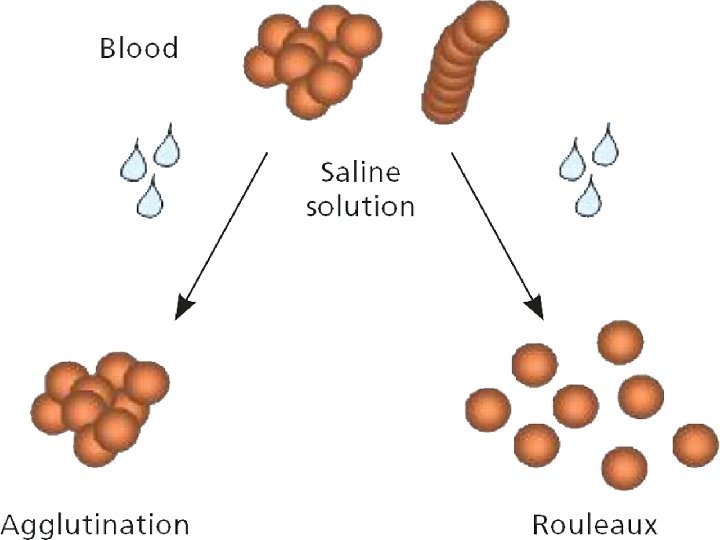

Rouloux Formation Agglutination
: Morphology: Sickle shaped red")
II-Variation of red cells shape (Poikilocytosis 14 - Sickle Cells(Drepanocytes): Morphology: Sickle shaped red cells, are formed as a result of the presence of hemoglobin S in the red cell. -As the red cell ages, it becomes less flexible or deformable and becomes rigid as it passes through the low oxygen tension atmosphere of the small capillaries in the body. -In the absence of oxygen, hemoglobin S polymerizes into rods, causing the sickle cell shape. Sickle cells can be somewhat pointed at the ends, Found in: Hb-S disease

-Most sickled cells can revert back to the discoid shape when oxygenated. -About 10% of sickled cells are unable to revert back to their original shape after repeated sickling episodes.
 15 - Nucleated red blood cells. These red")
II-Variation of red cells shape (Poikilocytosis) 15 - Nucleated red blood cells. These red blood cells are released from the bone marrow early into the blood stream, due to the need for oxygen. Normal red blood cells do not contain a nucleus on a peripheral smear

16 -Envelope Form Cell Found in Thalassaemia Sickle cell anemia

17 -Knizocyte -A streak of hemoglobin through the • centre of the cell. -In some hemolytic anemia cases •

Terminology Description Condition Target Cells Central Hemoglobin; target shaped Liver Disease; Thalassaemia, Abnormal Hb; Iron Deficiency Echinocyte Short specula's, equallyspaced Uremia, Hypokalemia, Artifact Acanthocyte Speculated, Irregular Liver disease (Alcohol), Postspleenoctomy. Spherocyte Spherical, no central pallor HS, immune Hemolytic anemia Shistocyte Fragmented RBC, Helmet cells MAHA, burns Ovalocyte Oval / Elliptical shaped Hereditary elliptocytosis, Megaloblastic anemia. Sickle Cell Bipolar speculated shape “ banana” shaped Hb S-containing hemoglobinopathy Teardrop cell Single elongated extremity Myelophthistic changes Bite cells Irregular gap in membrane G 6 PD deficiency

Erythrocyte Inclusions with Wright’s Inclusion Composition Appearance Stain Condition Basophilic stippling Precipitated ribosomes Evenly dispersed fine or coarse granules - Lead poisoning Thalassaemia , other anemia. Howell-Jolly bodies DNA in origin Nuclear Fragment Dense, round blue granule Post – Splenectomy Pappenheimer bodies Iron-containing granules Small blue granules in clusters Anemia's Heinz bodies Denatured Hemoglobin Round blue precipitates G 6 PD Cabot Rings Remnants of Nuclear membrane Reddish-blue thread Severe anemia, like rings Lead poisoning. Organism Small blue inclusion Malaria Babesiosis

1 -Howell-Jolly Bodies Small round cytoplasmic red cell inclusion with same staining characteristics as nuclei, may be seen in sickle cell anemia. These bodies are DNA that stain dark purple within the red blood cell Found in Post splenectomy Megaloblastic anemia
 These are iron containing granules in red blood cells")
2 -Siderotic Granules (Pappenheimer Bodies) These are iron containing granules in red blood cells that are seen because the iron is aggregated with mitochondria and ribosomes. They appear as faint violet or magenta specks, often in small clusters, due to staining of the associated protein. They are associated with severe anemia's and thalassemia's. Pappenheimer bodies can be increased in hemolytic anemia, infections and post-splenectomy.

3 -Basophilic stippling Considerable numbers of small basophilic inclusions in red cells. is granules of RNA seen within the red blood cell. The granules stain blue to purple Found in Thalassaemia Megaloblastic anemia Hemolytic anemia Liver disease Heavy metal poisoning
 within a cell. With a supravital")
4 -Heinz Bodies Represent denatured hemoglobin (methemoglobin Fe+++) within a cell. With a supravital stain like crystal violet, Heinz bodies appear as round blue precipitates. Presence of Heinz bodies indicates red cell injury and is usually associated with G 6 PD-deficiency.

5 -Cabot Rings Reddish-blue threadlike rings in RBCs of severe anemia's. These are remnants of the nuclear membrane or remnants of microtubules and appear as a ring or figure 8 pattern. Very rare finding in patients with Megaloblastic anemia. severe anemia's. lead poisoning. Dyserythropoiesis A - Cabot ring B - Howell-Jolly body

6 -Parasites of Red Cell. Two organisms are have a tendency to invade the RBCs 1 -All 4 species of the malaria parasite will invade RBCs. We will see the Plasmodium of different species in. RBCs 2 -Theileria microti (Bebesia microti)

Malaria

III -Variation In Erythrocyte Color -A normal erythrocyte has a pinkish-red color with a slightly lighter-colored center (central pallor) when stained with a blood stain, such as Wright. -The color of the erythrocyte is representative of hemoglobin concentration in the cell. -Under normal conditions, when the color, central pallor, and hemoglobin are proportional, the erythrocyte is referred to as Normochromic.
 is white, while buff-colored hemoglobin")
RBC Color -the central area (1/3 of the cell) is white, while buff-colored hemoglobin is visible in the outer 2/3 of the cell. -The MCHC (32 -36 gm/dl) is the index value which is used to verify the presence of adequate hemoglobin concentration in the cells visible on the peripheral smear. -A decreased amount of hemoglobin is referred to as hypochromasia or hypochromia. -MCHC values of 30% or less reflect this condition. -Hyperchromasia and hyperchromia, refer to a hypothetical situation rather than an actual occurrence.

Variation in color Hypochromic: A descriptive term applied to a red blood cell with a decreased concentration of hemoglobin. Normochromic: A descriptive term applied to a red blood cell with a normal concentration of hemoglobin.

1 -Hypochromia Increased central pallor and decreased hemoglobin concentration, the central pallor occupies more than the normal third of the red cell diameter. Found in -Iron deficiency -Thalassaemia -any of the conditions leading to Microcytosis

2 - HyperchromiaPolychromasia Red cells stain shades of blue-gray as a consequence of uptake of both eosin (by hemoglobin) and basic dyes (by residual ribosomal RNA). Often slightly larger than normal red cells and round in shape - round macrocytosis. Found in Any situation with reticulocytosis – for example bleeding, hemolysis or response to heamatinic factor replacement.

What Abnormal Results Mean This test is used to diagnose the cause of anemia. The following are the types of anemia and their causes: -Normocytic/ normochromic (NC/NC) anemia is caused by sudden blood loss, prosthetic heart valves, sepsis, tumor, longterm disease or aplastic anemia. -Microcytic/ hypochromic anemia is caused by iron deficiency, lead poisoning, or thalassemia. -Microcytic/ normochromic anemia results from a deficiency of the hormone erythropoietin from kidney failure. -Macrocytic /normochromic anemia results from chemotherapy, folate deficiency, or vitamin B-12 deficiency.
, a")
G 6 PD: This enzyme participates in the pentose phosphate pathway (see image), a metabolic pathway that supplies reducing energy to cells (such as erythrocytes) by maintaining the level of the co-enzyme nicotinamide adenine dinucleotide phosphate (NADPH). The NADPH in turn maintains the level of glutathione in these cells that helps protect the red blood cells against oxidative damage from compounds like hydrogen peroxide. [1] Of greater quantitative importance is the production of NADPH for tissues involved in biosynthesis of fatty acids or isoprenoids, such as the liver, mammary glands, adipose tissue, and the adrenal glands. G 6 PD reduces NADP+ to NADPH while oxidizing glucose-6 -phosphate

G 6 PD is remarkable for its genetic diversity. Many variants of G 6 PD, mostly produced from missense mutations, have been described with wide-ranging levels of enzyme activity and associated clinical symptoms. Two transcript variants encoding different isoforms have been found for this gene. [18]Glucose-6 -phosphate dehydrogenase deficiency is very common worldwide, and causes acute hemolytic anemia in the presence of simple infection, ingestion of fava beans, or reaction with certain medicines, antibiotics, antipyretics, and antimalarials. [3]

Q 1/Fill in the blanks : 1 - Red cells are smaller than a nucleus of the lymphocyte called ____. 2 -pathological conditions for Macrocytosis _____ , _____. 3 -Red cells have long axis is twice the short axis called _____. 4 -The index value which used to adequate hemoglobin concentration ____. 5 -cause of rouleaux formation _______ , ______. Q 2/Give one feature of morphology for the cells: 1 -Keratocytes 2 - Blister cell 3 -spherocyte 4 - knizocyte Q 3/Write causes of the following : 1 -G-6 PD 2 -Siderotic granules
- Slides: 53