Anemia in Childhood Laura Rooms M D Definition

- Slides: 51
Anemia in Childhood Laura Rooms, M. D.
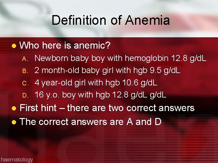
Definition of Anemia l Who here is anemic? A. B. C. D. Newborn baby boy with hemoglobin 12. 8 g/d. L 2 month-old baby girl with hgb 9. 5 g/d. L 4 year-old girl with hgb 10. 6 g/d. L 16 y. o. boy with hgb 12. 8 g/d. L First hint – there are two correct answers l The correct answers are A and D l
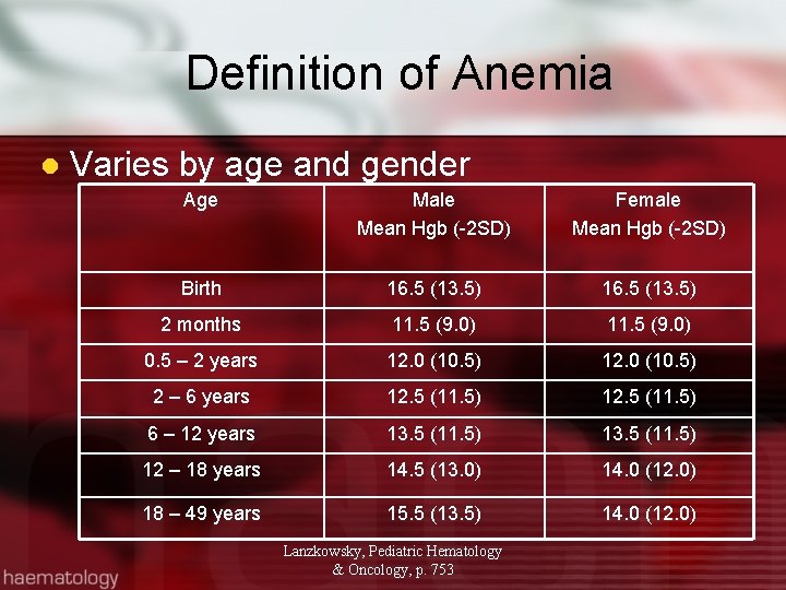
Definition of Anemia l Varies by age and gender Age Male Mean Hgb (-2 SD) Female Mean Hgb (-2 SD) Birth 16. 5 (13. 5) 2 months 11. 5 (9. 0) 0. 5 – 2 years 12. 0 (10. 5) 2 – 6 years 12. 5 (11. 5) 6 – 12 years 13. 5 (11. 5) 12 – 18 years 14. 5 (13. 0) 14. 0 (12. 0) 18 – 49 years 15. 5 (13. 5) 14. 0 (12. 0) Lanzkowsky, Pediatric Hematology & Oncology, p. 753
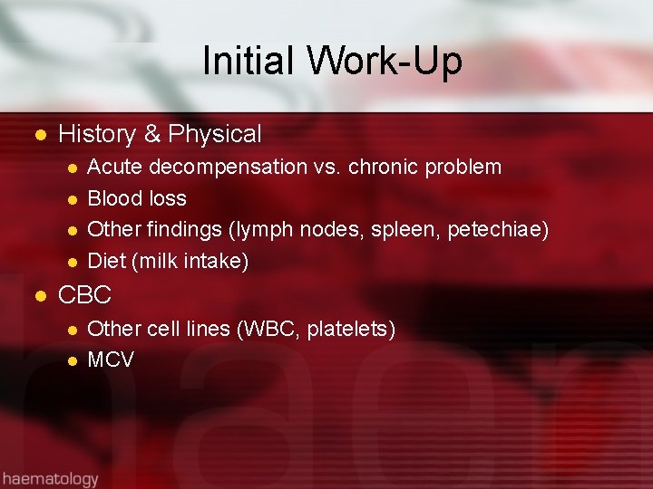
Initial Work-Up l History & Physical l l Acute decompensation vs. chronic problem Blood loss Other findings (lymph nodes, spleen, petechiae) Diet (milk intake) CBC l l Other cell lines (WBC, platelets) MCV
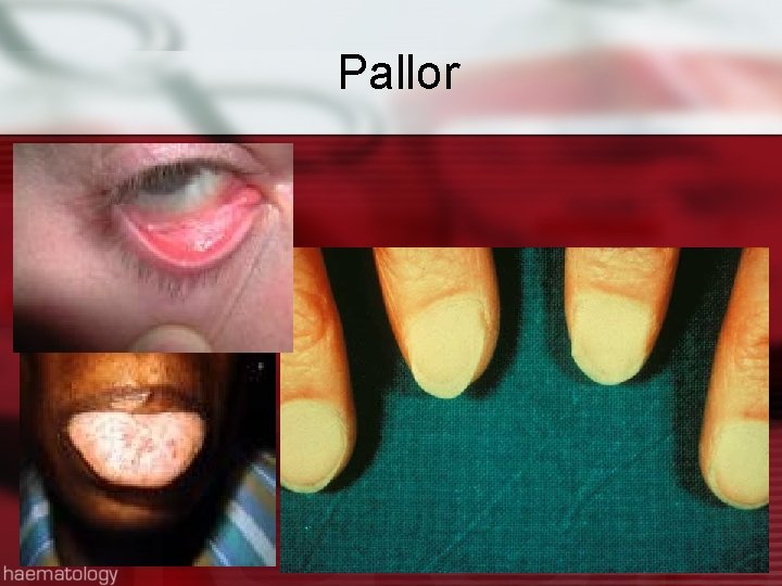
Pallor
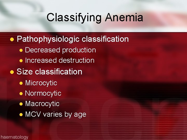
Classifying Anemia l Pathophysiologic classification Decreased production l Increased destruction l l Size classification Microcytic l Normocytic l Macrocytic l MCV varies by age l
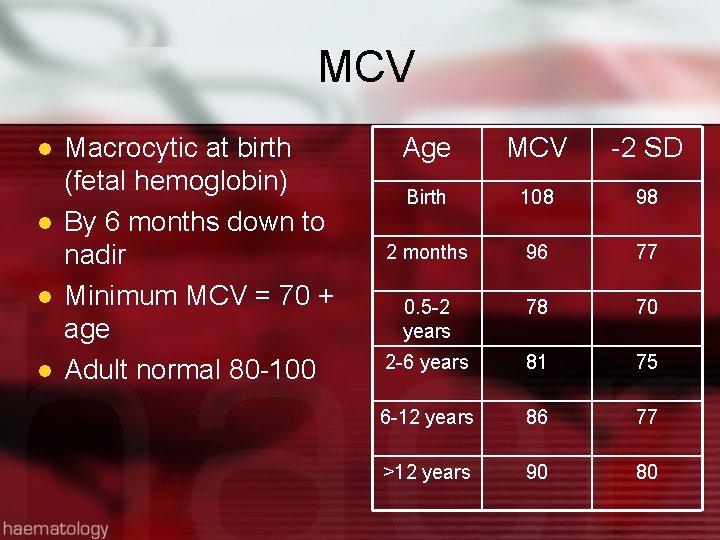
MCV l l Macrocytic at birth (fetal hemoglobin) By 6 months down to nadir Minimum MCV = 70 + age Adult normal 80 -100 Age MCV -2 SD Birth 108 98 2 months 96 77 0. 5 -2 years 78 70 2 -6 years 81 75 6 -12 years 86 77 >12 years 90 80
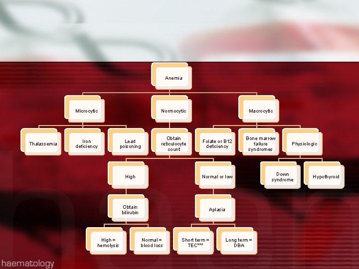
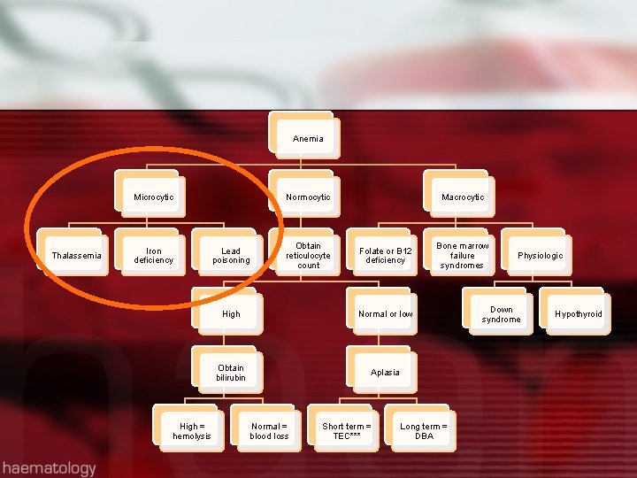
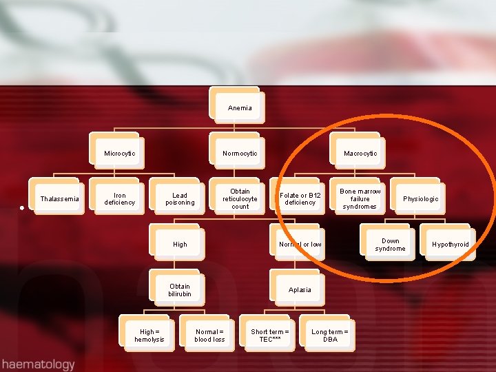
Anemia Microcytic Thalassemia Iron deficiency High = hemolysis Normocytic Lead poisoning Obtain reticulocyte count Macrocytic Folate or B 12 deficiency High Normal or low Obtain bilirubin Aplasia Normal = blood loss Short term = TEC*** Bone marrow failure syndromes Long term = DBA Physiologic Down syndrome Hypothyroid
Anemia Microcytic Thalassemia Iron deficiency High = hemolysis Normocytic Lead poisoning Obtain reticulocyte count Macrocytic Folate or B 12 deficiency High Normal or low Obtain bilirubin Aplasia Normal = blood loss Short term = TEC*** Bone marrow failure syndromes Long term = DBA Physiologic Down syndrome Hypothyroid
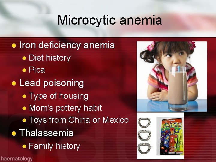
Microcytic anemia l Iron deficiency anemia Diet history l Pica l l Lead poisoning Type of housing l Mom’s pottery habit l Toys from China or Mexico l l Thalassemia l Family history
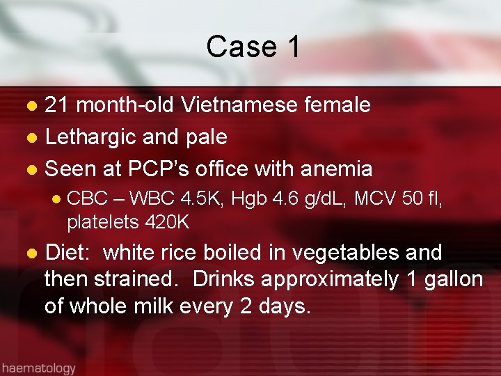
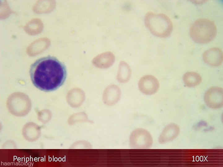
Case 1 21 month-old Vietnamese female l Lethargic and pale l Seen at PCP’s office with anemia l l l CBC – WBC 4. 5 K, Hgb 4. 6 g/d. L, MCV 50 fl, platelets 420 K Diet: white rice boiled in vegetables and then strained. Drinks approximately 1 gallon of whole milk every 2 days.
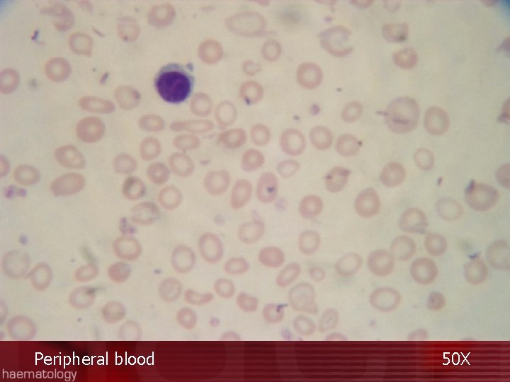
Peripheral blood 50 X
Peripheral blood 100 X
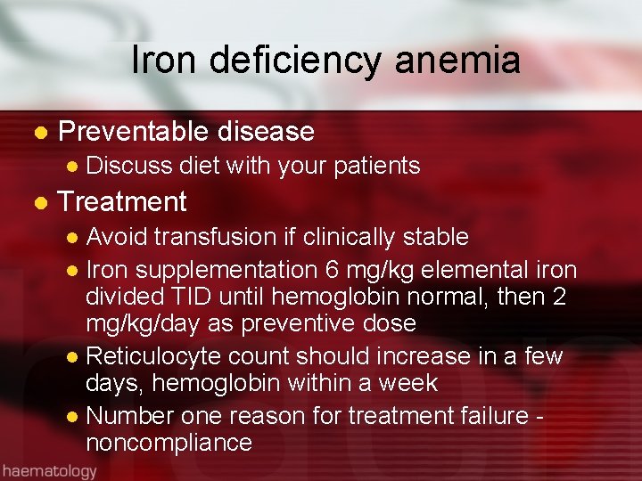
Iron deficiency anemia l Preventable disease l l Discuss diet with your patients Treatment Avoid transfusion if clinically stable l Iron supplementation 6 mg/kg elemental iron divided TID until hemoglobin normal, then 2 mg/kg/day as preventive dose l Reticulocyte count should increase in a few days, hemoglobin within a week l Number one reason for treatment failure - noncompliance l
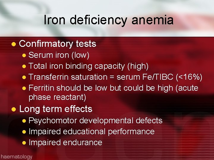
Iron deficiency anemia l Confirmatory tests Serum iron (low) l Total iron binding capacity (high) l Transferrin saturation = serum Fe/TIBC (<16%) l Ferritin should be low but could be high (acute phase reactant) l l Long term effects Psychomotor developmental defects l Impaired educational performance l Impaired endurance l
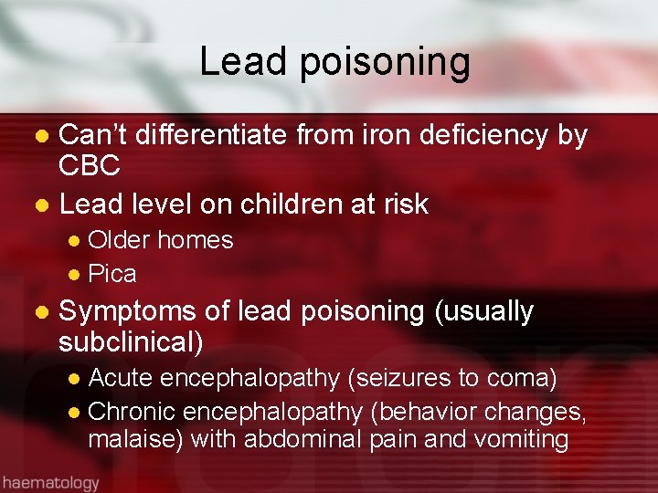
Lead poisoning Can’t differentiate from iron deficiency by CBC l Lead level on children at risk l Older homes l Pica l l Symptoms of lead poisoning (usually subclinical) Acute encephalopathy (seizures to coma) l Chronic encephalopathy (behavior changes, malaise) with abdominal pain and vomiting l
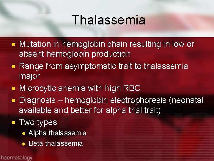
Thalassemia l l l Mutation in hemoglobin chain resulting in low or absent hemoglobin production Range from asymptomatic trait to thalassemia major Microcytic anemia with high RBC Diagnosis – hemoglobin electrophoresis (neonatal available and better for alpha thal trait) Two types l l Alpha thalassemia Beta thalassemia
Thalassemia
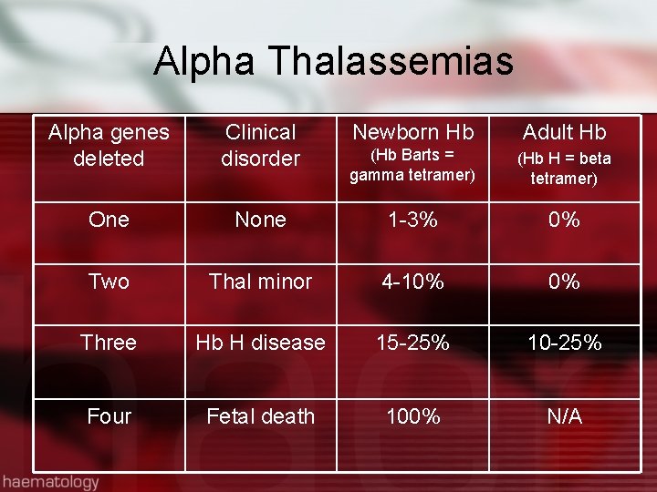
Alpha Thalassemias Alpha genes deleted Clinical disorder Newborn Hb Adult Hb (Hb Barts = gamma tetramer) (Hb H = beta tetramer) One None 1 -3% 0% Two Thal minor 4 -10% 0% Three Hb H disease 15 -25% 10 -25% Four Fetal death 100% N/A
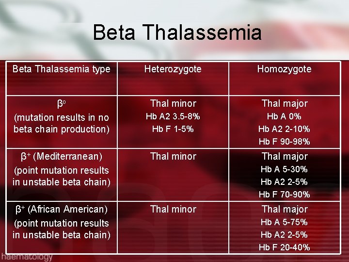
Beta Thalassemia type Heterozygote Homozygote βo (mutation results in no beta chain production) Thal minor Thal major Hb A 2 3. 5 -8% Hb F 1 -5% Hb A 0% Hb A 2 2 -10% Hb F 90 -98% β+ (Mediterranean) (point mutation results in unstable beta chain) Thal minor Thal major β+ (African American) (point mutation results in unstable beta chain) Thal minor Hb A 5 -30% Hb A 2 2 -5% Hb F 70 -90% Thal major Hb A 5 -75% Hb A 2 2 -5% Hb F 20 -40%
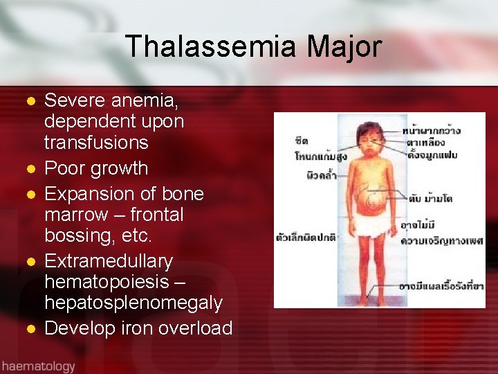
Thalassemia Major l l l Severe anemia, dependent upon transfusions Poor growth Expansion of bone marrow – frontal bossing, etc. Extramedullary hematopoiesis – hepatosplenomegaly Develop iron overload
Anemia Microcytic Thalassemia Iron deficiency High = hemolysis Normocytic Lead poisoning Obtain reticulocyte count Macrocytic Folate or B 12 deficiency High Normal or low Obtain bilirubin Aplasia Normal = blood loss Short term = TEC*** Bone marrow failure syndromes Long term = DBA Physiologic Down syndrome Hypothyroid
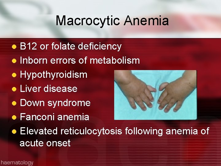
Macrocytic Anemia B 12 or folate deficiency l Inborn errors of metabolism l Hypothyroidism l Liver disease l Down syndrome l Fanconi anemia l Elevated reticulocytosis following anemia of acute onset l
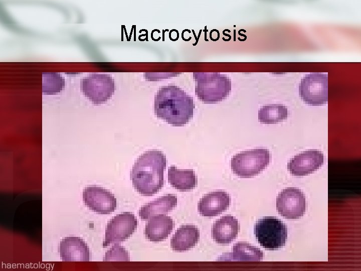
Macrocytosis
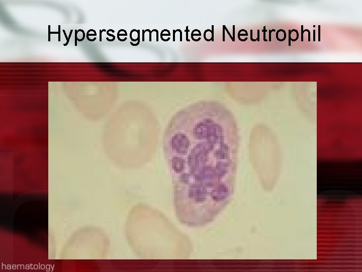
Hypersegmented Neutrophil
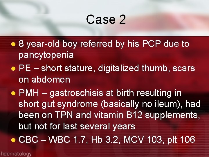
Case 2 8 year-old boy referred by his PCP due to pancytopenia l PE – short stature, digitalized thumb, scars on abdomen l PMH – gastroschisis at birth resulting in short gut syndrome (basically no ileum), had been on TPN and vitamin B 12 supplements, but not for last several years l CBC – WBC 1. 7, Hb 3. 2, MCV 103, plt 106 l
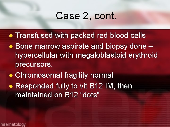
Case 2, cont. Transfused with packed red blood cells l Bone marrow aspirate and biopsy done – hypercellular with megaloblastoid erythroid precursors. l Chromosomal fragility normal l Responded fully to vit B 12 IM, then maintained on B 12 “dots” l
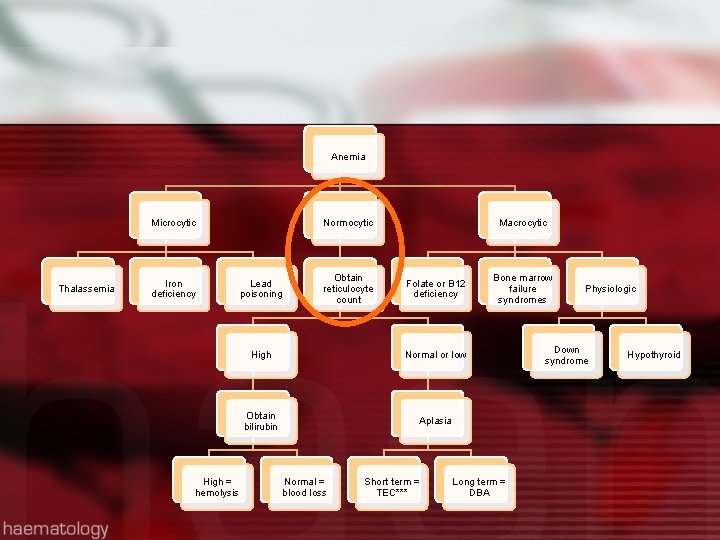
Anemia Microcytic Thalassemia Iron deficiency High = hemolysis Normocytic Lead poisoning Obtain reticulocyte count Macrocytic Folate or B 12 deficiency High Normal or low Obtain bilirubin Aplasia Normal = blood loss Short term = TEC*** Bone marrow failure syndromes Long term = DBA Physiologic Down syndrome Hypothyroid
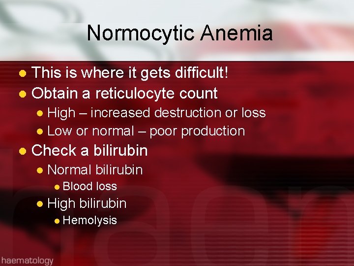
Normocytic Anemia This is where it gets difficult! l Obtain a reticulocyte count l High – increased destruction or loss l Low or normal – poor production l l Check a bilirubin l Normal bilirubin l Blood loss l High bilirubin l Hemolysis
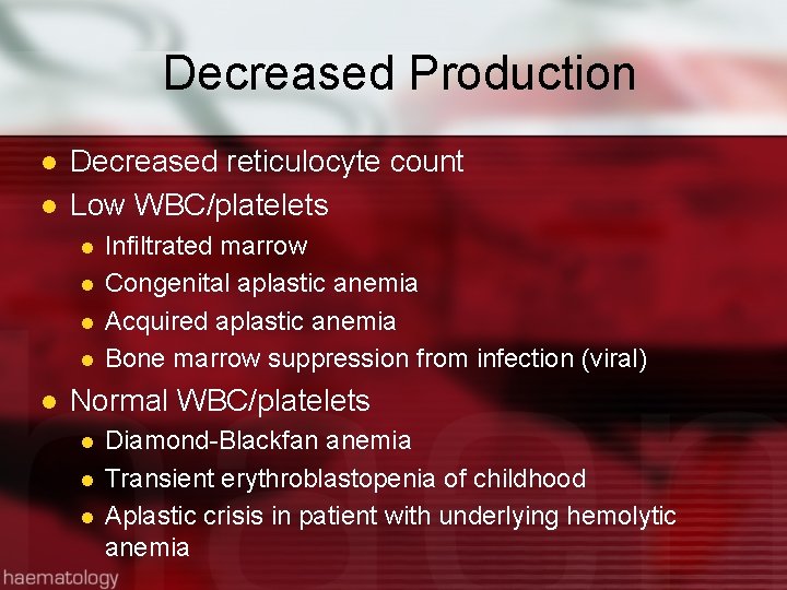
Decreased Production l l Decreased reticulocyte count Low WBC/platelets l l l Infiltrated marrow Congenital aplastic anemia Acquired aplastic anemia Bone marrow suppression from infection (viral) Normal WBC/platelets l l l Diamond-Blackfan anemia Transient erythroblastopenia of childhood Aplastic crisis in patient with underlying hemolytic anemia
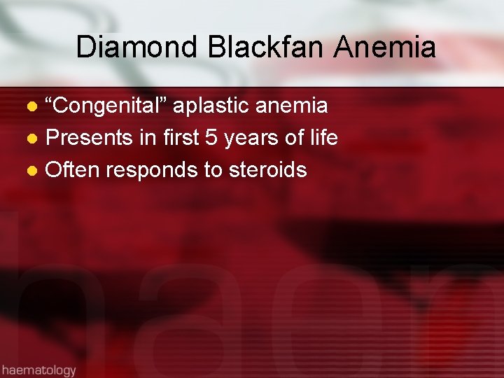
Diamond Blackfan Anemia “Congenital” aplastic anemia l Presents in first 5 years of life l Often responds to steroids l
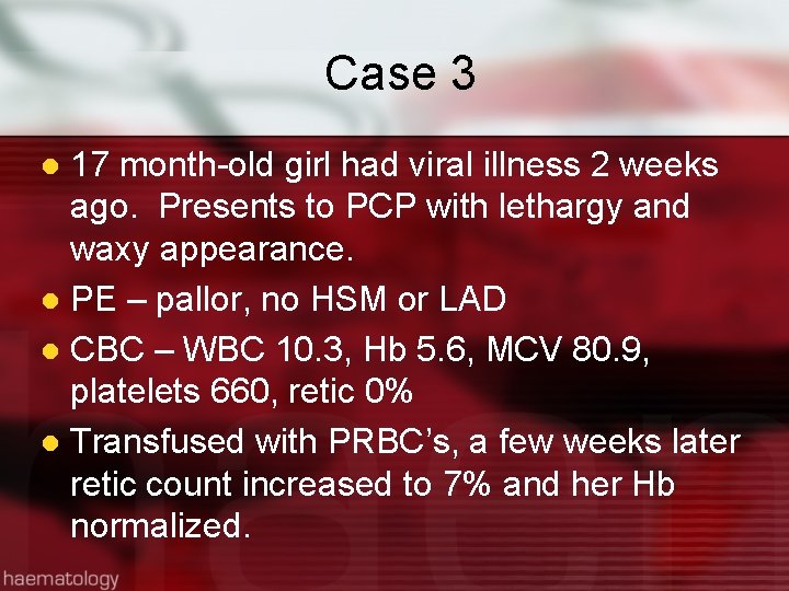
Case 3 17 month-old girl had viral illness 2 weeks ago. Presents to PCP with lethargy and waxy appearance. l PE – pallor, no HSM or LAD l CBC – WBC 10. 3, Hb 5. 6, MCV 80. 9, platelets 660, retic 0% l Transfused with PRBC’s, a few weeks later retic count increased to 7% and her Hb normalized. l
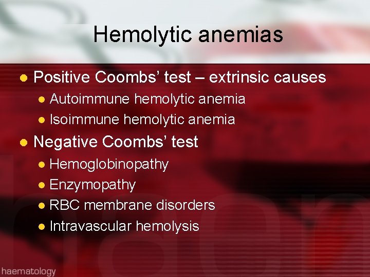
Hemolytic anemias l Positive Coombs’ test – extrinsic causes Autoimmune hemolytic anemia l Isoimmune hemolytic anemia l l Negative Coombs’ test Hemoglobinopathy l Enzymopathy l RBC membrane disorders l Intravascular hemolysis l
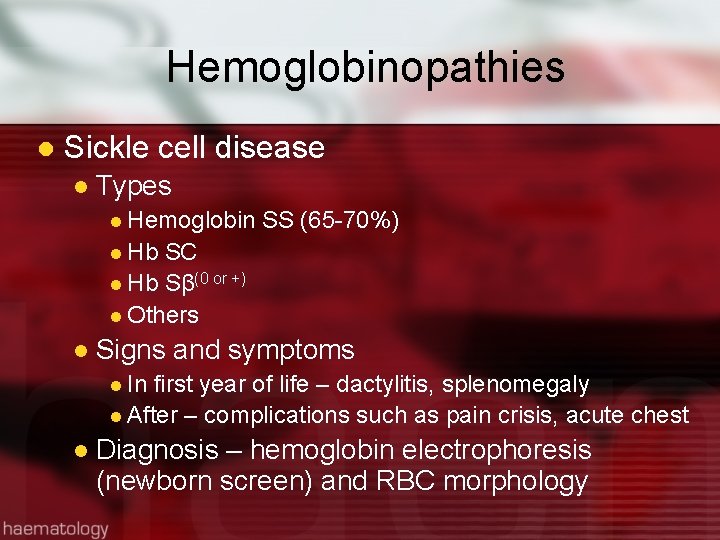
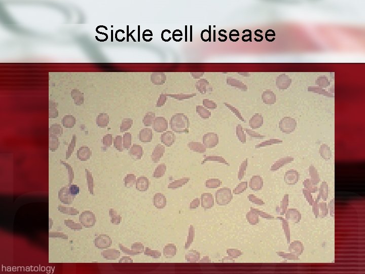
Hemoglobinopathies l Sickle cell disease l Types l Hemoglobin SS (65 -70%) l Hb SC l Hb Sβ(0 or +) l Others l Signs and symptoms l In first year of life – dactylitis, splenomegaly l After – complications such as pain crisis, acute chest l Diagnosis – hemoglobin electrophoresis (newborn screen) and RBC morphology
Sickle cell disease
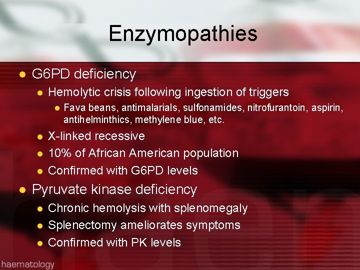
Enzymopathies l G 6 PD deficiency l Hemolytic crisis following ingestion of triggers l l l Fava beans, antimalarials, sulfonamides, nitrofurantoin, aspirin, antihelminthics, methylene blue, etc. X-linked recessive 10% of African American population Confirmed with G 6 PD levels Pyruvate kinase deficiency l l l Chronic hemolysis with splenomegaly Splenectomy ameliorates symptoms Confirmed with PK levels
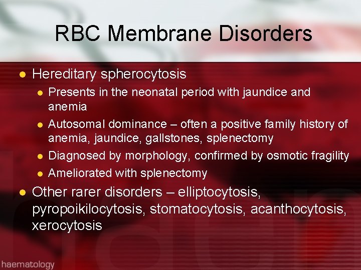
RBC Membrane Disorders l Hereditary spherocytosis l l l Presents in the neonatal period with jaundice and anemia Autosomal dominance – often a positive family history of anemia, jaundice, gallstones, splenectomy Diagnosed by morphology, confirmed by osmotic fragility Ameliorated with splenectomy Other rarer disorders – elliptocytosis, pyropoikilocytosis, stomatocytosis, acanthocytosis, xerocytosis
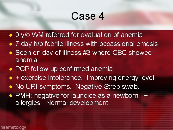
Case 4 l l l l 9 y/o WM referred for evaluation of anemia 7 day h/o febrile illness with occassional emesis Seen on day of illness #3 where CBC showed anemia. PCP follow up confirmed anemia + exercise intolerance. Improving energy level. No URI symptoms. Negative Strep swab. PMH: negative for jaundice as a newborn. + allergies. Normal development
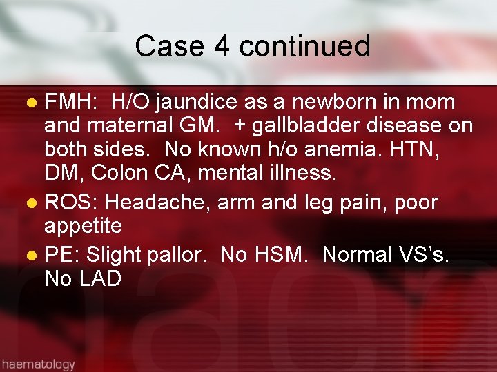
Case 4 continued FMH: H/O jaundice as a newborn in mom and maternal GM. + gallbladder disease on both sides. No known h/o anemia. HTN, DM, Colon CA, mental illness. l ROS: Headache, arm and leg pain, poor appetite l PE: Slight pallor. No HSM. Normal VS’s. No LAD l
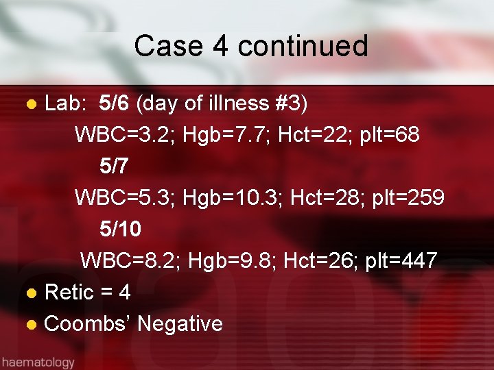
Case 4 continued Lab: 5/6 (day of illness #3) WBC=3. 2; Hgb=7. 7; Hct=22; plt=68 5/7 WBC=5. 3; Hgb=10. 3; Hct=28; plt=259 5/10 WBC=8. 2; Hgb=9. 8; Hct=26; plt=447 l Retic = 4 l Coombs’ Negative l
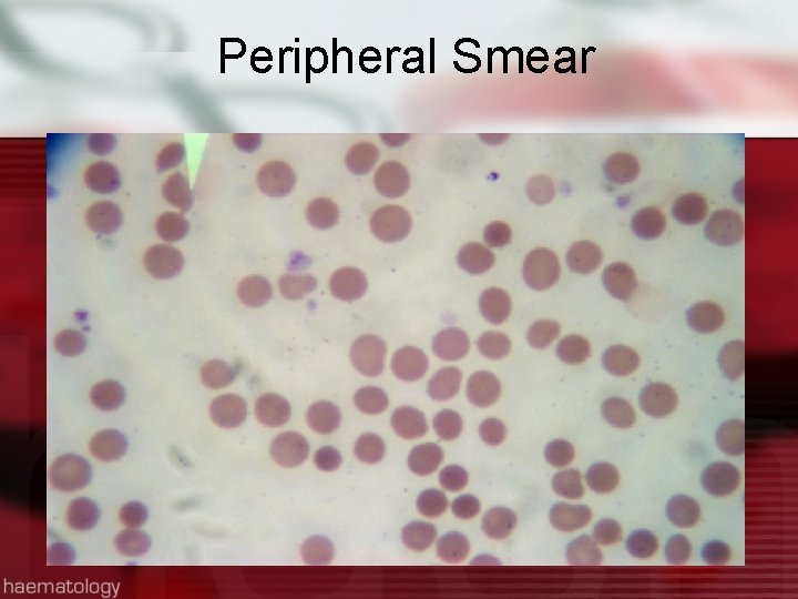
Peripheral Smear
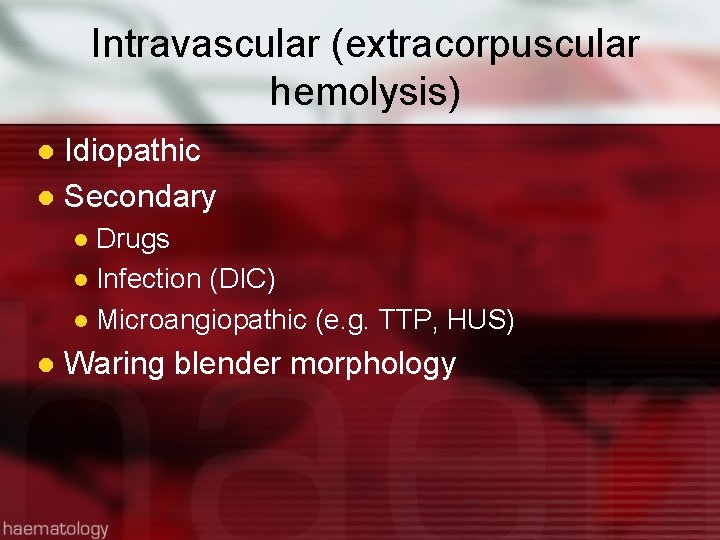
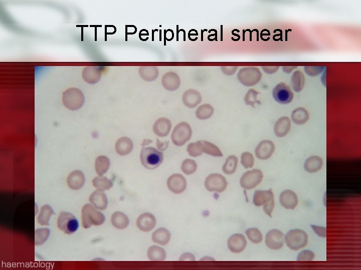
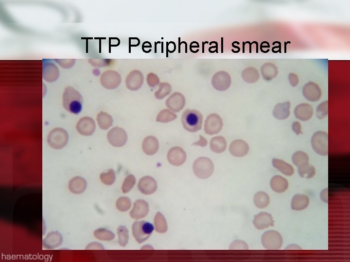
Intravascular (extracorpuscular hemolysis) Idiopathic l Secondary l Drugs l Infection (DIC) l Microangiopathic (e. g. TTP, HUS) l l Waring blender morphology
TTP Peripheral smear
TTP Peripheral smear
Cases to Work Out If Time Permits
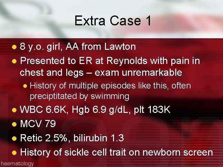
Extra Case 1 8 y. o. girl, AA from Lawton l Presented to ER at Reynolds with pain in chest and legs – exam unremarkable l l History of multiple episodes like this, often preciptitated by swimming WBC 6. 6 K, Hgb 6. 9 g/d. L, plt 183 K l MCV 79 l Retic 2. 5%, bilirubin 1. 3 l History of sickle cell trait on newborn screen l
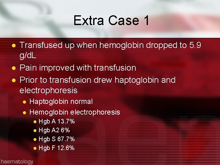
Extra Case 1 l l l Transfused up when hemoglobin dropped to 5. 9 g/d. L Pain improved with transfusion Prior to transfusion drew haptoglobin and electrophoresis l l Haptoglobin normal Hemoglobin electrophoresis l l Hgb A 13. 7% Hgb A 2 6% Hgb S 67. 7% Hgb F 12. 6%
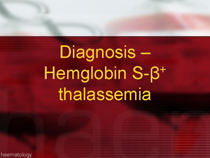
Diagnosis – + Hemglobin S-β thalassemia
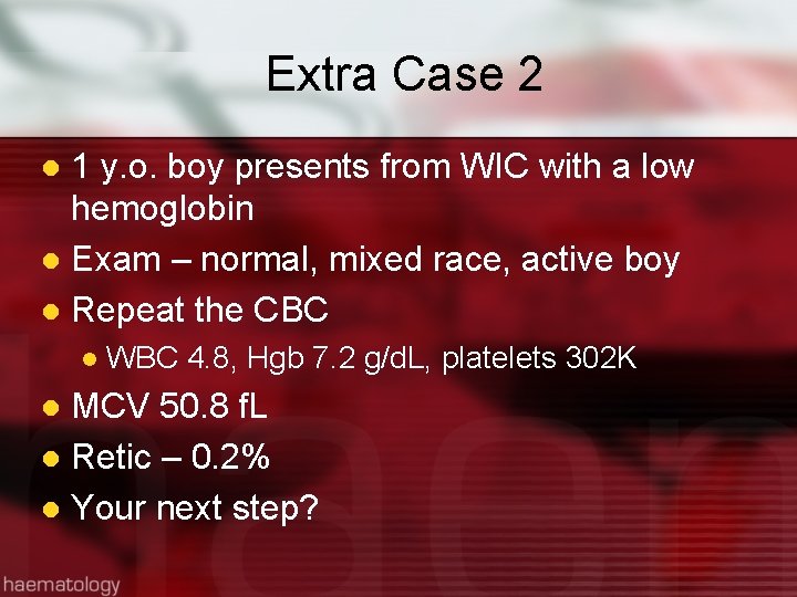
Extra Case 2 1 y. o. boy presents from WIC with a low hemoglobin l Exam – normal, mixed race, active boy l Repeat the CBC l l WBC 4. 8, Hgb 7. 2 g/d. L, platelets 302 K MCV 50. 8 f. L l Retic – 0. 2% l Your next step? l
Extra Case 2 l RTC in 1 week l l CBC virtually identical, retic 0. 4% RTC in 1 month l Hgb 6. 2 g/d. L, retic 0. 5% Refractory to iron? l Iron deficiency or something else? l
References l l Khan, L. Anemia in Childhood. Pediatr Annals. 2018, Feb 1; 47(2): e 42 -e 47. Yawn, BP et al. Management of sickle Cell Disease: summary of the 2014 evidence-based report by expert panel members. JAMA. 2014 Sep 10; 312(10): 1033 -48. Wang, M. Iron Deficiency and other types of anemia in infants and children. Am Fam Phys 2016 Feb 15; 93(4): 2708. Nathan and Oski's Hematology of Infancy and Childhood - 7 th Edition