G Gene manipulation Gene manipulation DNA cloning an


,经随机酶切或 机械剪切为一定长度的片段再与适当的载体DNA 连接后转移到受体细胞中而形成的各种片段的重 组子克隆的总和。 • Genomic libraries are prepared from")


")


")


 of plasmid DNA")


![• Different concentrations of Gel [1%, 1. 5%(w/v), etc. ] will allow the](https://slidetodoc.com/presentation_image_h2/b6ef63947019fb8dadc1fd82bb32af67/image-58.jpg "• Different concentrations of Gel [1%, 1. 5%(w/v), etc. ] will allow the")


 of interest is")
 and religated vector")


- Slides: 118
G Gene manipulation
Gene manipulation • DNA cloning: an overview • Preparation of plasmid DNA • Restriction enzymes and electrophoresis • Ligation and transformation and analysis of recombinants
G 1 an overview • DNA cloning • Hosts and vectors • Subcloning • DNA libraries • Screening libraries • Analysis of a clone
DNA cloning • Place a relatively short fragment of a genome, which might contain the target gene or other sequence of interest, in an autonomously replicating piece of DNA, known as a vector, vector forming recombinant DNA, DNA which can be replicated independently of the original genome, and normally in another host species altogether. • Propagation of the host organism containing the recombinant DNA forms a set of genetically identical organisms, or a clone This process is DNA cloning • Genetic engineering
Genetic engineering • DNA sequencing, and hence the derivation of protein sequence. • Isolation and analysis of gene promoters and other control sequence. • Investigation of protein/enzyme/RNA function by large-scale production normal and altered forms. • Identification of mutations, for example gene defects leading to disease. • Biotechnology; the large-scale commercial production of proteins and other molecules of biological importance, for example human insulin and growth hormone. • Engineering animals and plants, and gene therapy. • Engineering proteins to alter their properties.
Hosts and vectors • Hosts: E. coli yeast • Vectors: ♦ Vectors must normally be capable of being replicated and isolated independently of the host’s genome. ( natural replicons) ♦ Vectors also incorporate a selectable marker, a gene which allows host cells containing the vector to be selected from amongst those which do not, usually by conferring resistance to a toxin, or enabling their survival under certain growth conditions. ♦ Familiar vectors
Familiar vectors • Plasmids • Bacteriophages ♦ Phage λ can be used to clone fragments larger than plasmid vectors. ♦ Phage M 13 allows cloned DNA to be isolated in single-stranded form. • More specialist vectors have been engineered to use aspects of plasmids and bacteriophages. ♦ Cosmids : plasmid-bacteriophage hybrids ♦ Bacterial artificial chromosomes (BACs) ♦ Yeast artificial chromosomes (YACs)
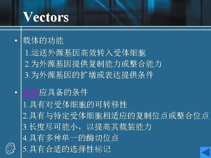
• Some phages may be used to incorporate DNA into the host genome ♦ Phage ♦ yeast episomal plasmids, ♦ Agrobacterium tumefaciens Ti plasmid, ♦ virus
Subcloning • The simplest kind of cloning experiment. • Conception: transfer of a fragment of cloned DNA from one vector to another • Subcloning may be used to investigate a short region of a large cloned fragment in more detail, or to transfer a gene to a vector designed to express it in a particular species. • The most common situation in E. coli
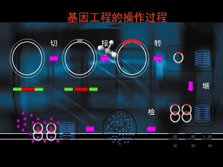
Process • Isolation of plasmid DNA containing the cloned sequence of interest • Digestion (cutting) of the plasmid into discrete fragments with restriction endonuleases • Separation of the fragments by agarose gel electrophoresis • Purification of the desired target fragment • Ligation of the fragment into a new plasmid vector, to form a new recombinant molecule • Transfer of the ligated plasmid into an E. coli strain (transformation) • Selection of transformed bacteria • Analysis of recombinant plasmids
DNA libraries • There are two main sources from which DNA is derived for cloning experiments designed to identify an unknown gene-bulk genomic DNA from the species of interest and, in the case of eukaryotes, bulk m. RNA from a cell or tissue where the gene is known to be expressed. They are used in the formation of genomic libraries and c. DNA libraries respectively. • Genomic libraries • c. DNA libraries
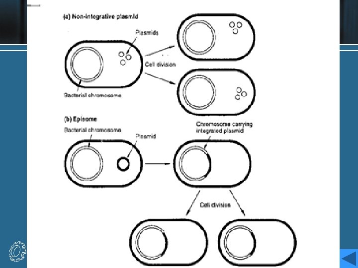
Genomic libraries • 基因组文库是指用分子克隆手段将提取的某一生 物完整的DNA序列(全染色体),经随机酶切或 机械剪切为一定长度的片段再与适当的载体DNA 连接后转移到受体细胞中而形成的各种片段的重 组子克隆的总和。 • Genomic libraries are prepared from random fragements of genomic DNA. • Genomic libraries may be an inefficient method of finding a gene, particularly in large eukaryotic genomes, where much of the DNA is noncoding.
c. DNA libraries • Other way to find target gene is to use as the source of the library the m. RNA from a cell or tissue which is known to express the gene. • DNA copies (c. DNA) are synthesized from the m. RNA by reverse transcription and are then inserted into a vector to form a c. DNA library. • c. DNA libraries are efficient for cloning a gene sequence, but yield only the coding region, and not the surrounding genomic sequences.
Screening libraries • We can use a radioactively or fluorescently labeled DNA probe which is complementary or partially complementary to a region of the gene sequence, and which can be used to detect it by hybridization. The probe sequence might be an oligonucleotide derived from the sequence of the protein product of the gene, if it is available, or from a related gene from another species. ★ An increasingly important method for the generation of probes is the polymerase chain reaction. ★ • Other screening methods rely on the expression of the coding sequences of the clones in the library and identification of the protein product from its activity,
Analysis of a clone • Once a clone containing a target gene is identified, the structure of the cloned fragment may be investigated further using restriction mapping, the analysis of the fragmentation of the DNA with restriction enzymes, or ultimately by the sequencing of the entire fragment. • The sequence can then be analyzed by comparison with other known sequences from databases, and the complete sequence is then available for manipulation in any of the applications of cloning described above.
G 2 Preparation of plasmid DNA • Plasmids as vectors • Plasmid minipreparation • Alkaline lysis • Phenol extraction • Ethanol precipitation • Cesium chloride gradient • Example of preparation of plasmid DNA
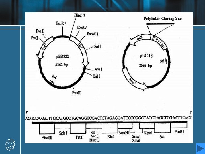
Plasmids as vectors • The first cloning vectors in the mid 1970 s, were naturally occurring bacterial plasmids from E. coli. • Plasmids are small, extrachromosomal circular molecules, from 2 to around 200 kb in size, which exist in multiple copies within the host E. coli cell. • They contain an origin of replication (ori), which enables them to be replicated independently, although this normally relies on polymerases and other components of the hose cell’s machinery. • They usually carry a few genes, one of which may confer resistance to antibacterial substances. The most widely known resistance gene is the bla, or ampr gene and tet. A gene.
Plasmid minipreparation • The first step in a subcloning procedure is the isolation of plasmid DNA. • Since plasmids are so much smaller than E. coli chromosomal DNA, they can be separated from the latter by physico-chemical methods. • It is normally possible to isolate sufficient plasmid DNA for initial manipulation from a few milliliters of bacterial culture. Such an isolation is normally known as a minipreparation or miniprep. • A sample of an E. coli strain harboring the required plasmid is inoculated into a few milliliters of culture broth. After growth to stationary phase (overnight), the suspension is centrifuged to yield a cell pellet.
Alkaline lysis • The most commonly used method • The cell pellet is resuspended in a buffer solution which may optionally contain lysozyme to digest the cell wall. The cell lysis solution, which contains SDS in an alkaline sodium hydroxide solution, is then added. • The preparation is then neutralized with a concentrated solution of potassium acetate (KAc) at p. H 5. This has the effect of precipitating the denatured proteins, along with the chromosomal DNA and most of the detergent. • The sample is centrifuged again, and the resulting supernatant (lysate) now contains plasmid DNA and a lot of small RNA and some proteins. • Plasmid DNA which being small and closed-circular, is easily renatured after the alkali treatment.
Phenol extraction • There are now innumerable proprietary method for the isolation of pure plasmid DNA from the lysate above, many of which involve the selective binding of DNA to a resin or membrane and the washing away of protein and RNA. • The classical method, which is slower but perfectly effective, involves the extraction of the lysate with phenol or a phenol-chloroform mixture. The phenol is immiscible with the aqueous layer but, when mixed vigorously with it, then allowed to separate, denatures the remaining proteins , which form a precipitate at the interface between the layers.
Ethanol precipitation • The DNA and RNA remaining in the aqueous layer are now concentrated by ethanol precipitation. • If sodium acetate is added to the solution until the Na+ concentration is more than 0. 3 M, the DNA and/or RNA may be precipitated by the addition of 2 -3 volumes of ethanol. • Centrifugation will pellet the nucleic acid, which may then be resuspended in a smaller volume, or in a buffer with new constituents, etc. • In the case of a minipreparation, ethanol is added directly to the phenol-extracted lysate, and the pellet is taken up in Tris-EDTA solution, the nomal solution for the storage of DNA.
• This solution contains Tris-hydrochloride to buffer the solution (usually p. H 8) and a low concentration of EDTA, which chelates any Mg 2+ ions in the solution, protecting the DNA against degradation by nucleases, most of which require magnesium. • Ribonuclease A (RNase A) may also be added to the solution to digest away any remaining RNA contamination. The enzyme digests RNA but leaves DNA untouched and does not require Mg 2+.
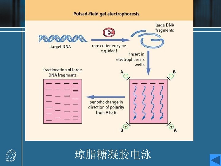
Cesium chloride gradient • The alkaline lysis method may also be used on a larger scale to prepare up to milligram quantities of plasmid DNA. • In this case, Cs. Cl density gradient centrifugation may be used as a final purification step. • Laborious, best method , supercoiled plasmid DNA • If a crude lysate is fractionated on a Cs. Cl gradient in the presence of EB, the various constituents may all be separated. • Supercoiled DNA binds less EB than linear or nicked DNA, and has a higher density. • The solution containing the plasmid band is removed from the centrifuge tube and may then be concentrated using ethanol precipitation after removal of EB.
Restriction enzymes and electrophoresis • Restriction endonucleases • Recognition sequences • Cohesive ends • Restriction digests • Agarose gel electrophoresis • Isolation of fragments
Restriction endonucleases • To incorporate fragments of foreign DNA into a plasmid vector, methods for the cutting and rejoining of ds. DNA are required. • The identification and manipulation of restriction endonucleases was the key of DNA cloning. • Restriction-modification systems occur in many bacterial species, and constitute a defense mechanism against the introduction of foreign DNA into the cell. • They consist of two components: ♦ Restriction endonuclease ♦ Methylase
• Restriction endonuclease It recognizes a short, symmetrical DNA sequence and cut (hydrolyzes) the DNA backbone in each strand at a specific site within that sequence. Foreign DNA will hence be degraded to relatively short fragments. • Methylase It adds a methyl group to a C or A base within the same recognition sequences in the cellular DNA. This modification renders the host DNA resistant to degradation by the endonuclease.
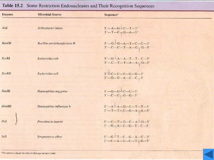
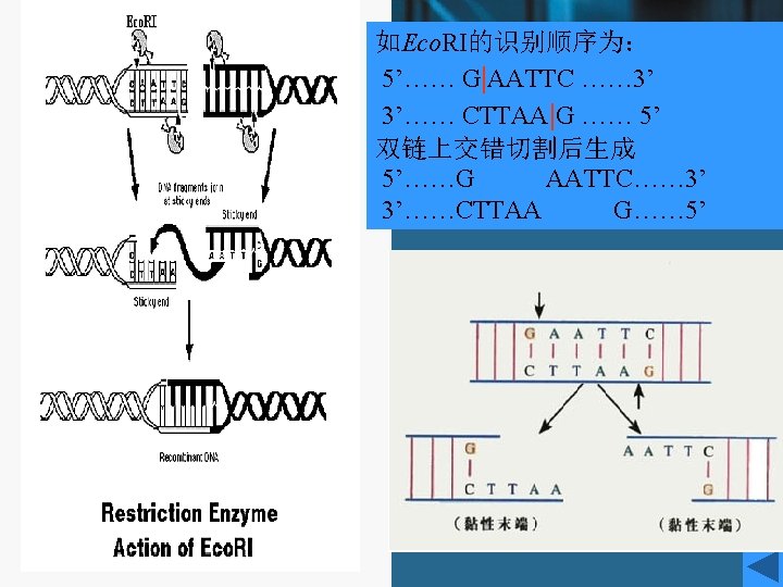
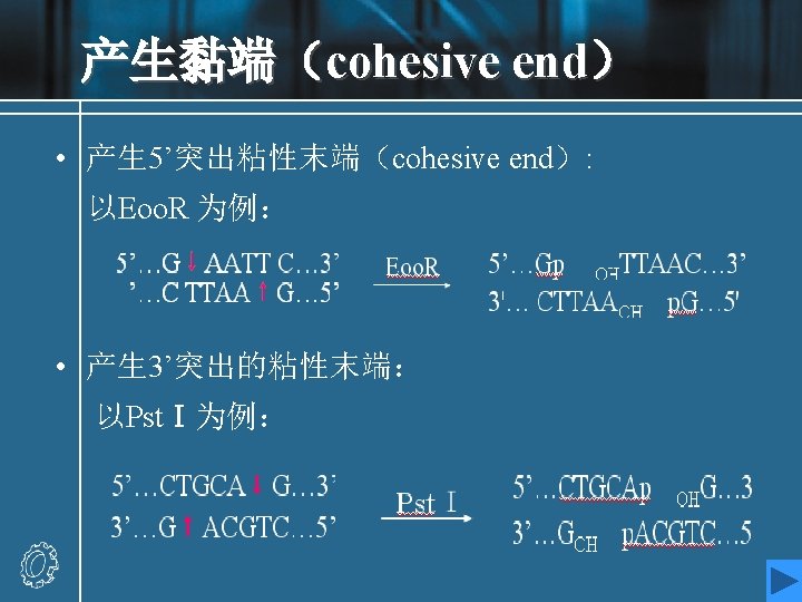
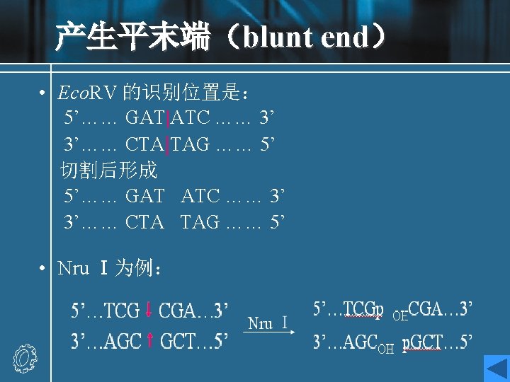
Recognition sequences • Eco. RI • Hundreds of restriction enzymes are now known, and a large number are commercially available. • They recognize sites ranging in size from 4 to 8 bp or more, and may give products with protruding 5`or 3`- tails or blunt ends. The newly formed 5`- ends always retain the phosphate groups. • Type of recognition sequence • The extremely high specificity of restriction enzymes for their sites of action allows large DNA molecules and vectors to be cut reproducibly into defined fragments.
Eco. RI • This enzyme acts as a dimer. • Eco. R I will only recognize a 6 bp palindromic sequence (the sequence is the same, reading 5`→ 3`, on each strand). • The product of the cutting reaction at this site on a linear DNA is two double-stranded fragments (restriction fragments), each with an identical protruding singlestranded 5`- end with a phosphate group attached. The 3` - ends have free hydroxyl groups. • A 6 bp recognition sequence will occur on average every 46=4096 bp in random sequence DNA; hence, a very large DNA molecule will be cut into specific fragments averaging 4 kb by such an enzyme.
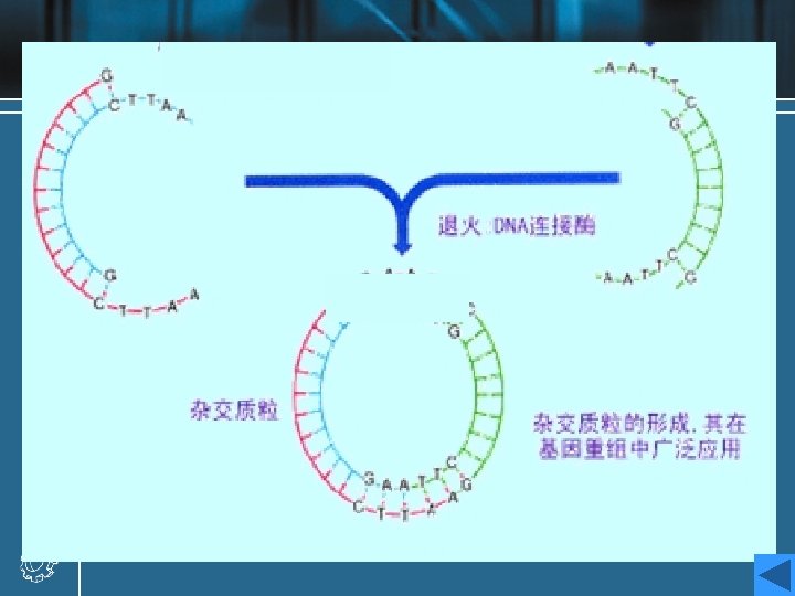
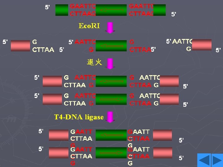
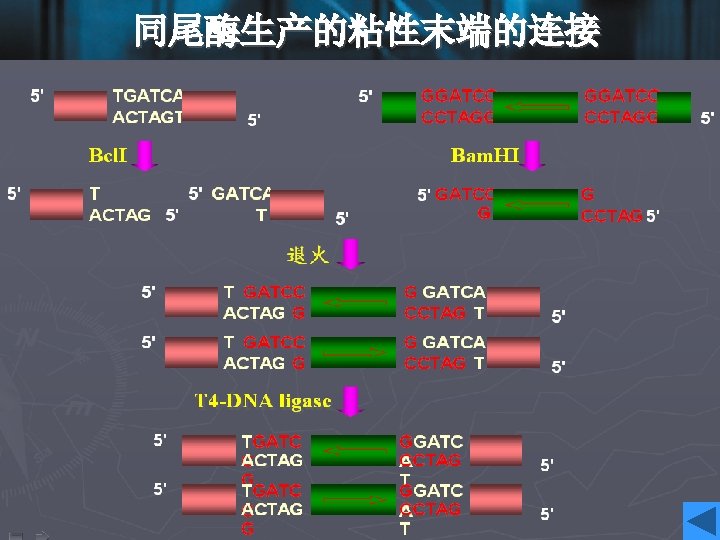
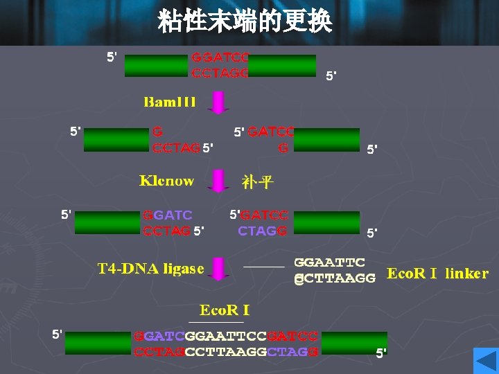
Cohesive ends • Those products of restriction enzyme digestion with protruding ends have a further property; these ends are known as cohesive, or ‘sticky’ ends, since they can bind to any other end with the same overhanging sequence, by base pairing (annealing) of the single-stranded tails. • Hence, for example, any fragment formed by an Eco. RI cut can anneal to any other fragment formed in the same way, and may subsequently be joined covalently by ligation. • In fact, in some cases, DNA ends formed by enzymes with different recognition sequences may be compatible, provided the single-stranded tails can base-pair together.
Restriction digests • Digestion of plasmid or genomic DNA is carried out with restriction enzymes for analytical or preparative purposes, using commercial enzymes and buffer solutions. • All restriction enzymes require Mg 2+, usually at a concentrations of up to 10 m. M, but different enzymes require different p. Hs, Na. Cl concentrations or other solution constituents for optimum activity. • The buffer solution required for a particular enzyme is supplied with it as a concentrate. The digestion of a sample plasmid with two different restriction enzymes, Bam. HI and Eco. RI, is illustrated in Fig. 2.
• The digestion of a few hundred nanograms (<1 µg) of plasmid DNA is sufficient for analysis by agarose gel electrophoresis; preparative purposes may require a few micrograms. The former amount corresponds to a few percent of a miniprep sample, as described in Topic G 2. • The DNA is incubated with the enzyme and the appropriate buffer at the optimum temperature (usually 37℃), in a volume of perhaps 20 µl. A dye mixture is then added to the solution, and the sample is loaded on to an agarose gel.
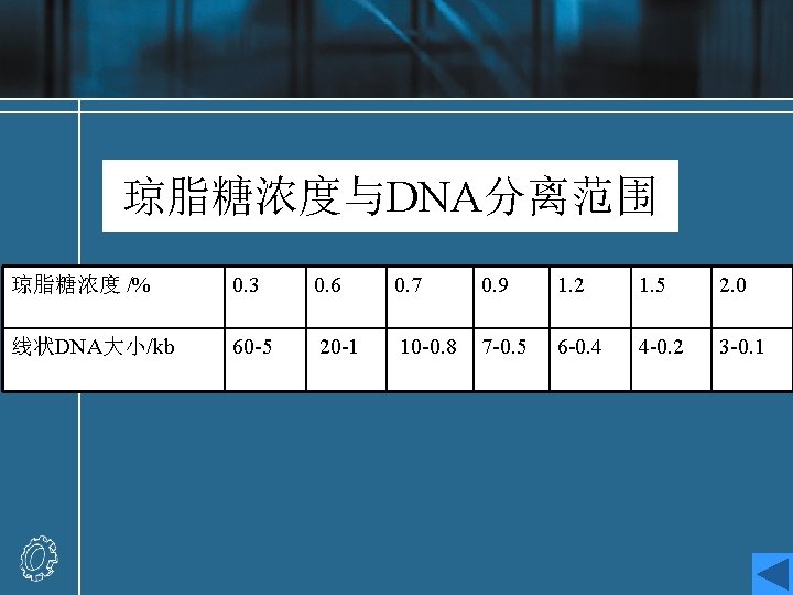
Agarose gel electrophoresis • Agarose is a polysaccharide derived from seaweed, which forms a solid gel when dissolved in aqueous solution at concentrations between 0. 5 and 2%(w/v). • Agarose used for electrophoresis is a more purified form of the agar used to make bacterial culture plates. • Agarose gel electrophoresis procedure • Analysis of electrophoresis result
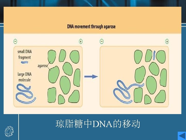
Agarose gel electrophoresis procedure • When an electric field is applied to an agarose gel in the presence of a buffer solution which will conduct electricity, DNA fragments move through the gel towards the positive electrode (DNA is highly negatively charged) at a rate which is dependent on their size and shape. • Small linear fragments move more quickly than large ones, which are retarded by entanglement with the network of agarose fibers forming the gel. Hence, this process of electrophoresis may be used to separate mixtures of DNA fragments on the basis of size.
• Different concentrations of Gel [1%, 1. 5%(w/v), etc. ] will allow the optimal resolution of fragments in different size ranges. • The DNA samples are placed in wells in the gel surface, the power supply is switched on and the DNA is allowed to migrate through the gel in separate lanes or tracks. The added dye also migrates, and is used to follow the progress of electrophoresis. • The DNA is stained by the inclusion of ethidium bromide in the gel , or by soaking the gel in a solution of ethidium bromide after electrophoresis. The DNA shows up as an orange band on illumination by UV
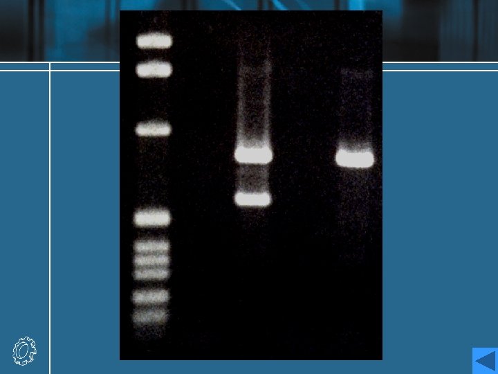
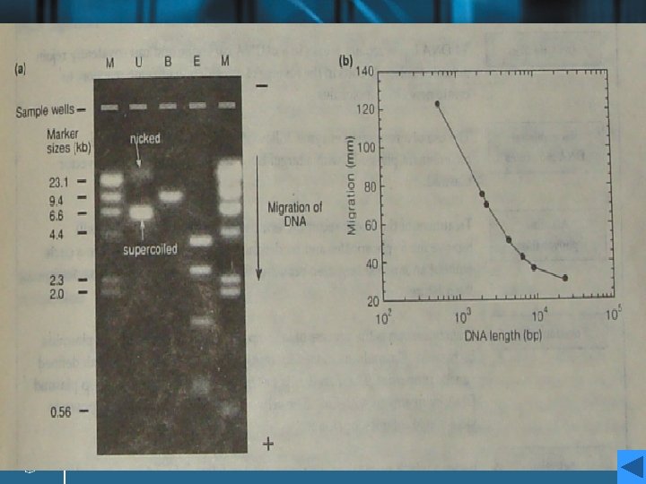
Analysis of electrophoresis result • The plasmid has been run on the gel without digestion (track U), and after digestion with Bam. HI (track B) and Eco. RI (track E). • A set of linear marker DNA fragments of known sizes (track M) have been run alongside the samples at two different concentrations; the sizes are marked on the figure. A number of points may be noted. • Analysis
Analysis • Track U consists of two bands. the lower: negatively supercoiled plasmid DNA the upper: open-circular or nicked DNA • The lanes containing the digested DNA clearly reveal a single fragment (track B) and five fragments (track E), whose sizes can be estimated by marker. • A more accurate determination of the sizes of the linear fragments can be made by plotting a calibration curve of the log of the size of the known fragments in track M against the distance migrated by each fragment.
Isolation of fragments • Agarose gels may also be used preparatively to isolate specific fragments for use in subsequent ligation and other cloning experiments. • Fragments are excised from the gel, and treated by one of a number of procedures to purify the DNA away from the contaminating agarose and ethidium bromide stain. • If we assume that the Eco. RI fragment containing the gene X is the target DNA for a subcloning experiment, then the third largest fragment in track E could be purified from the gel ready for ligation into a new vector.
G 4 Ligation transformation and analysis of recombinants • DNA ligation • Recombinant DNA molecules • Transformation
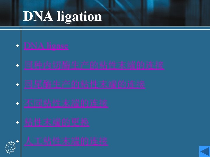
DNA ligase • To insert a target DNA fragment into a vector, a method for the covalent joining of DNA molecules is essential. DNA ligase enzymes perform just this function. • DNA ligase will repair (ligate) a break in one strand of a ds. DNA molecule, provide the 5’-end has a phosphate group. • They require an adenylating agent to activate the phosphate group for attack by the 3’-OH; the E. coli enzyme used NAD+, and the more commonly used enzyme from bacteriophage T 4 used ATP.
• Ligases are efficient at sealing the broken phosphodieaster bonds in an annealed pair of cohesive ends, essentially the reverse of a restriction enzyme reaction, and T 4 ligase can even ligate one blunt end to another, albeit with rather lower efficiency. • DNA ligase of E. coli • DNA ligase of T 4 phage • DNA ligase of eukaryotes
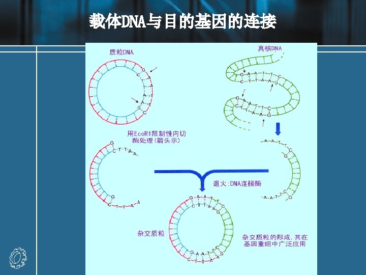
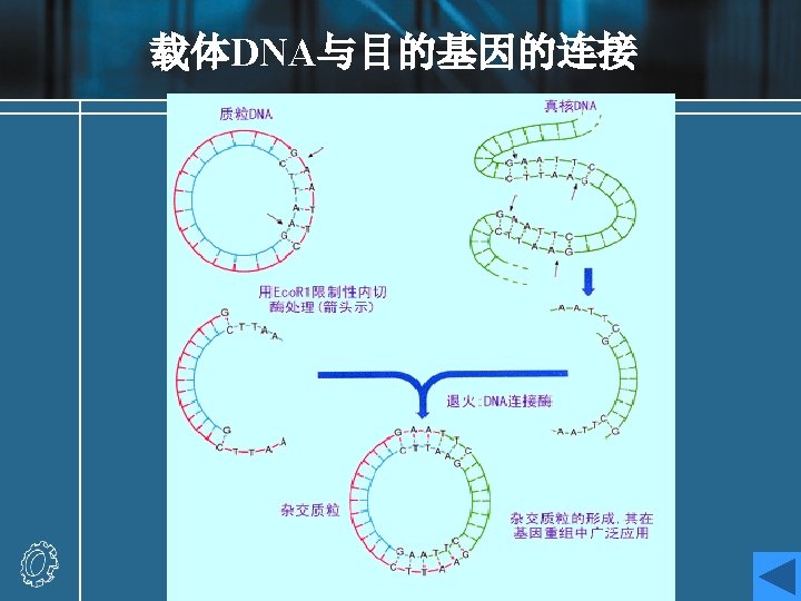
Recombinant DNA molecules Experiment: a DNA fragment containing a gene (X) of interest is inserted into a plasmid vector. • The target DNA may be a single fragment isolated from an agarose gel, or a mixture of many fragments from, for example, genomic DNA. • If the target has been prepared by digestion with Eco. RI, then the fragment can be ligated with vector DNA cut with the same enzyme. • The vector should have only one site for cleavage with the relevant enzyme.
• Recombinant result: recombinant A, recombinant B (with either orientation) and religated vector • Recombination fraction = recombinant molecules / total vectors recombination fraction : 25%-75% • Methods of increasing recombination fraction
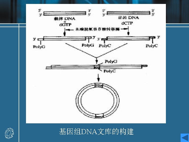
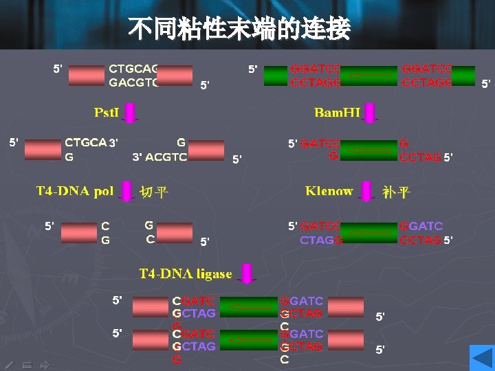
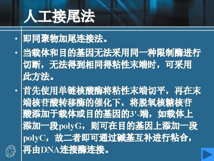
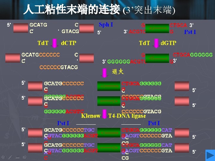
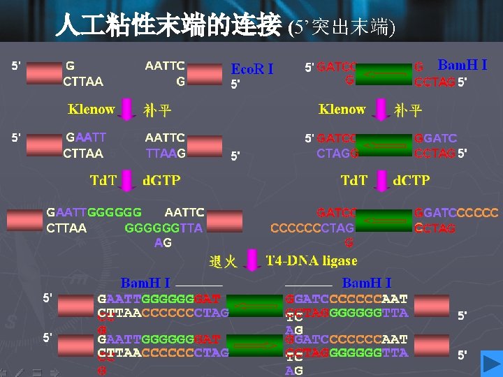
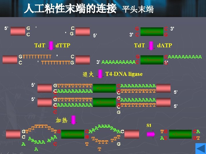
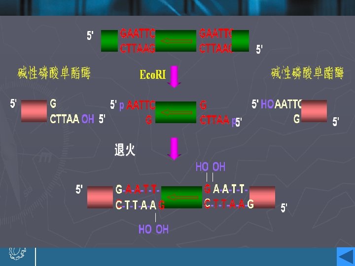
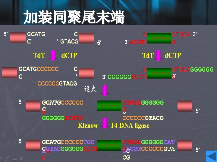
Increasing recombination fraction • Increasing the rate of foreign DNA fragments and vectors: 5: 1 -10: 1 • One solution is to prepare both the target and the vector using a pair of distinct restriction enzymes, such that they have noncompatible cohesive ends at either end. • If it is inconvenient to use two restriction enzymes, then the linear vector fragment may be treat with the enzyme alkaline phosphatase after restriction enzyme digestion. • 加装同聚尾末端
Alkaline phosphatase • Alkaline phosphatase removes phosphate groups from the 5’-ends of vector DNA molecules. • The linear vector will hence be unable to ligate into a circle, since no phosphates are available for the ligation reaction. • A ligation with a target DNA insert can still proceed, since one phosphate is present to ligate one strand at each cut site. • The remaining nicks in the other strands will be repaired by cellular mechanisms after transformation.
Transformation • Transformation theory • Transformation technology • Transformation efficiency • Selection • Growth and storage of transformants
Transformation theory • E. coli cells treated with solutions containing Ca 2+ ions were rendered susceptible to take up exogenous DNA, a process knows as transformation. • Cells pre-treated with Ca 2+ (and sometimes more exotic metal such as Rb 2+ and Mn 2+ ), in order to render them able to take up DNA, are known as competent cells. • A solution of a plasmid molecule, or a mixture of molecules formed in a ligation reaction, is combined with a suspension of competent cells for a period, to allow the DNA to be taken up. • The precise mechanism of the transfer of DNA into the cell is obscure.
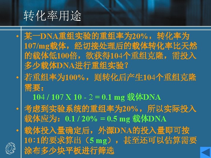
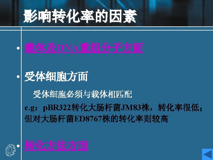
Transformation efficiency • The quality of a given preparation of competent cells may be measured by determining the transformation efficiency, defined as the number of colonies formed (on a selective plate) per microgram of input DNA, where that DNA is a pure plasmid, most commonly the vector to be used in a cloning experiment. • 每毫克载体DNA转化后,受体细胞接纳DNA的 个数,即克隆数 • 转化率的用途:利用转化率和重组率等参数可以帮助设计 DNA重组实验规模 • 转化率的影响因素
Growth and storage of transformants • Single colonies from a transformation plate are transferred to culture broth and grown overnight to stationary phase. • The broth must include the antibiotic used to select the transformants on the original plate, to maintain the selection for the presence of the plasmid. • The plasmid are then prepared from the cultures by the minipreparation technique. • The culture were subpackaged in glycerol stock.