Clinical Applications of Nuclear Medicine in GU Tract


 Ao-to-Kid ~")
 function DRF (Differential Renal Function)")


 full bladder, no catheter")
 with catheter in bladder")


")

 in acute UTI")


 Techniques: Ø Post Captopril scan ( 50 mg")


- Slides: 116
Clinical Applications of Nuclear Medicine in GU Tract a brief revew a brief review V. R. Dabbagh Kakhki, M. D. Nuclear Medicine Specialist Associate Professor DSNMC Nuclear Medicine Research Center (NMRC; MUMS)
Dynamic Renography : DTPA Nuclear Medicine & GU Renal Cortical Imaging: DMSA Direct Radionuclide Cystography (DRC): VCUG with Radioisotope
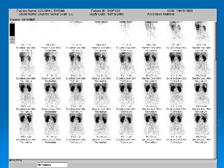
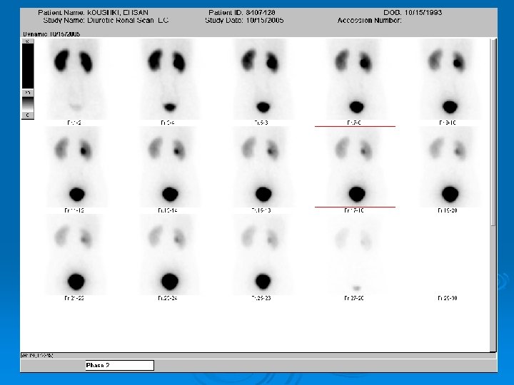
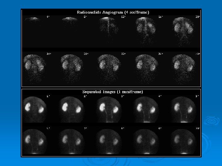
Dynamic Renography Technique: Ø Bolus injection of tracer Ø Obtained serial images l l Supine posterior view Imaging: Ø Perfusion: 1 -2 sec/view for 1 -2 min Ø Functional : 30 sec/view for 30 min
Dynamic Renography Technique: Imaging: Ø Perfusion: 1 -2 sec/view for 1 -2 min Ø Functional : 30 sec/view for 30 min
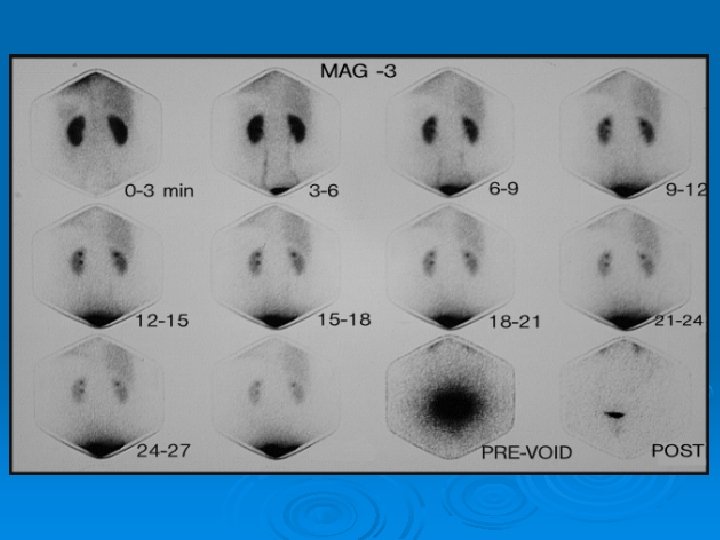
Dynamic Renography Radiotracers Glomerular Filtration Tubular Secretion Ø 99 m. Tc-DTPA >95% Ø 99 m. Tc-MAG 3 <5% 95% Ø 99 m. Tc-EC <10% 90% Ø 131 I-OIH 20% 80% Extraction 20% 40 -50% ~100%
DTPA: Diethylenetriaminepentaacetic acid MAG 3: Mercaptoacetylglycine EC: ethylenedicysteine OIH: Orthoiodohipuric acid
Dynamic Renography; Evaluates: Clinical applications: Ø Renal ØObstructive nephrouropathy perfusion Ø Renal function, renal morphology and size ØReflux uropathy ØRenal failure Ø GFR Ø ERPF ØRenal transplant ØRVH
DTPA normal
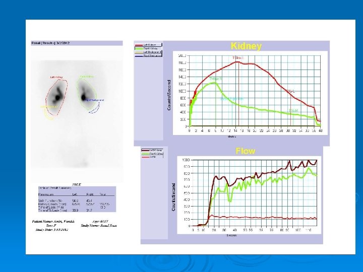
A curve can be generated that represents the perfusion only Mean counts/second Renal perfusion time-activity for Tc-99 m DTPA
Renogram
2 1 3 1 - Blood flow phase (20 - 40 sec) Ao-to-Kid ~ 3” 2 - Concentration phase (3 -5 min) Tpeak < 5’ 3 - Excretory(washout) phase
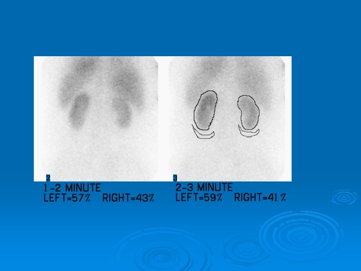
Relative (split) function DRF (Differential Renal Function)
DTPA normal
DTPA flow + Functional Phases GFR = 29 ml/’ Creat = 2. 0 DRF: LK= 33% RK= 67%
Renal artery occlusion
Rt renal infarct
Normal Renogram
Renal Function Ø Creatinine Clearance: l No accurate due to tubular excretion l Overestimates GFR in chronic renal disease and decreased muscle mass l No measure individual renal function unless catheterization of each kidney Ø Clearance of DTPA l Plasma-sample : more accurate l Camera-based Ø Relative uptake: DRF l Normal: 50/50 to 56/44 l 57/43 to 59/41: borderline l 60/40: abnormal
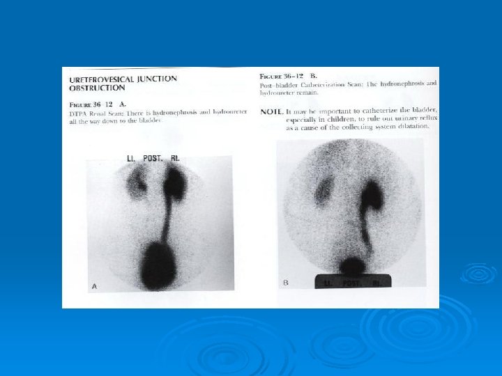
Obstructive uropathy Ø Diuresis renography for l l l Diagnosis Assesment of parenchymal damage Postsurgical evaluation Ø Anatomical imaging methods rely on demonstrating the structural abnormalities Ø Diuretic renography : Very useful : • evaluate renal function and urodynamics in a single test • but contributes little to determining etiology.
Obstructive uropathy Ø Ø There are two protocols in use: l F+20 : demonstrates a diuretic response l F-15 : more decisive in detecting minor degrees of obstruction If SKGFR<16 ml/min only Whitaker test can be done
No Obstruction
F+20
F-15
T 1/2 washout cts 100% 50% T 1/2 min
Quantitative t 1/2 diuretic response clearance Ø Washout half time l <10 min No obstruction l Between 10 and 20 min Indeterminate l >20 min Urinary obstruction
Diuretic Renal Scan Indications Ø Evaluate functional significance of hydronephrosis Ø Determine need for surgery Ø l obstructive hydronephrosis - surgical Rx l non-obstructive hydronephrosis - medical Rx Monitor effect of therapy
Diuretic Renal Scan Requirements Ø Rapidly cleared tracer Ø Well hydrated patient Ø Good renal function
pre-Lasix
post-Lasix No Obstruction
Lt hydronephrosis 3 -wk old baby 3164897
Lt UPJ obstruction 3164897
Lt UPJ obstruction 3164897
Diuretic Renal Scan Interpretation Ø Interpret whole study, not T 1/2 Ø Visual (dynamic images) Ø Washout curve shape Ø T 1/2 alone
Diuretic Renal Scan Pitfalls Ø False l l Distended bladder Gross hydronephrosis Poorly functioning / immature kidney Dehydration Ø False l l positive for obstruction negative Low grade obstruction Poorly functioning / immature kidney
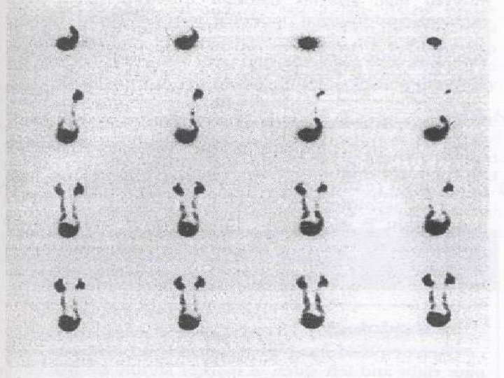
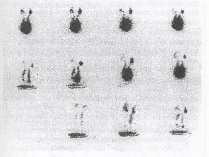
Effect of catheterization (1) full bladder, no catheter
Effect of catheterization (2) with catheter in bladder
Whitaker test Ø Standard for obstruction Ø Invasive Ø Infusion l l l : pressure> 22 cm H 2 O to achieve a pelvoureteral flow rate of 10 ml/min : Obs Indeterminate <15 cm H 2 O : No Obs
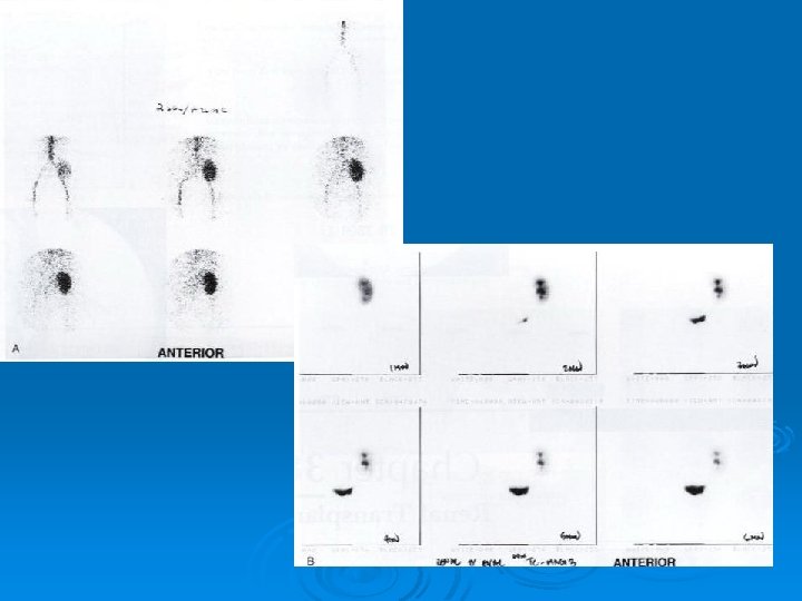
Renal transplant scan; Ø Essential part of transplant department. Ø Best tracer: 99 m. Tc-MAG 3. Ø Perfusion and function analysis Ø Useful in DDx of transplant complications: ATN, Rejection, Cyclosporine toxicity, Obstructive disease, Urinoma, Lymphocele, …
Complications after renal transplantation ATN Minutes to hours Ø Rejection l Hyperacute Minutes to hours l Accelerated 1 -5 days l Acute After 5 days (first 3 months) l Chronic Months to years Ø Cyclosporine toxicity Months Ø Surgical l Urine leak, Hematoma, Wound infection, l Obstruction, Lymphocele, RAS Ø
Differential diagnosis of various complications Requires correlation of scintigraphic findings wi Pt’s clinical course , physical findings, laborator values, current therapy, prior scintigraphic findings, and results of other imaging tests.
Ø Since many of the complications are diagnosed from the patterns of changes in time, it is important to obtain a baseline study soon after transplantation. Follow-up studies should be performed always by the same technique.
Renal transplant scan: TRS Ø Two Aspects: l l Ø Perfusion phase Function Phase Choice; Tc 99 m-MAG 3 l l Good quality perfusion images High extraction rate • Excellent images l Evaluation of collecting system, ureter and bladder
TRS: Perfusion Ø Images Ø Curves
TRS: Functional Phase Ø Images Ø Curves l l l Uptake: Tc 99 m-MAG 3: Max: before : 5 min Parenchymal transit Excretion of the tracer
Nuclear medicine testing Ø Ø Ø Obtain a baseline study soon after transplantation ATN: Perfusion is better than function, decreased uptake, delayed transit, diminished clearance AR: Decreased perfusion, tracer uptake, delayed transit and decreased clearance CR: low uptake, normal parenchymal transit with absent or minimal cortical retention Cyc toxicity: l Mild : like CR l Severe: like ATN RVH caused by RAS can not differentiated from CR unless challenged by ACEIs. ( pattern of CR changes to a pattern of AR)
Acute rejection Functional phase Perfusion Phase
Surgical complications Ø US and Tc 99 m-MAG 3 images Ø Diuretic renography : Differentiate obstruction from simple pelvocalyceal dilatation
Lymphocele
Urinary Leak
Radionuclide Cystogram
Indications Ø Evaluation of children with recurrent UTI l 30 -50% have VUR Ø F/U after initial VCUG Ø Assess effect of therapy / surgery Ø Screening of siblings of reflux pts.
Methods Direct Ø Tc-99 m S. C. or Tc. O 4 Advant. Ø via Foley can do at any age Disadv. Ø VUR during filling Ø Ø catheterization Indirect Tc-99 m DTPA or Tc-99 m MAG 3 Ø i. v. Ø no catheter Ø info on kidneys Ø need pt cooperation Ø need good renal fct Ø
Direct Cystography Ø 1 m. Ci S. C. in saline via Foley Ø Fill bladder until reversal of flow l (bladder capacity = (age+2) x 30 Ø Continuous imaging during filling & voiding Ø Post void image Ø Record l l l volume instilled volume voided pre- and post- void cts
RN Cystogram vs. VCUG Advantages Disadvantages Lower radiation dose Ø Cannot detect distal (5 vs 300 mrad to ureteral reflux ovary) Ø No anatomic detail Ø Smaller amount of Ø Grading difficult reflux detectable Ø Quantitation of postvoid residual volume Ø
Normal cystogram filling voiding post-void
VUR - filling phase A
VUR - voiding phase & post- void B
Post void residual volume RV = voided vol x post-void cts pre-void cts - post void cts
Reflux nephropathy 16% 84%
Vesicoureteral reflux Conventional method : X-ray micturating cystography l Excellent delination of bladder and urethral anatomy l Grading of the reflux Ø An alternative method : radionuclide cystography Ø Technique: Ø l l l Direct & Indirect Direct: The same as radiological VCUG Indirect: At the end of DTPA or MAG 3 renal scan, lesser sensitivity, no for initial screening test, a positive study is reliable but a negative study should be confirmed by direct cystography
Radionuclide cystography: Ø Advantages: l l l Ø High sensitivity. Low radiation. (50 to 200 times less radiation to gonads comapred to the contrast cystography Quantification ( post voiding residue). Disadvantages: l l Poor grading ability. No anatomic detail of the urerthra * VCUG is reserved for the initial work up of male patients to exclude an anatomical abnormality, such as PUV.
Radionuclide cystography: Indications: Initial screening to detect reflux in girls with UTI Ø Follow up of patients with reflux. Ø Screening of siblings Ø Serial evaluation of children with neuropathic bladder who are at risk to develop reflux Ø
Renal infection Ø Radiopharmaceuticals: l 99 m. Tc-DMSA l 99 m. Tc-glucoheptonate l 67 Ga l 111 In-WBC or 99 m. Tc-WBC
Renal sonography Commonly used in the evaluation and management of UTI Ø Non-invasive imaging Ø Detection of hydronephrosis, and congenital anomalies Ø Detection of renal abscesses, pyeonephrosis and abnormalities of the perinephric space Ø Changes secondary to acute pyelonephritis may also be recognized Ø
Cortical imaging: 99 m. Tc-DMSA Cortical agent Ø Trapped in the cytoplasm of the proximal tubular cells Ø An indicator of functioning tubular renal mass. Ø Advantages : (over IVP and US) l Pyelonephritis and renal scars: more sensitive • Tc 99 m-DMSA : 94% • Intravenous pyelography : 76% • US: 65% l Defects on DMSA scan become apparent before on IVP or US l Lower radiation dose (as compared to the IVP) l Is not affected by overlying bowel gas or bones l Avoids possible allergic reaction Ø
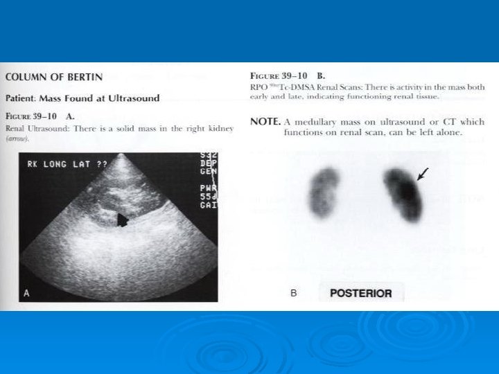
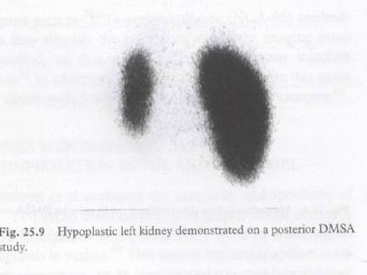
DMSA Renal scan; Clinical applications; Ø Ø Ø Ø Ø Renal size, shape and location Renal cortical assessment. Determining DRF (most accurate noninvasive method) Infectious disease and distinguish upper from lower UTI(Early detection and follow up of pyelonephritis; most sensitive, 95% Vs 76%) Follow up of patients(serial scans) Congenital renal anomaly(Ectopia, . . ) Vascular lesions (infarct) DDx of pseudomass from SOL. Renal trauma Confirm the total absence of function in dysplastic kidney
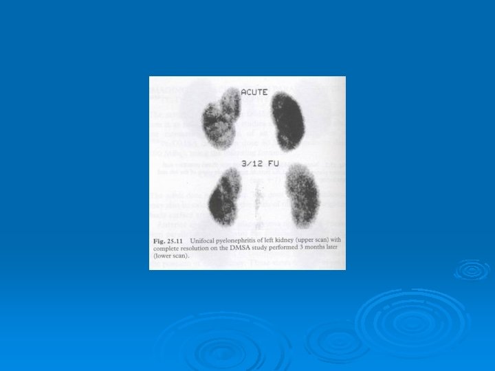
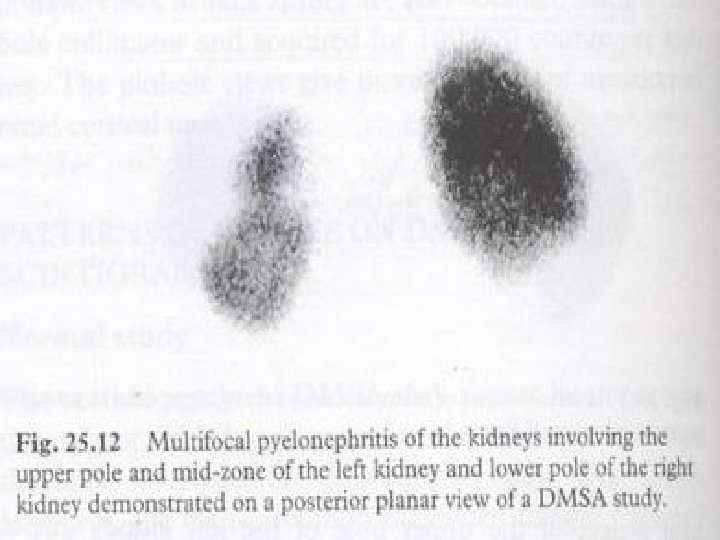
Acute pyelonephritis in DMSA scan Ø Single or multiple areas of decreased cortical uptake Ø No loss of volume Ø Diffusely decreased uptake in an enlarged kidney Diminished uptake may be due to both focal tubular cell dysfunction and ischemia l A mature cortical scar is usually associated with contraction, cortical thinning, loss of volume and marked reduction in uptake
Normal DMSA Renal Scan
Evaluation of Renal Infection Renal Morphology Scan (Renal Cortical Scintigraphy)
UTI Ø VUR l risk factor for PN, l not all pts w PN have VUR Ø PN may lead to scarring >>> ESRD, HTN l early Dx and Rx necessary Ø Clinical & laboratory Dx of renal involvement in UTI unreliable
Renal Cortical Scintigraphy Indications Ø Determine involvement of upper tract (kidney) in acute UTI (acute pyelonephritis) Ø Detect cortical scarring (chronic pyelonephr. ) Ø Follow-up post Rx
Renal Cortical Scintigraphy Procedure Ø Tracers l l Tc-99 m DMSA Tc-99 m GHA Ø Acquisition l l l 2 -4 hrs post-injection parallel hole posterior pinhole post. + post. oblique (or SPECT) Ø Processing: relative fct
Renal Cortical Scintigraphy Interpretation Ø Acute PN l l single or multiple “cold” defects renal contour not distorted diffuse decreased uptake diffusely enlarged kidney or focal bulging Ø Chronic PN l l volume loss, cortical thinning defects with sharp edges Ø Differentiation of Ac. PN vs. Ch. PN unreliable
Renal Cortical Scintigraphy “Cold Defect “ Ø Acute or chronic PN Ø Hydronephrosis Ø Cyst Ø Tumors Ø Trauma (contusion, laceration, rupture, hematoma) Ø Infarct
DMSA Normal
Normal DMSA pinhole LPO RPO
Acute pyelonephritis DMSA Defect in Right Kidney post L post R LEAP LPO RPO
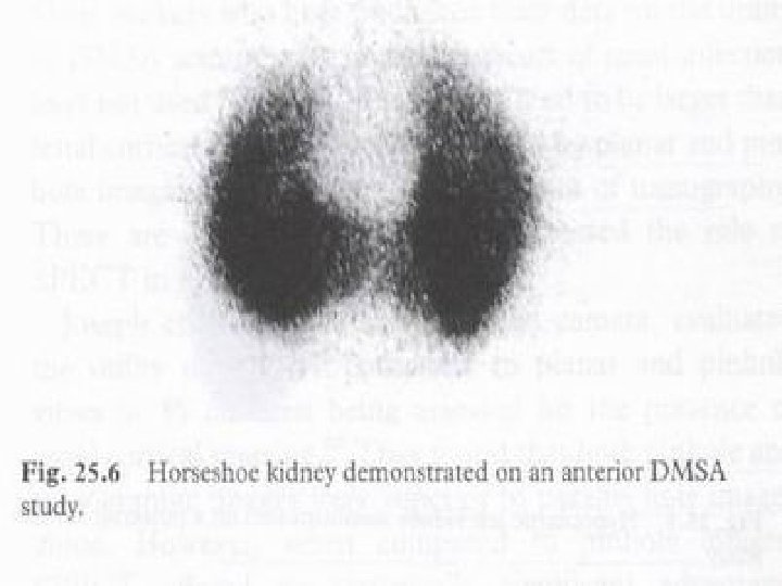
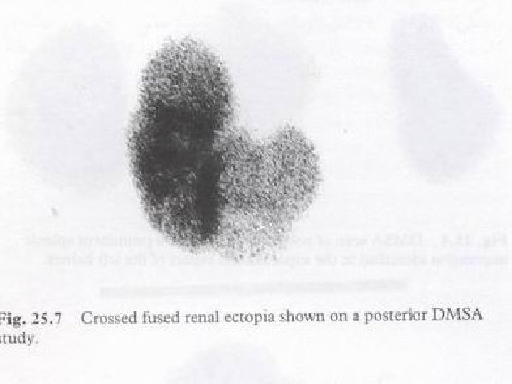
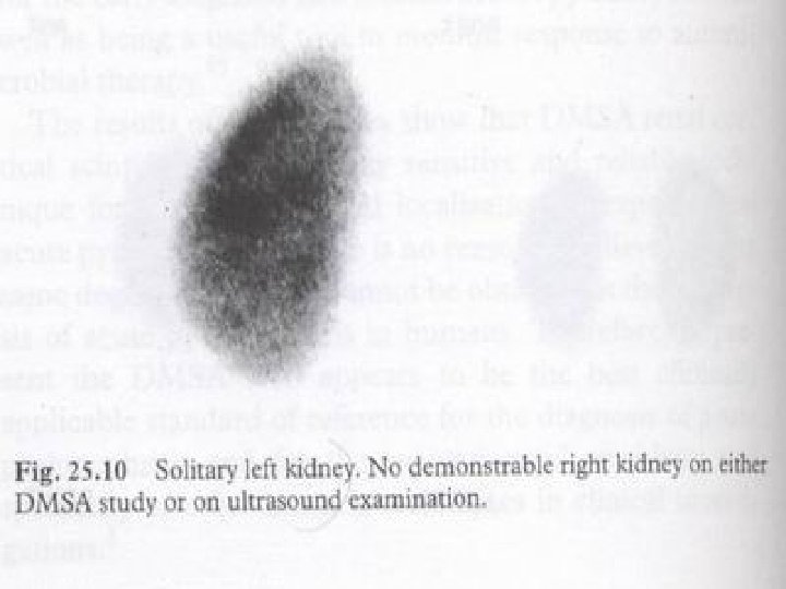
Renal Cortical Scintigraphy Congenital Anomalies Ø Agenesis Ø Ectopy Ø Fusion (horseshoe, crossed fused ectopia) Ø Polycystic kidney Ø Multicystic dysplastic kidney Ø Pseudomasses (fetal lobulation, hypertrophic column of Bertin)
DMSA horseshoe kidney
DMSA LK Agenesis
Crossed ectopia 74% 26%
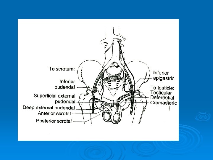
Scrotal scintigraphy; Ø Ø Ø Ø If the diagnosis of testicular torsion is established : surgery When the diagnosis is uncertain, imaging studies should be rapidly obtained Tracer: Technetium pertechnetate (99 m. Tc. O 4) Easy, simple and takes only 10 minutes DDx of acute torsion from other complications ( epididymitis) Scintigraphy can confirm the clinically suspected diagnosis of torsion and direct the patient to surgery Scintigraphy can minimize unnecessary exploration in patients with an inflammatory cause of their pain. For chronic or painless disorders of the scrotum US is the method of choice.
Acute testicular torsion Scintigraphic findings The findings depend on the time l Early torsion: Decreased activity in the region of the involved testicle l Late torsion: Increased scrotal activity but relatively decreased activity in the region of the ischemic testicle “ bull’s eye” Ø Acute epididymitis & epididymoorchitis: Increased activity Ø Scrotal scintigraphy is not the imaging of choice in most other conditions affecting the scrotal contents. Ø
Scrotal scintigraphy; Ø SS for acute testicular torsion l l Sensitivity and specificity > 95% False negative • Spontaneous detorsion • Incomplete twists • Inguinal testis
Normal Early torsion Late Torsion
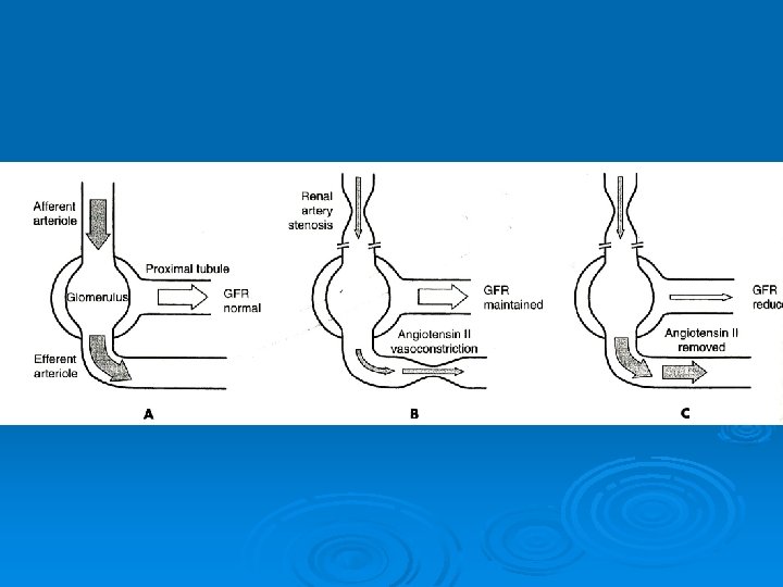
Renovascular Hypertension Ø Ø Ø Defined as stenotic lesions of the renal arteries that induce elevated BP and potentially improve after revascularization Anatomic stenosis is not equivalent to the diagnosis of RVH Significant morbidity from renal artery angioplasty or revascularization A functional diagnosis is needed before proceeding with therapy Discrepancies between scintigraphy and angiography l Involving the small vessels l RAS incidentally in a substantial proportion of elderly Pts The prevalence of RVH<1%
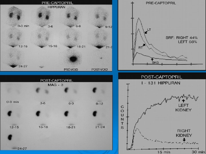
Captopril Renal Scan (ACE inhibition renography) Techniques: Ø Post Captopril scan ( 50 mg orally, . . ) Ø Base line scan Ø Positive if GFR or Curve worsening Mechanism: Ø Inhibition of ACE, releasing efferent arteriole constriction. Advantages: Ø Best screening test for RVH Ø Best predictor of response to surgery, angioplasty and captopril therapy Ø Determine which Pts have hemodynamically significant stenoses enough to cause HTN Ø Avoided unnecessary arteriograms Ø Detection of RVH whatever the level of arterial obstruction Ø High accuracy Ø Sensitivity: 80 -90% Ø Specificity: 90 -95%
Criteria of positivity Ø 10% rise in cortical retention ratio Ø Increase in the Time to peak Ø Worsening in DRF ( relative uptake) Ø MPTT
Criteria of positivity Ø Shape of curve: a worsening of at least one grade
Baseline Study Post-captopril Study Pre- and post captopril excretion curves of the right kideny
Selection of patients for evaluation Ø Diagnosis of RVH can be made only after the patient responds to revascularization Ø The tests can not be used as a mass screening procedure: l l The incidence of RVH is low and therefore post test probability of a positive test still has a low probability of RVH. CRS appears to have improved sensitivity and specificity to the point where it is practical to screen selected patients.
Indication of ACE inhibition renography Ø Ø Ø Accelerated or malignant HTN Abrupt or recent onset HTN Onset under age 30 or over age 55 Refractory HTN Abdominal or flank bruits Unexplained azotemia Worsening renal function during therapy with ACEIs End organ damage ( LVH, retinopathy) Occlusive disease in other vascular beds Previous hypertensive urogram suggestive of RAS Unilateral small kidney
Aspirin Renography Renal blood flow PGE 2 Renin Ø Ø - Inhibition of PG synthesis would decrease renin : An effect similar to captopril Aspirin reduces both renal blood flow and glomerular filtration Similar sensitivities to ACEI scintigraphy
V. R. Dabbagh; DSNMC; www. DSNMC. ir