Function and Structure in Nuclear Medicine Nuclear Medicine


")


")


 scattered photon (scatter and collimator response ) absorbed")
 scattered photon (scatter and collimator response ) absorbed")


- Slides: 65
Function and Structure in Nuclear Medicine
Nuclear Medicine • Radioactive material is administered into the patient body • Photons emitted in the patients are detected by a scintillator • Distribution of the radioactivity in the patient body is reconstructed from projections
Radioactivity Maria Składowska-Curie (1867 -1934)
Radioactivity Process that involves the nucleus
Radioactivity Energy state of nucleus changes
Radioactivity Decay scheme diagram for 14 C Transition energy • Numbers of protons and neutrons change • b- particle is emitted
Radioactivity Decay scheme diagram for 14 C is not good for imaging because the decay does not produce photons
Nuclear Medicine • Radioactive material that emits photons is administered into the patient body • Photons coming in the patients are detected by a scintillator • Distribution of the radioactivity in the patient body is reconstructed from projections
Radioactivity Decay scheme diagram for 14 C not only does not produce useful photons but also its half-life is 5730 years
Nuclear Medicine • Radioactive material with half-life in order of few hours that emits photons is administered into the patient body • Photons coming in the patients are detected by a scintillator • Distribution of the radioactivity in the patient body is reconstructed from projections
Decay scheme diagram for 133 Xe
Decay scheme diagram for 133 Xe Half-life 5. 2 days
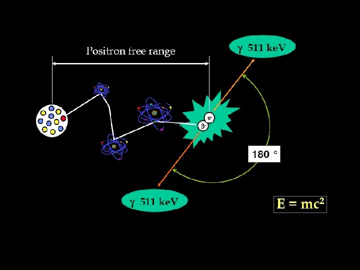
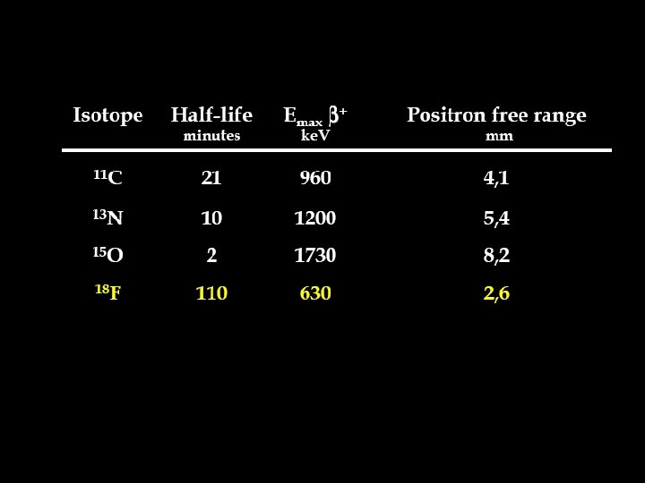
What about positron emitters ?
Decay scheme diagram for 18 F
Nuclear Medicine • Radioactive material with half-life in order of few hours that emits gamma photons is administered into the patient body • Photons coming in the patients are detected by a scintillator • Distribution of the radioactivity in the patient body is reconstructed from projections
Detection • Photon energy cannot be too high because photons would have too high penetration • Hard to detect • Safety issues • Photon energy cannot be too low because photons would have too low penetration
Nuclear Medicine • Radioactive material with half-life in order of few hours that emits gamma photons is administered into the patient body • Photons with energies ranging from 50 ke. V to 600 ke. V coming in the patients are detected by a scintillator • Distribution of the radioactivity in the patient body is reconstructed from projections
Gas Filled Detectors
Solid State Detectors
Scintillation Detectors Nuclear Medicine uses scintillation detectors Interaction of a gamma photon with the crystal produces visible light (several hundred photons)
Crystals Commonly used in NM
Scintillation Detectors
Direction of incoming photon Photomultiplier tubes Crystal
Direction of incoming photon Photomultiplier tubes Crystal Lead collimator
Direction of incoming photon Photomultiplier tubes Crystal Lead collimator The ambiguity is resolved by performing tomography i. e. rotating detector around patient
Single Photon Emission Tomography
Single Photon Emission Computed Tomography SPECT Typically: • 60 projections are acquired around patient over at least 180° • It takes 1 minute per projection to acquire enough photon counts
2 detector camera
3 detector camera
SPECT is not a very efficient technique
SPECT is not a very efficient technique • Only 1 in every 10, 000 gamma photons is detected • Resolution is only about 1 cm
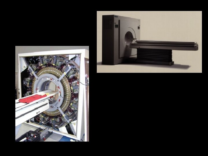
Positron Emission Tomography (PET)
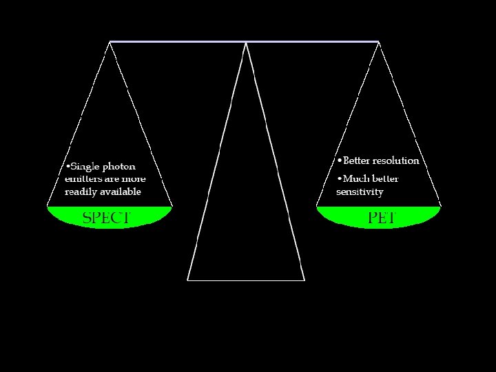
SPECT PET
PET
PET SPECT
Examples of NM images SPECT Cardiac Imaging • Perfusion • Viability
Cardiac SPECT
Cardiac SPECT
Brain SPECT primary photon (collimator response) scattered photon (scatter and collimator response ) absorbed photon (absorption)
Brain SPECT primary photon (collimator response) scattered photon (scatter and collimator response ) absorbed photon (absorption)
Brain SPECT
Brain SPECT Frontal Lobe Dementia
Oncology PET – 18 FDG 57 years old male, Lung Nodule D 1 cm Hypermetabolic nodule : cancer No adenopathy, no metastasis : possible surgery FDG : 15 m. Ci ; 15 min
Oncology PET – 18 FDG 57 years old male, lung cancer Ganglionary hepatic and bony metastases FDG : 5 m. Ci ; 7 x 7, 5 min
Oncology PET – 18 FDG 68 year old male, pancreatic cancer and melanoma Ganglion, bone and diffuse metastases FDG : 10 m. Ci ; 13 min
Is there an ideal Nuclear Medicine camera ?
Is there an ideal Nuclear Medicine camera ? • Electronic collimation • Single photon emitter
Is there an ideal Nuclear Medicine camera ? • Electronic collimation • Single photon emitter Yes, there is
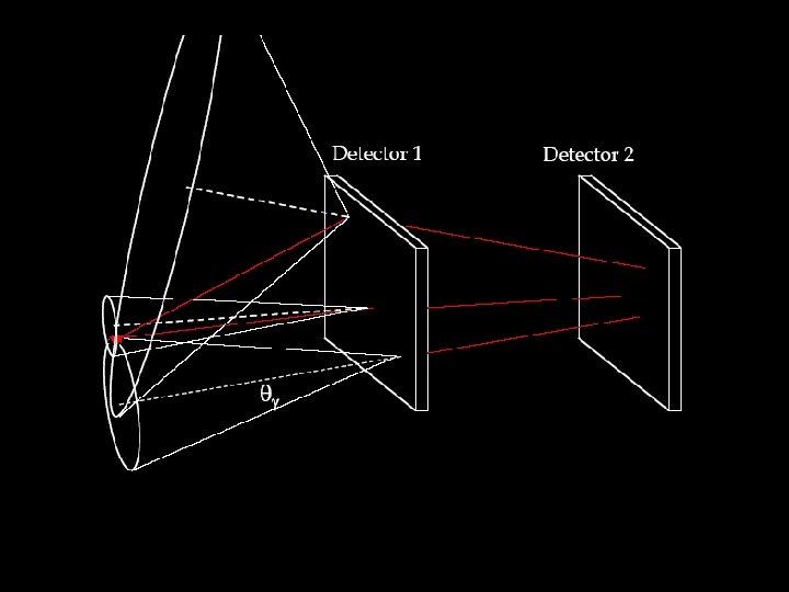
Compton Camera
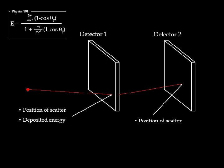
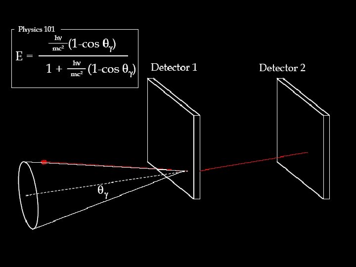
Physics 101 -Compton Scattering
Physics 101 -Compton Scattering
Gamma photon is scattered and deposits energy E Scattered gamma photon is detected
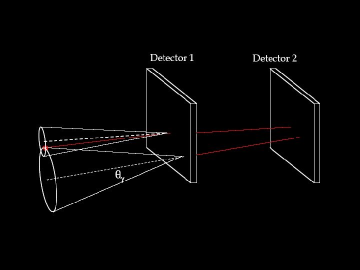
Challenges still to overcome • Detector 1 need to have VERY good energy resolution Solid state (semiconductor) detectors need to be used
Challenges still to overcome • In order to reconstruct the distribution of radioactivity from “Compton cones” HUGE inverse problem needs to be solved