
Brain Structure and Function If the human brain

Brain Structure and Function
“If the human brain were so simple that we could understand it, we would be so simple that we couldn’t” -Emerson Pugh, The Biological Origin of Human Values (1977)

The Brain • Brainstem – responsible for automatic survival functions • Medulla – controls heartbeat and breathing
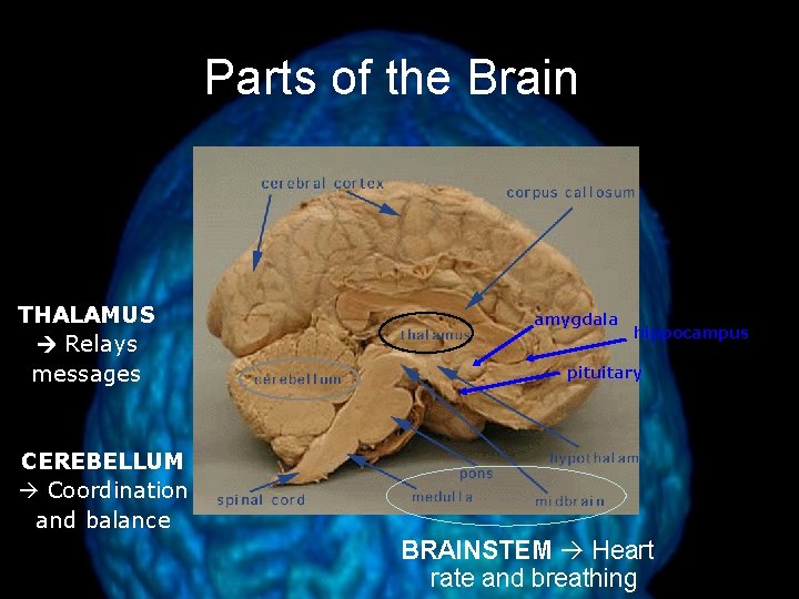
Parts of the Brain THALAMUS Relays messages amygdala hippocampus pituitary CEREBELLUM Coordination and balance BRAINSTEM Heart rate and breathing
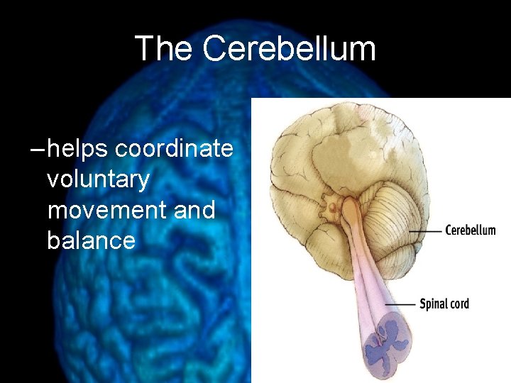
The Cerebellum – helps coordinate voluntary movement and balance
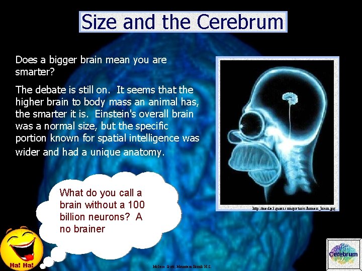
Size and the Cerebrum Does a bigger brain mean you are smarter? The debate is still on. It seems that the higher brain to body mass an animal has, the smarter it is. Einstein's overall brain was a normal size, but the specific portion known for spatial intelligence was wider and had a unique anatomy. What do you call a brain without a 100 billion neurons? A no brainer http: //media 3. guzer. com/pictures/homers_brain. jpg Cerebrum Ha! Melissa Scott, Mountain Brook H. S.

The Limbic System • Hypothalamus, pituitary, amygdala, and hippocampus all deal with basic drives, emotions, and memory • Hippocampus Memory processing • Amygdala Aggression (fight) and fear (flight) • Hypothalamus Hunger, thirst, body temperature, pleasure; regulates pituitary gland (hormones)
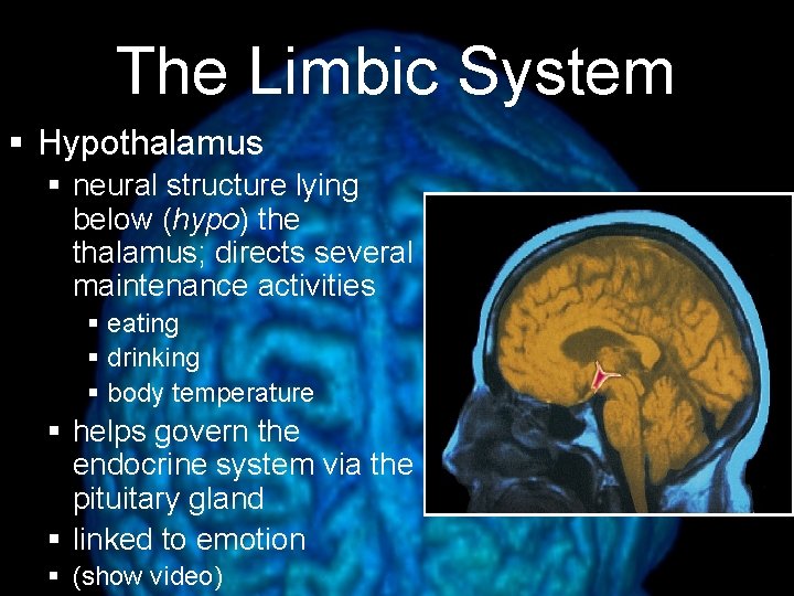
The Limbic System § Hypothalamus § neural structure lying below (hypo) the thalamus; directs several maintenance activities § eating § drinking § body temperature § helps govern the endocrine system via the pituitary gland § linked to emotion § (show video)
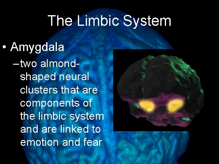
The Limbic System • Amygdala – two almondshaped neural clusters that are components of the limbic system and are linked to emotion and fear
August 1 st, 1966 Charles Whitman
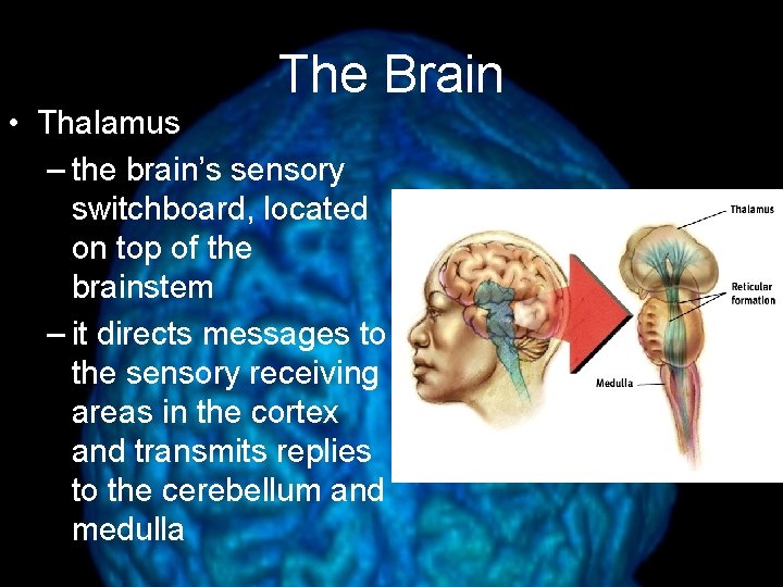
The Brain • Thalamus – the brain’s sensory switchboard, located on top of the brainstem – it directs messages to the sensory receiving areas in the cortex and transmits replies to the cerebellum and medulla

The Cerebral Cortex • Cerebral Cortex – the body’s ultimate control and information processing center
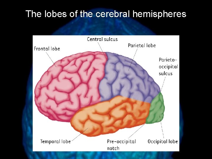
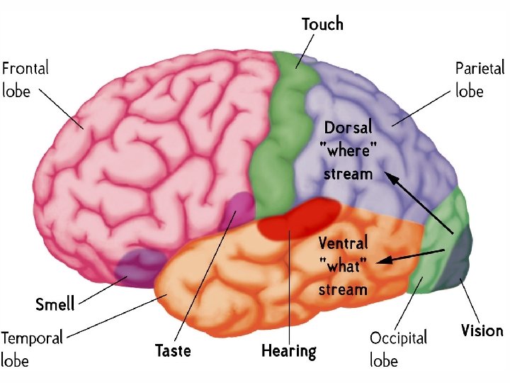
The lobes of the cerebral hemispheres
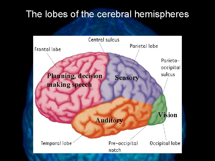
The lobes of the cerebral hemispheres Planning, decision making speech Sensory Auditory Vision
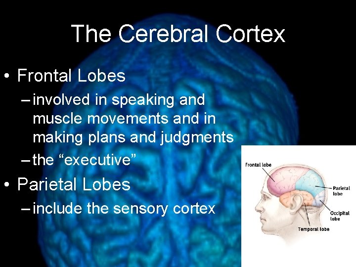
The Cerebral Cortex • Frontal Lobes – involved in speaking and muscle movements and in making plans and judgments – the “executive” • Parietal Lobes – include the sensory cortex
The Cerebral Cortex • Occipital Lobes – include the visual areas, which receive visual information from the opposite visual field • Temporal Lobes – include the auditory areas, each of which receives auditory information primarily from the opposite ear
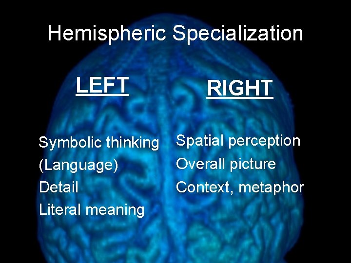
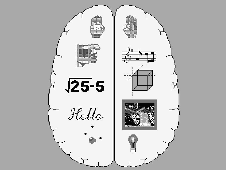
Hemispheric Specialization LEFT RIGHT Symbolic thinking (Language) Detail Literal meaning Spatial perception Overall picture Context, metaphor
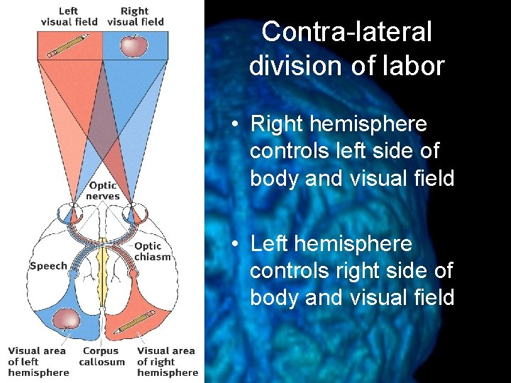
Contra-lateral division of labor • Right hemisphere controls left side of body and visual field • Left hemisphere controls right side of body and visual field
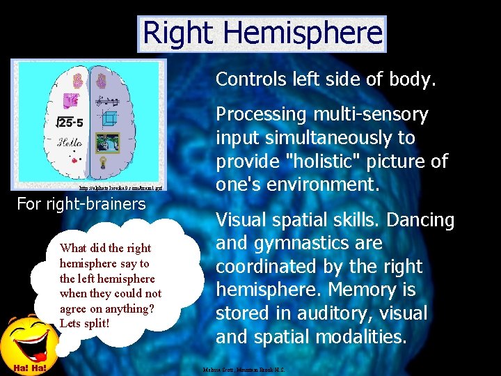
Right Hemisphere Controls left side of body. http: //alphatel. waika 9. com/brain 1. gif For right-brainers What did the right hemisphere say to the left hemisphere when they could not agree on anything? Lets split! Ha! Processing multi-sensory input simultaneously to provide "holistic" picture of one's environment. Visual spatial skills. Dancing and gymnastics are coordinated by the right hemisphere. Memory is stored in auditory, visual and spatial modalities. Melissa Scott, Mountain Brook H. S.
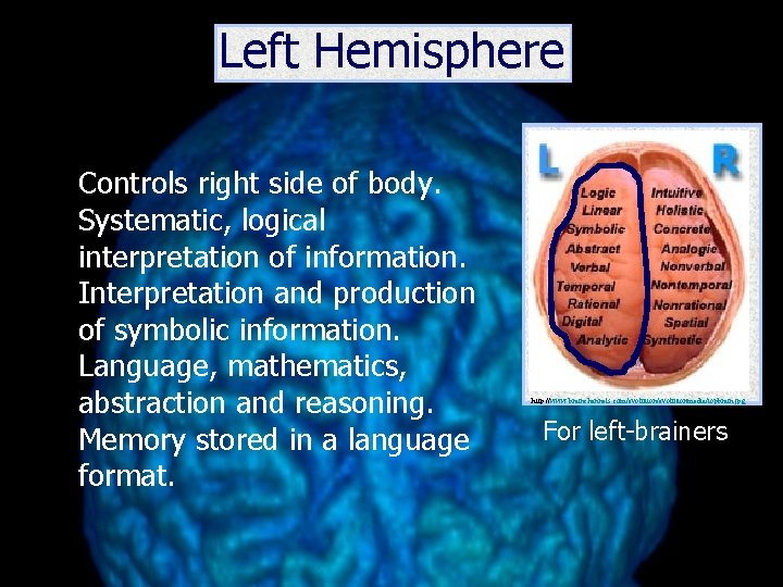
Left Hemisphere Controls right side of body. Systematic, logical interpretation of information. Interpretation and production of symbolic information. Language, mathematics, abstraction and reasoning. Memory stored in a language format. http: //www. brainchannels. com/evolutionmedia/topbrain. jpg For left-brainers
Brain Plasticity • The ability of the brain to reorganize neural pathways based on new experiences • Persistent functional changes in the brain represent new knowledge • Age dependent component • Brain injuries
Sensation • The process by which the central nervous system receives input from the environment via sensory neurons • Bottom up processing
Perception • The process by which the brain interprets and organizes sensory information • Top-down processing
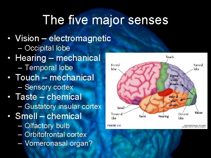
The five major senses • Vision – electromagnetic – Occipital lobe • Hearing – mechanical – Temporal lobe • Touch – mechanical – Sensory cortex • Taste – chemical – Gustatory insular cortex • Smell – chemical – Olfactory bulb – Orbitofrontal cortex – Vomeronasal organ?
The sixth sense And the seventh…and eighth…and ninth… • Vestibular balance and motion – Inner ear • Proprioceptive relative position of body parts – Parietal lobe • Temperature heat – Thermoreceptors throughout the body, sensory cortex • Nociception pain – Nociceptors throughout the body, sensory cortex
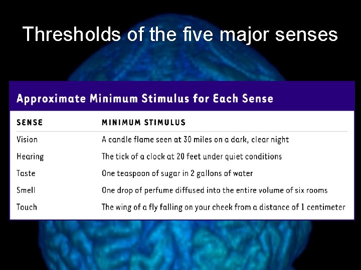
Thresholds of the five major senses
The Human Eye and Vision • The structure of the • The Retina eye – Photoreceptors – Iris – Cornea – Lens • Focusing – Cornea – Accommodation – Processing time – Sensitivity
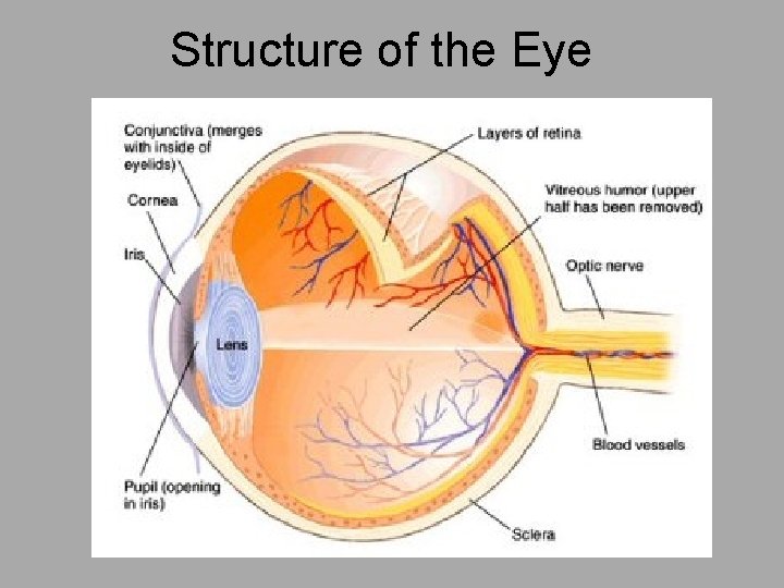
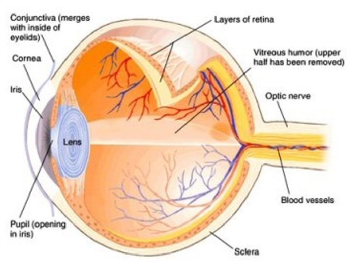
Structure of the Eye
The Human Eye and Vision • The structure of the • The Retina eye – Photoreceptors – Iris – Cornea – Lens • Focusing – Cornea – Accommodation – Processing time – Sensitivity
Parts of the Eye • Sclera: a tough white layer of connective tissue that covers all of the eyeball except the cornea. – Conjunctiva: external cover of the sclera — keeps the eye moist. • Cornea: transparent covering of the front of the eye. – Allows for the passage of light into the eye and functions as a fixed lens.
• Choroid: thin, pigmented layer lining the interior surface of the sclera. – Prevents light rays from scattering and distorting the image. – Anteriorly it forms the iris. • The iris regulates the size of the pupil. • Retina: lines the interior surface of the choroid. – Contains photoreceptors. • Except at the optic disk (where the optic nerve attaches).
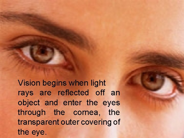
Vision begins when light rays are reflected off an object and enter the eyes through the cornea, the transparent outer covering of the eye.
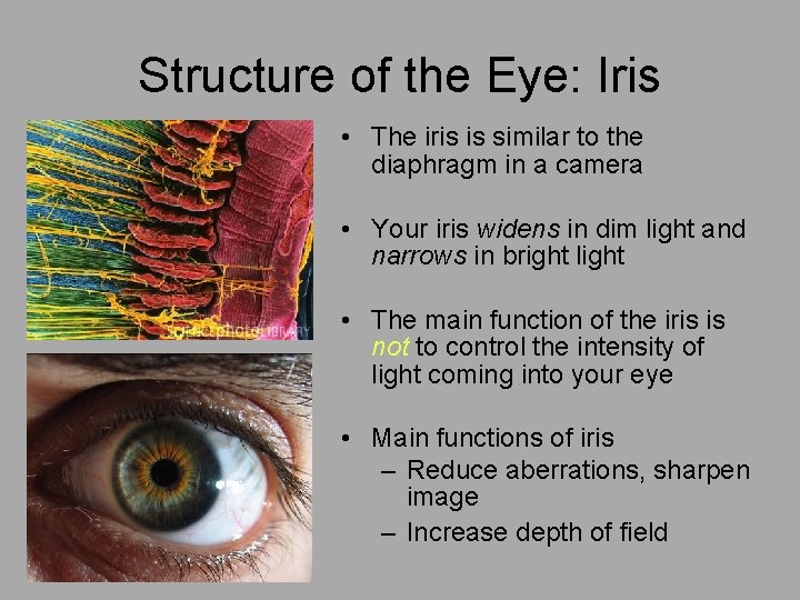
Structure of the Eye: Iris • The iris is similar to the diaphragm in a camera • Your iris widens in dim light and narrows in bright light • The main function of the iris is not to control the intensity of light coming into your eye • Main functions of iris – Reduce aberrations, sharpen image – Increase depth of field
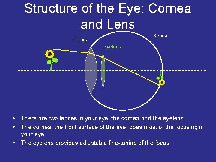
Structure of the Eye: Cornea and Lens Retina Cornea Eyelens • There are two lenses in your eye, the cornea and the eyelens. • The cornea, the front surface of the eye, does most of the focusing in your eye • The eyelens provides adjustable fine-tuning of the focus
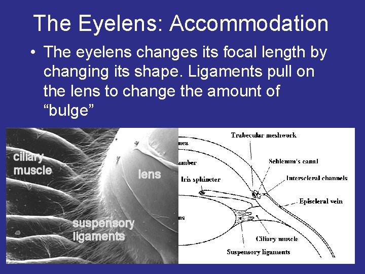
The Eyelens: Accommodation • The eyelens changes its focal length by changing its shape. Ligaments pull on the lens to change the amount of “bulge”
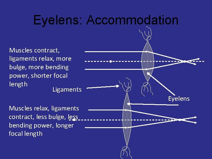
Eyelens: Accommodation Muscles contract, ligaments relax, more bulge, more bending power, shorter focal length Ligaments Eyelens Muscles relax, ligaments contract, less bulge, less bending power, longer focal length
How Your Eyelens Focuses • Your eyelens has a small depth of field – You can't see something close and far with both objects in focus at the same time • Hold out your thumb about a foot away from your eye – Then, alternately focus on thumb and me (right above your thumb) • Note that you cannot see both me and your thumb sharply (in focus) at the same time – You focus on one or the other by changing the bulge of your eyelens
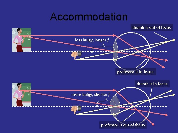
Accommodation thumb is out of focus less bulgy, longer f professor is in focus thumb is in focus more bulgy, shorter f professor is out of focus
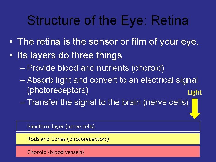
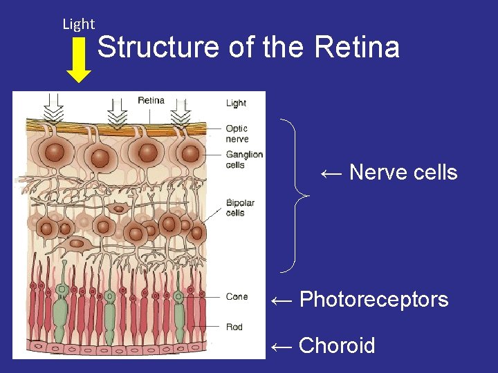
Structure of the Eye: Retina • The retina is the sensor or film of your eye. • Its layers do three things – Provide blood and nutrients (choroid) – Absorb light and convert to an electrical signal (photoreceptors) Light – Transfer the signal to the brain (nerve cells) Plexiform layer (nerve cells) Rods and Cones (photoreceptors) Choroid (blood vessels)
Light Structure of the Retina ← Nerve cells ← Photoreceptors ← Choroid
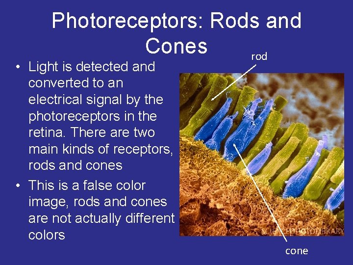
Photoreceptors: Rods and Cones rod • Light is detected and converted to an electrical signal by the photoreceptors in the retina. There are two main kinds of receptors, rods and cones • This is a false color image, rods and cones are not actually different colors cone
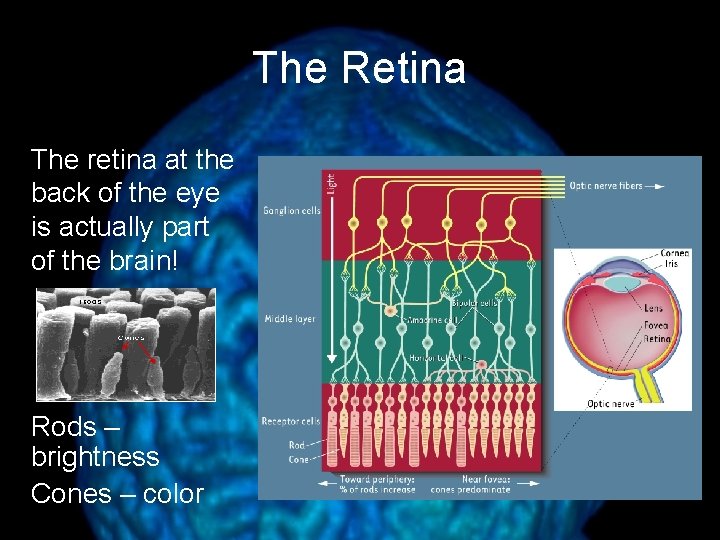
The Retina The retina at the back of the eye is actually part of the brain! Rods – brightness Cones – color
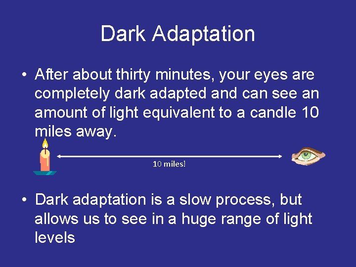
Dark Adaptation • After about thirty minutes, your eyes are completely dark adapted and can see an amount of light equivalent to a candle 10 miles away. 10 miles! • Dark adaptation is a slow process, but allows us to see in a huge range of light levels
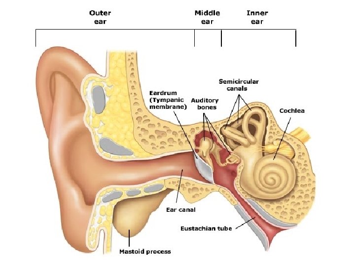
The Human Ear
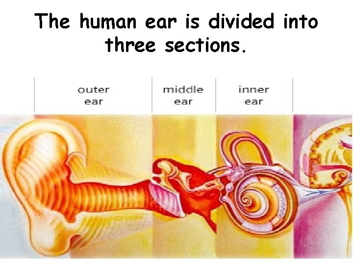
The human ear is divided into three sections.
Each part of the ear serves a specific purpose in the task of detecting and interpreting sound.
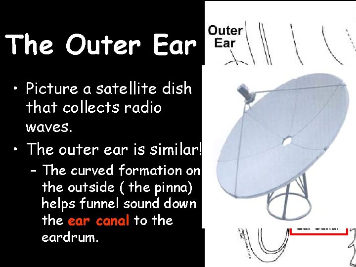
The Outer Ear • Picture a satellite dish that collects radio waves. • The outer ear is similar! – The curved formation on the outside ( the pinna) helps funnel sound down the ear canal to the eardrum.
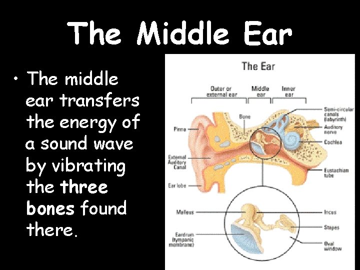
The Middle Ear • The middle ear transfers the energy of a sound wave by vibrating the three bones found there.
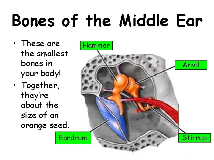
Bones of the Middle Ear • These are the smallest bones in your body! • Together, they’re about the size of an orange seed. Hammer Eardrum Anvil Stirrup
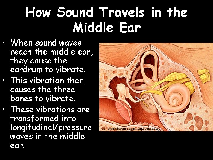
How Sound Travels in the Middle Ear • When sound waves reach the middle ear, they cause the eardrum to vibrate. • This vibration then causes the three bones to vibrate. • These vibrations are transformed into longitudinal/pressure waves in the middle ear.
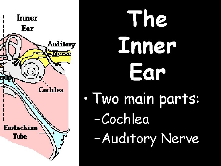
The Inner Ear • Two main parts: – Cochlea – Auditory Nerve
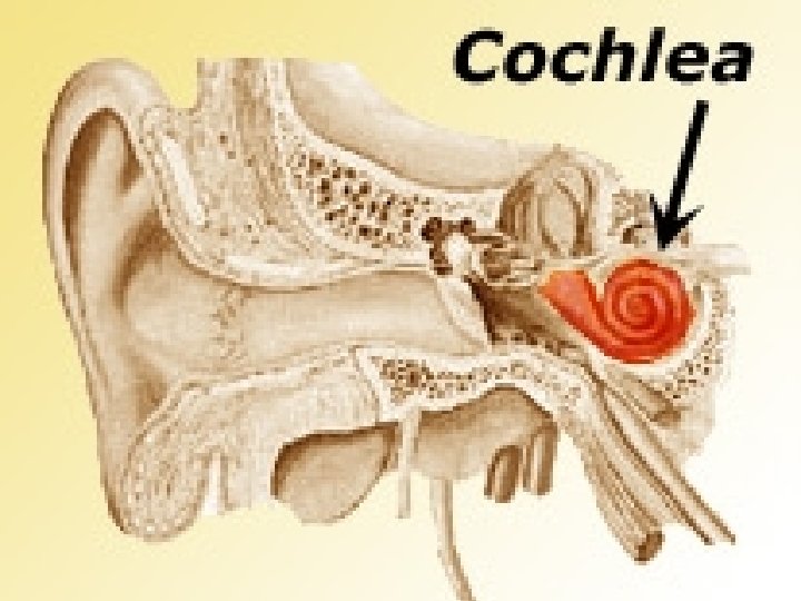
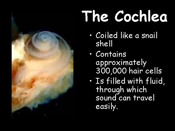
The Cochlea • Coiled like a snail shell • Contains approximately 300, 000 hair cells • Is filled with fluid, through which sound can travel easily.
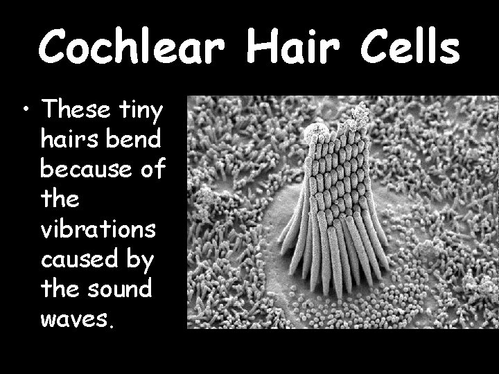
Cochlear Hair Cells • These tiny hairs bend because of the vibrations caused by the sound waves.
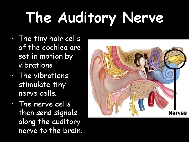
The Auditory Nerve • The tiny hair cells of the cochlea are set in motion by vibrations • The vibrations stimulate tiny nerve cells. • The nerve cells then send signals along the auditory nerve to the brain.
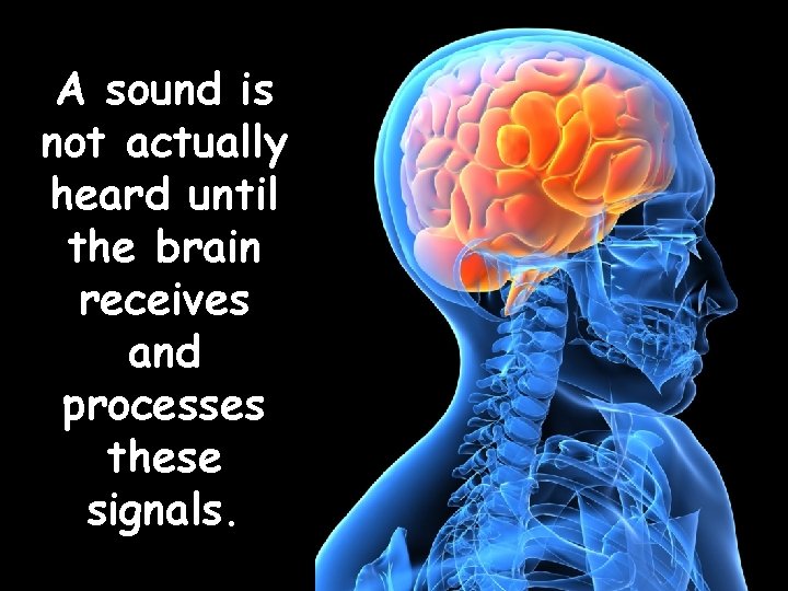
A sound is not actually heard until the brain receives and processes these signals.
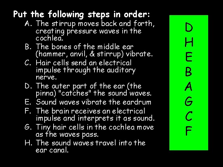
Put the following steps in order: A. The stirrup moves back and forth, creating pressure waves in the cochlea. B. The bones of the middle ear (hammer, anvil, & stirrup) vibrate. C. Hair cells send an electrical impulse through the auditory nerve. D. The outer part of the ear (the pinna) "catches" the sound waves. E. Sound waves vibrate the eardrum F. The brain receives an electrical impulse and interprets it as sound. G. Tiny hair cells in the cochlea move as the waves pass. H. The sound waves travel into the ear canal. D H E B A G C F
- Slides: 61