Chapter 4 Functional Anatomy of Prokaryotic and Eukaryotic
- Slides: 83
Chapter 4 Functional Anatomy of Prokaryotic and Eukaryotic Cells Copyright © 2010 Pearson Education, Inc. Lectures prepared by Christine L.
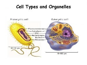
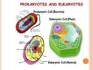
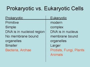
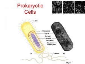
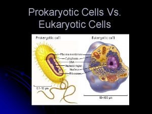
Prokaryotic and Eukaryotic Cells § Prokaryote comes from the Greek words for prenucleus. § Eukaryote comes from the Greek words for true nucleus. Copyright © 2010 Pearson Education, Inc.
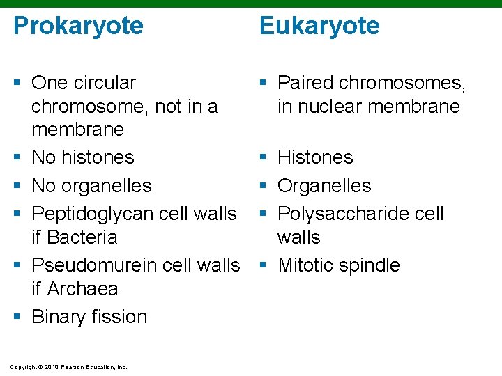
Prokaryote Eukaryote § One circular chromosome, not in a membrane § No histones § No organelles § Peptidoglycan cell walls if Bacteria § Pseudomurein cell walls if Archaea § Binary fission § Paired chromosomes, in nuclear membrane Copyright © 2010 Pearson Education, Inc. § Histones § Organelles § Polysaccharide cell walls § Mitotic spindle
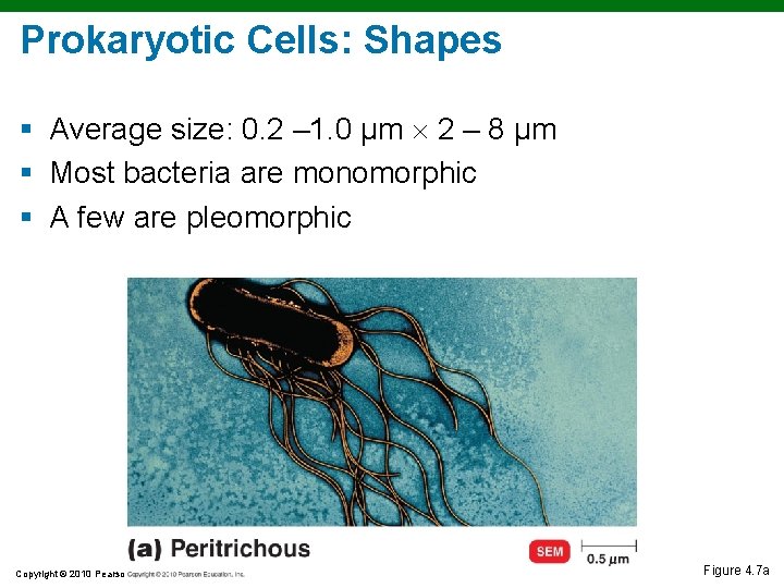
Prokaryotic Cells: Shapes § Average size: 0. 2 – 1. 0 µm 2 – 8 µm § Most bacteria are monomorphic § A few are pleomorphic Copyright © 2010 Pearson Education, Inc. Figure 4. 7 a
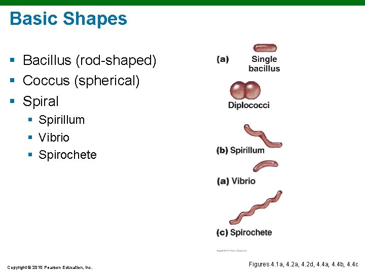
Basic Shapes § Bacillus (rod-shaped) § Coccus (spherical) § Spiral § Spirillum § Vibrio § Spirochete Copyright © 2010 Pearson Education, Inc. Figures 4. 1 a, 4. 2 d, 4. 4 a, 4. 4 b, 4. 4 c
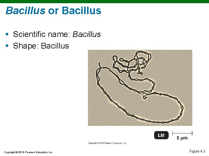
Bacillus or Bacillus § Scientific name: Bacillus § Shape: Bacillus Copyright © 2010 Pearson Education, Inc. Figure 4. 3
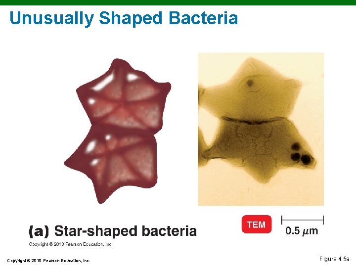
Unusually Shaped Bacteria Copyright © 2010 Pearson Education, Inc. Figure 4. 5 a
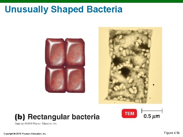
Unusually Shaped Bacteria Copyright © 2010 Pearson Education, Inc. Figure 4. 5 b
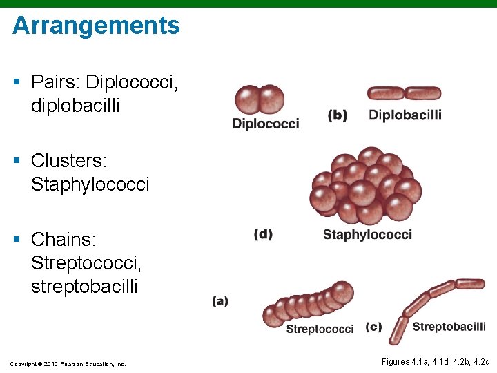
Arrangements § Pairs: Diplococci, diplobacilli § Clusters: Staphylococci § Chains: Streptococci, streptobacilli Copyright © 2010 Pearson Education, Inc. Figures 4. 1 a, 4. 1 d, 4. 2 b, 4. 2 c
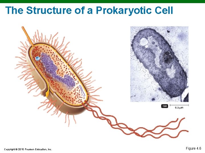
The Structure of a Prokaryotic Cell Copyright © 2010 Pearson Education, Inc. Figure 4. 6
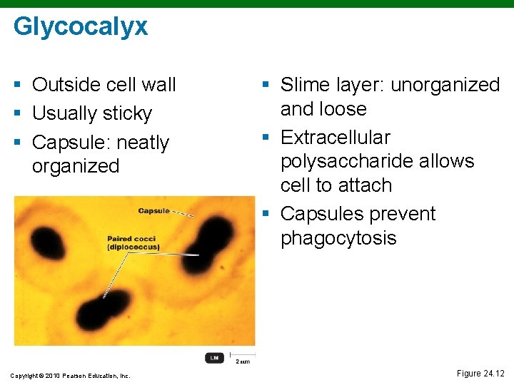
Glycocalyx § Outside cell wall § Usually sticky § Capsule: neatly organized Copyright © 2010 Pearson Education, Inc. § Slime layer: unorganized and loose § Extracellular polysaccharide allows cell to attach § Capsules prevent phagocytosis Figure 24. 12
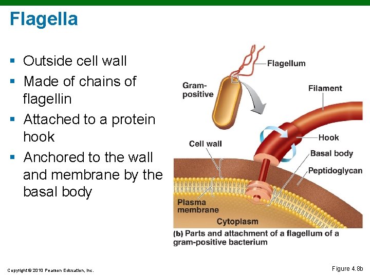
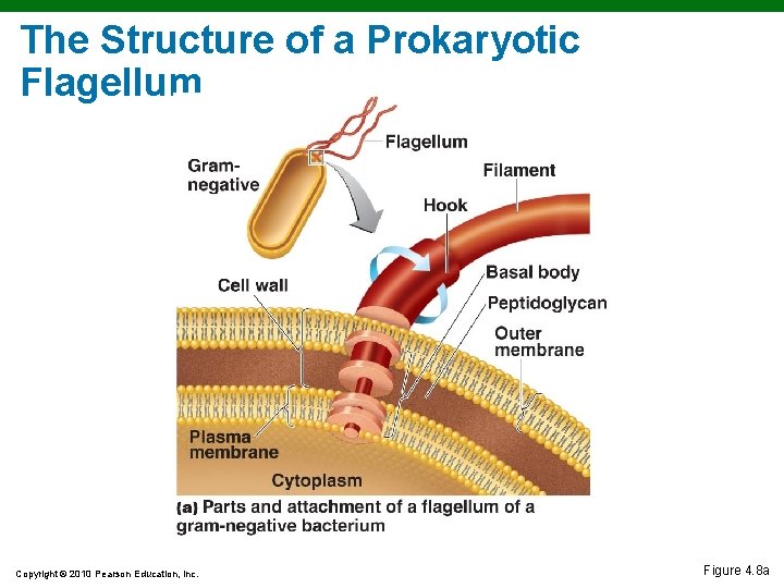
Flagella § Outside cell wall § Made of chains of flagellin § Attached to a protein hook § Anchored to the wall and membrane by the basal body Copyright © 2010 Pearson Education, Inc. Figure 4. 8 b
The Structure of a Prokaryotic Flagellum Copyright © 2010 Pearson Education, Inc. Figure 4. 8 a
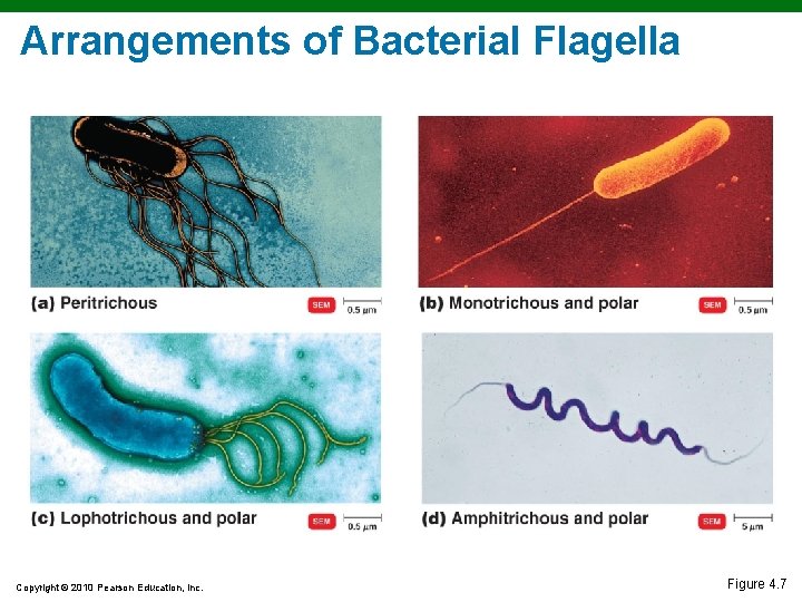
Arrangements of Bacterial Flagella Copyright © 2010 Pearson Education, Inc. Figure 4. 7
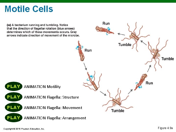
Motile Cells § Rotate flagella to run or tumble § Move toward or away from stimuli (taxis) § Flagella proteins are H antigens (e. g. , E. coli O 157: H 7) Copyright © 2010 Pearson Education, Inc.
Motile Cells ANIMATION Motility ANIMATION Flagella: Structure ANIMATION Flagella: Movement ANIMATION Flagella: Arrangement Copyright © 2010 Pearson Education, Inc. Figure 4. 9 a
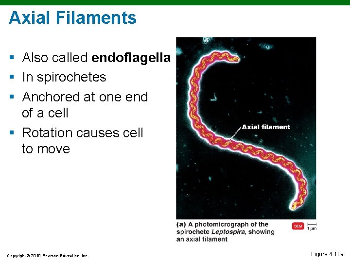
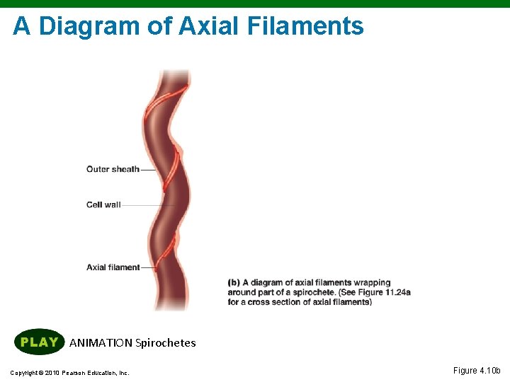
Axial Filaments § Also called endoflagella § In spirochetes § Anchored at one end of a cell § Rotation causes cell to move Copyright © 2010 Pearson Education, Inc. Figure 4. 10 a
A Diagram of Axial Filaments ANIMATION Spirochetes Copyright © 2010 Pearson Education, Inc. Figure 4. 10 b
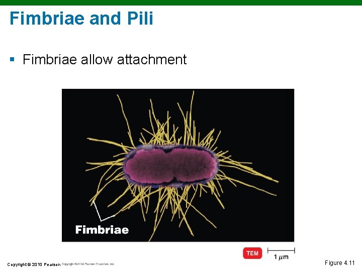
Fimbriae and Pili § Fimbriae allow attachment Copyright © 2010 Pearson Education, Inc. Figure 4. 11
Fimbriae and Pili § Facilitate transfer of DNA from one cell to another § Gliding motility § Twitching motility Copyright © 2010 Pearson Education, Inc.
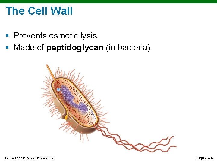
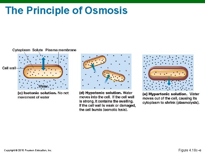
The Cell Wall § Prevents osmotic lysis § Made of peptidoglycan (in bacteria) Copyright © 2010 Pearson Education, Inc. Figure 4. 6
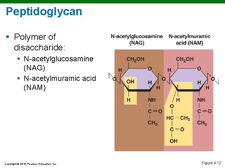
Peptidoglycan § Polymer of disaccharide: § N-acetylglucosamine (NAG) § N-acetylmuramic acid (NAM) Copyright © 2010 Pearson Education, Inc. Figure 4. 12
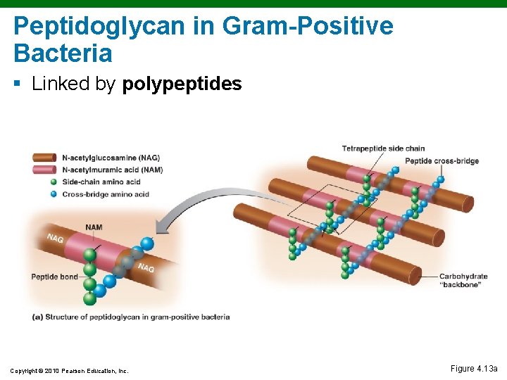
Peptidoglycan in Gram-Positive Bacteria § Linked by polypeptides Copyright © 2010 Pearson Education, Inc. Figure 4. 13 a
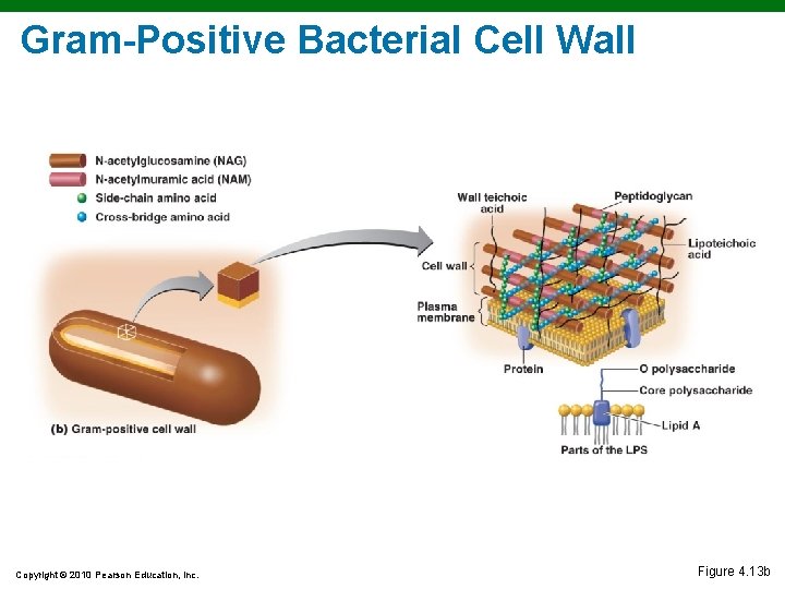
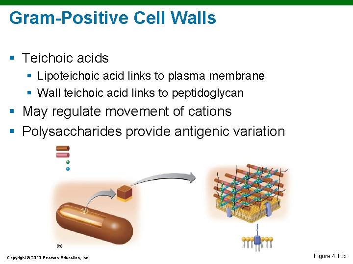
Gram-Positive Bacterial Cell Wall Copyright © 2010 Pearson Education, Inc. Figure 4. 13 b
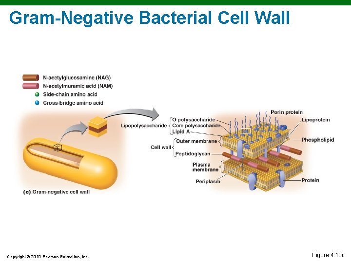
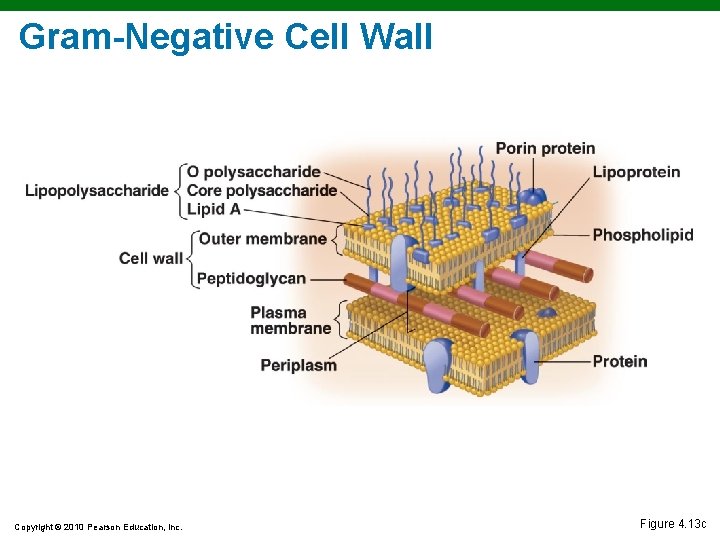
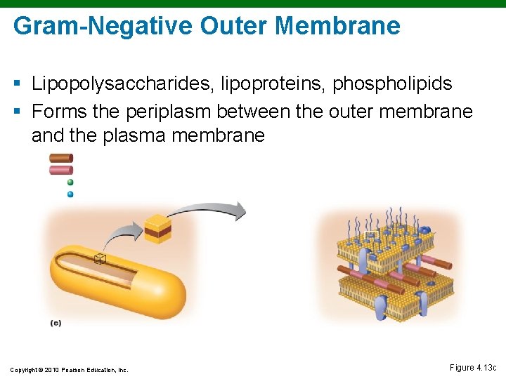
Gram-Negative Bacterial Cell Wall Copyright © 2010 Pearson Education, Inc. Figure 4. 13 c
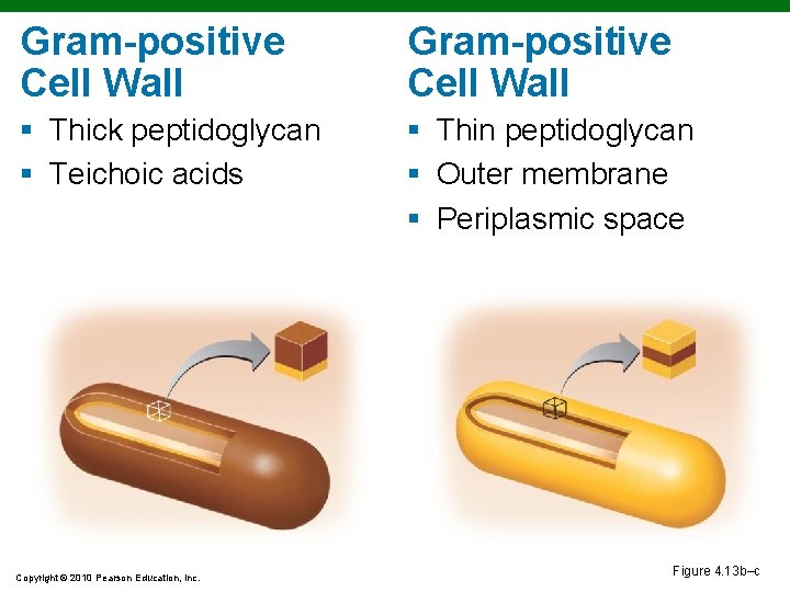
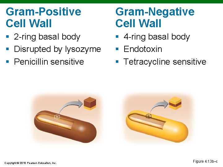
Gram-positive Cell Wall § Thick peptidoglycan § Teichoic acids § Thin peptidoglycan § Outer membrane § Periplasmic space Copyright © 2010 Pearson Education, Inc. Figure 4. 13 b–c
Gram-Positive Cell Walls § Teichoic acids § Lipoteichoic acid links to plasma membrane § Wall teichoic acid links to peptidoglycan § May regulate movement of cations § Polysaccharides provide antigenic variation Copyright © 2010 Pearson Education, Inc. Figure 4. 13 b
Gram-Negative Cell Wall Copyright © 2010 Pearson Education, Inc. Figure 4. 13 c
Gram-Negative Outer Membrane § Lipopolysaccharides, lipoproteins, phospholipids § Forms the periplasm between the outer membrane and the plasma membrane Copyright © 2010 Pearson Education, Inc. Figure 4. 13 c
Gram-Negative Outer Membrane § Protection from phagocytes, complement, and antibiotics § O polysaccharide antigen, e. g. , E. coli O 157: H 7 § Lipid A is an endotoxin § Porins (proteins) form channels through membrane Copyright © 2010 Pearson Education, Inc.
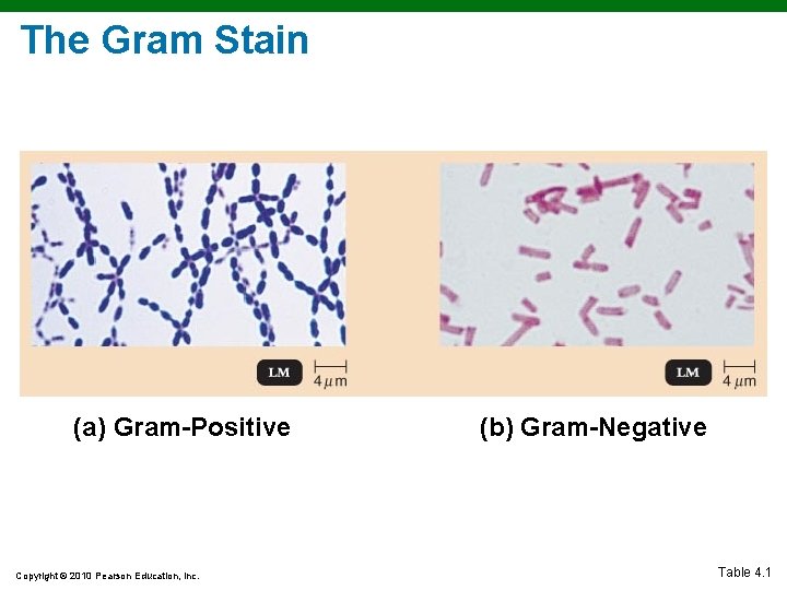
The Gram Stain (a) Gram-Positive Copyright © 2010 Pearson Education, Inc. (b) Gram-Negative Table 4. 1
The Gram Stain Mechanism § Crystal violet-iodine crystals form in cell § Gram-positive § Alcohol dehydrates peptidoglycan § CV-I crystals do not leave § Gram-negative § Alcohol dissolves outer membrane and leaves holes in peptidoglycan § CV-I washes out Copyright © 2010 Pearson Education, Inc.
Gram-Positive Cell Wall Gram-Negative Cell Wall § 2 -ring basal body § Disrupted by lysozyme § Penicillin sensitive § 4 -ring basal body § Endotoxin § Tetracycline sensitive Copyright © 2010 Pearson Education, Inc. Figure 4. 13 b–c
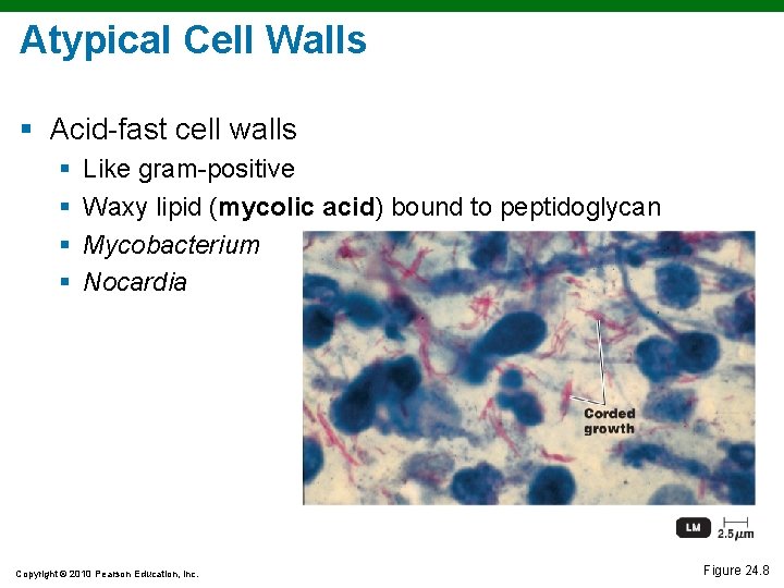
Atypical Cell Walls § Acid-fast cell walls § § Like gram-positive Waxy lipid (mycolic acid) bound to peptidoglycan Mycobacterium Nocardia Copyright © 2010 Pearson Education, Inc. Figure 24. 8
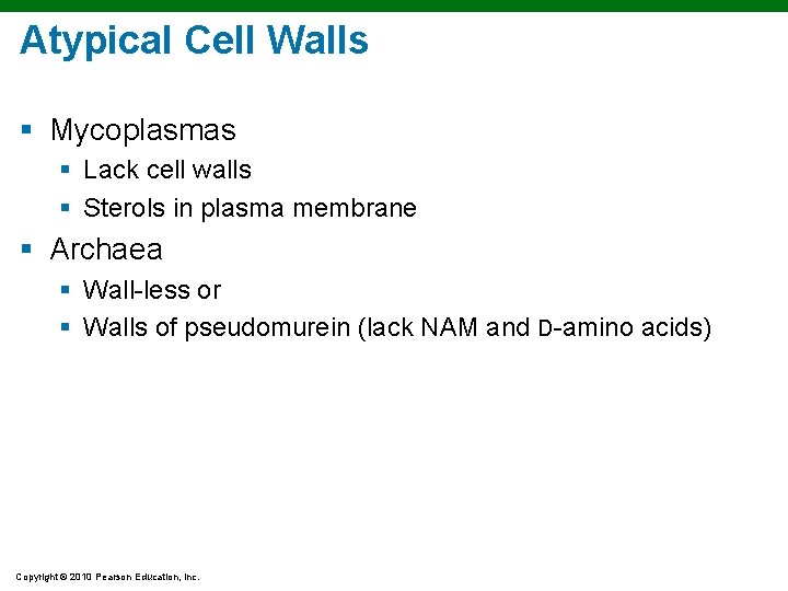
Atypical Cell Walls § Mycoplasmas § Lack cell walls § Sterols in plasma membrane § Archaea § Wall-less or § Walls of pseudomurein (lack NAM and D-amino acids) Copyright © 2010 Pearson Education, Inc.
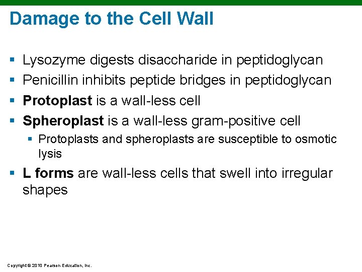
Damage to the Cell Wall § § Lysozyme digests disaccharide in peptidoglycan Penicillin inhibits peptide bridges in peptidoglycan Protoplast is a wall-less cell Spheroplast is a wall-less gram-positive cell § Protoplasts and spheroplasts are susceptible to osmotic lysis § L forms are wall-less cells that swell into irregular shapes Copyright © 2010 Pearson Education, Inc.
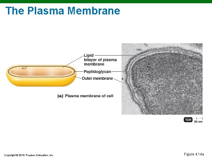
The Plasma Membrane Copyright © 2010 Pearson Education, Inc. Figure 4. 14 a
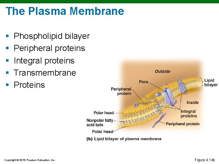
The Plasma Membrane § § § Phospholipid bilayer Peripheral proteins Integral proteins Transmembrane Proteins Copyright © 2010 Pearson Education, Inc. Figure 4. 14 b
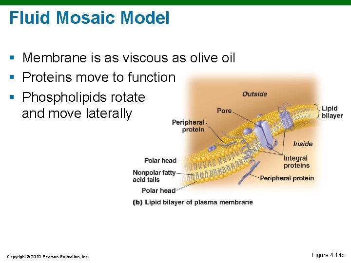
Fluid Mosaic Model § Membrane is as viscous as olive oil § Proteins move to function § Phospholipids rotate and move laterally Copyright © 2010 Pearson Education, Inc. Figure 4. 14 b
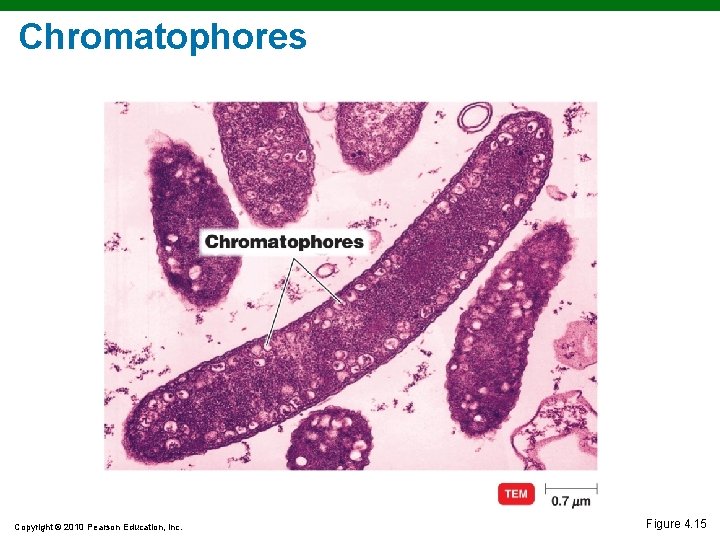
The Plasma Membrane § Selective permeability allows passage of some molecules § Enzymes for ATP production § Photosynthetic pigments on foldings called chromatophores or thylakoids Copyright © 2010 Pearson Education, Inc.
Chromatophores Copyright © 2010 Pearson Education, Inc. Figure 4. 15
The Plasma Membrane § Damage to the membrane by alcohols, quaternary ammonium (detergents), and polymyxin antibiotics causes leakage of cell contents ANIMATION Membrane Structure ANIMATION Membrane Permeability Copyright © 2010 Pearson Education, Inc.
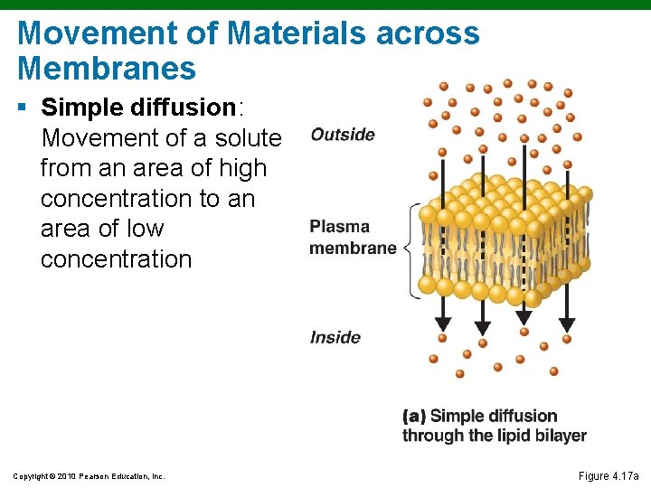
Movement of Materials across Membranes § Simple diffusion: Movement of a solute from an area of high concentration to an area of low concentration Copyright © 2010 Pearson Education, Inc. Figure 4. 17 a
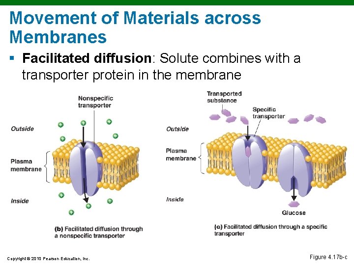
Movement of Materials across Membranes § Facilitated diffusion: Solute combines with a transporter protein in the membrane Copyright © 2010 Pearson Education, Inc. Figure 4. 17 b-c
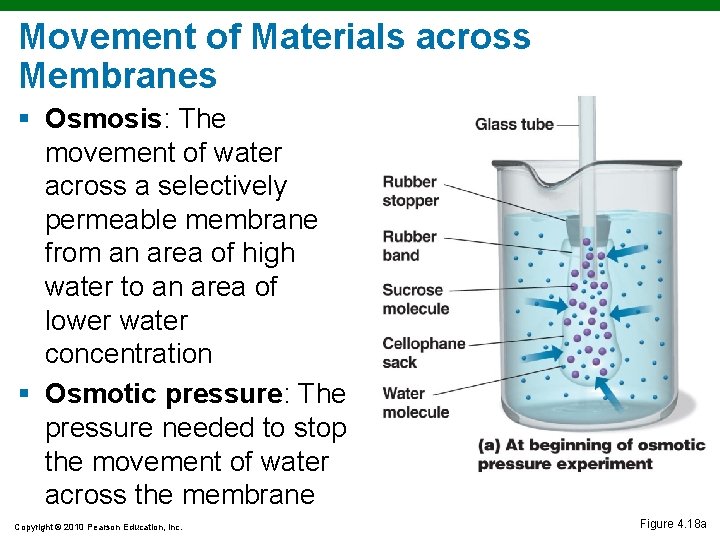
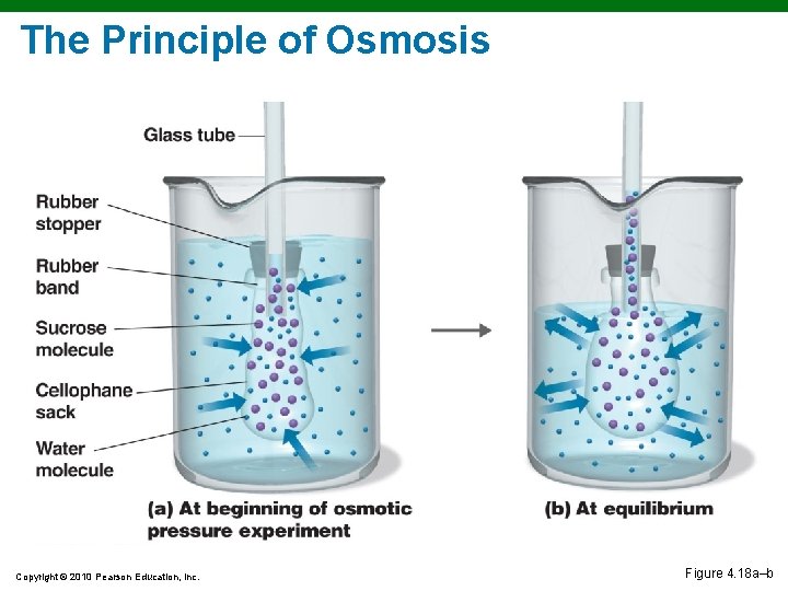
Movement of Materials across Membranes § Osmosis: The movement of water across a selectively permeable membrane from an area of high water to an area of lower water concentration § Osmotic pressure: The pressure needed to stop the movement of water across the membrane Copyright © 2010 Pearson Education, Inc. Figure 4. 18 a
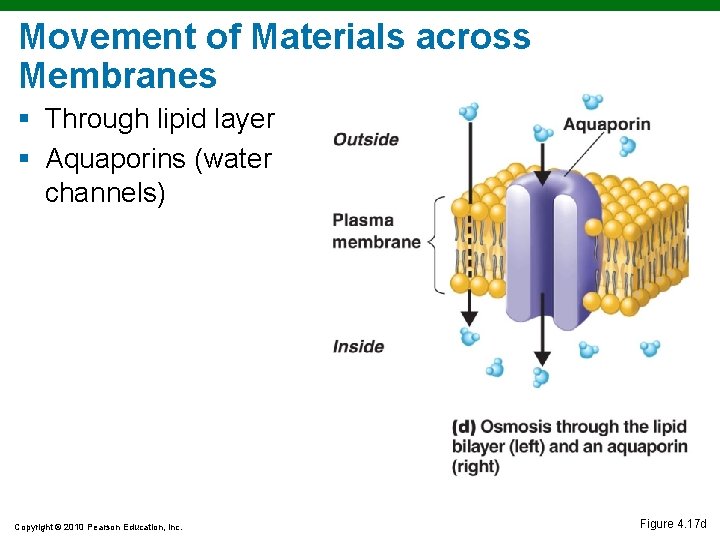
Movement of Materials across Membranes § Through lipid layer § Aquaporins (water channels) Copyright © 2010 Pearson Education, Inc. Figure 4. 17 d
The Principle of Osmosis Copyright © 2010 Pearson Education, Inc. Figure 4. 18 a–b
The Principle of Osmosis Copyright © 2010 Pearson Education, Inc. Figure 4. 18 c–e
Movement of Materials across Membranes § Active transport: Requires a transporter protein and ATP § Group translocation: Requires a transporter protein and PEP ANIMATION Active Transport: Types ANIMATION Active Transport: Overview Copyright © 2010 Pearson Education, Inc.
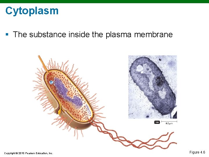
Cytoplasm § The substance inside the plasma membrane Copyright © 2010 Pearson Education, Inc. Figure 4. 6
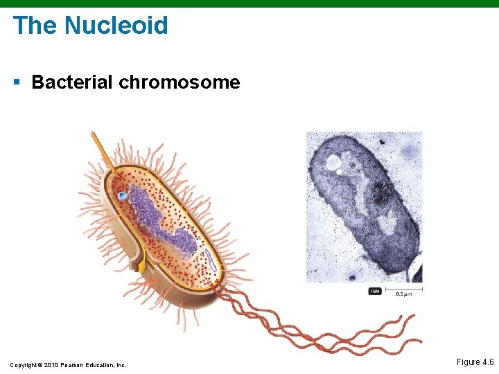
The Nucleoid § Bacterial chromosome Copyright © 2010 Pearson Education, Inc. Figure 4. 6
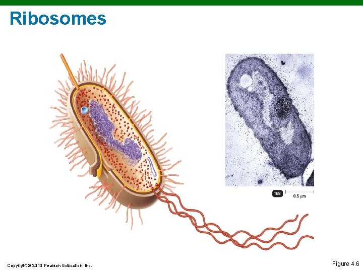
Ribosomes Copyright © 2010 Pearson Education, Inc. Figure 4. 6
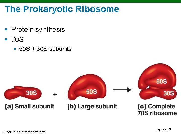
The Prokaryotic Ribosome § Protein synthesis § 70 S § 50 S + 30 S subunits Copyright © 2010 Pearson Education, Inc. Figure 4. 19
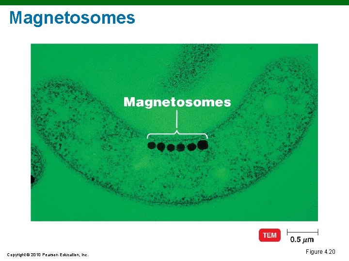
Magnetosomes Copyright © 2010 Pearson Education, Inc. Figure 4. 20
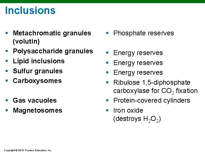
Inclusions § Metachromatic granules (volutin) § Polysaccharide granules § Lipid inclusions § Sulfur granules § Carboxysomes § Gas vacuoles § Magnetosomes Copyright © 2010 Pearson Education, Inc. § Phosphate reserves § § Energy reserves Ribulose 1, 5 -diphosphate carboxylase for CO 2 fixation § Protein-covered cylinders § Iron oxide (destroys H 2 O 2)
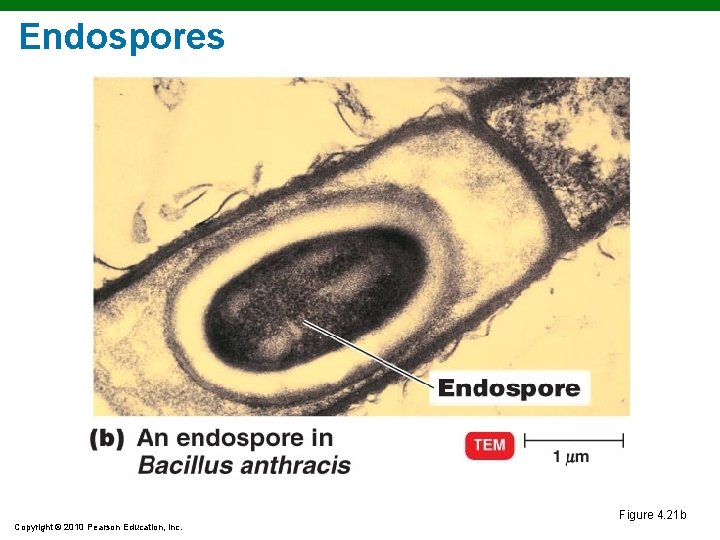
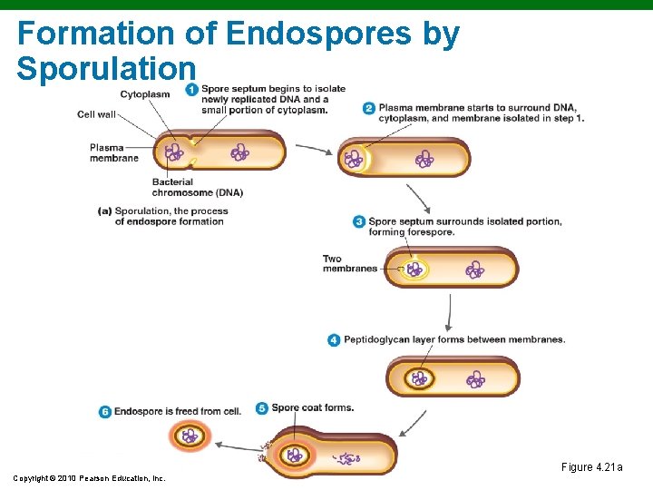
Endospores § § § Resting cells Resistant to desiccation, heat, chemicals Bacillus, Clostridium Sporulation: Endospore formation Germination: Return to vegetative state Copyright © 2010 Pearson Education, Inc.
Endospores Figure 4. 21 b Copyright © 2010 Pearson Education, Inc.
Formation of Endospores by Sporulation Figure 4. 21 a Copyright © 2010 Pearson Education, Inc.
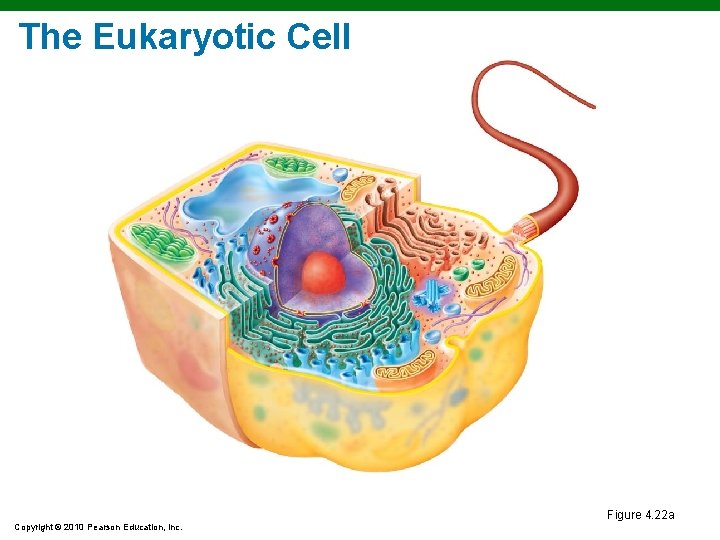
The Eukaryotic Cell Figure 4. 22 a Copyright © 2010 Pearson Education, Inc.
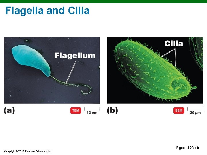
Flagella and Cilia Figure 4. 23 a-b Copyright © 2010 Pearson Education, Inc.
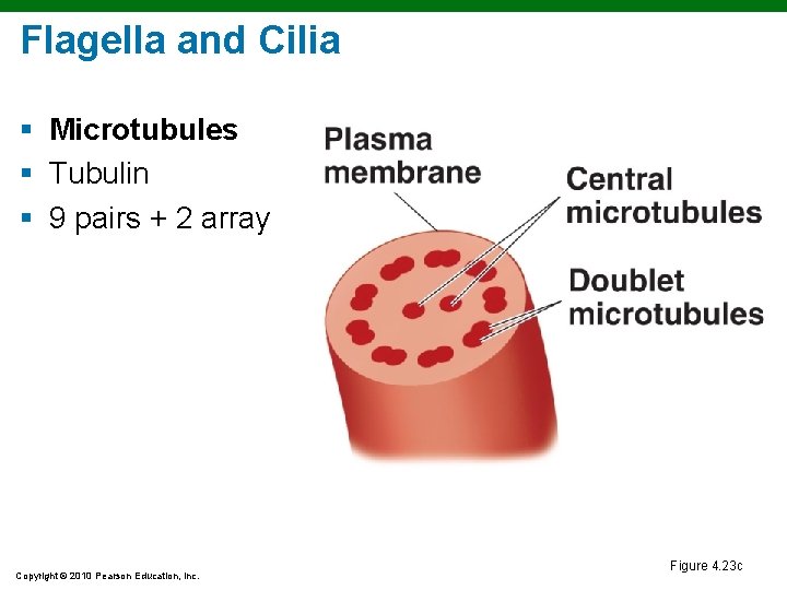
Flagella and Cilia § Microtubules § Tubulin § 9 pairs + 2 array Copyright © 2010 Pearson Education, Inc. Figure 4. 23 c
The Cell Wall and Glycocalyx § Cell wall § Plants, algae, fungi § Carbohydrates § Cellulose, chitin, glucan, mannan § Glycocalyx § Carbohydrates extending from animal plasma membrane § Bonded to proteins and lipids in membrane Copyright © 2010 Pearson Education, Inc.
The Plasma Membrane § § § Phospholipid bilayer Peripheral proteins Integral proteins Transmembrane proteins Sterols Glycocalyx carbohydrates Copyright © 2010 Pearson Education, Inc.
The Plasma Membrane § Selective permeability allows passage of some molecules § Simple diffusion § Facilitative diffusion § Osmosis § Active transport § Endocytosis § Phagocytosis: Pseudopods extend and engulf particles § Pinocytosis: Membrane folds inward, bringing in fluid and dissolved substances Copyright © 2010 Pearson Education, Inc.
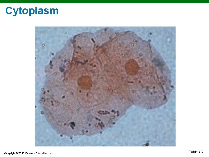
Cytoplasm Copyright © 2010 Pearson Education, Inc. Table 4. 2
Cytoplasm § Cytoplasm membrane: Substance inside plasma and outside nucleus § Cytosol: Fluid portion of cytoplasm § Cytoskeleton: Microfilaments, intermediate filaments, microtubules § Cytoplasmic streaming: Movement of cytoplasm throughout cells Copyright © 2010 Pearson Education, Inc.
Ribosomes § Protein synthesis § 80 S § Membrane-bound: Attached to ER § Free: In cytoplasm § 70 S § In chloroplasts and mitochondria Copyright © 2010 Pearson Education, Inc.
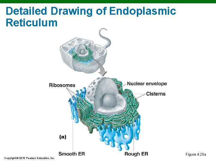
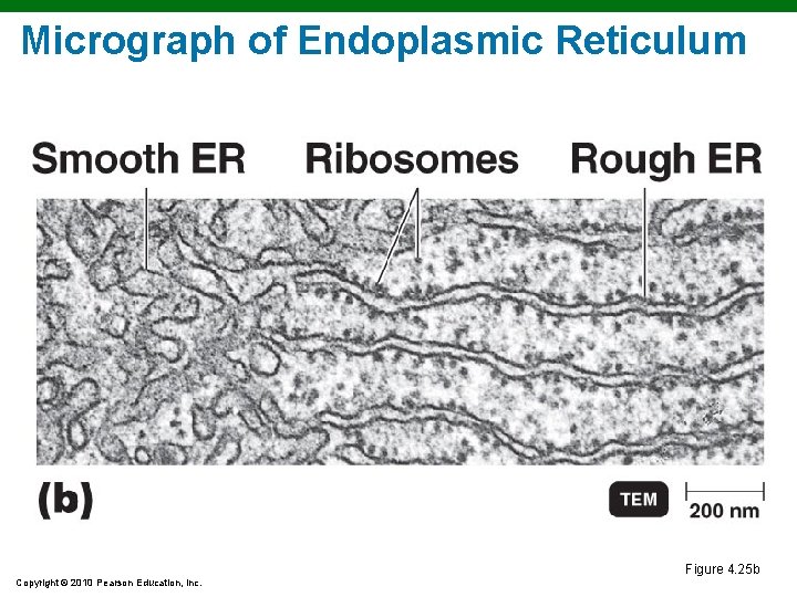
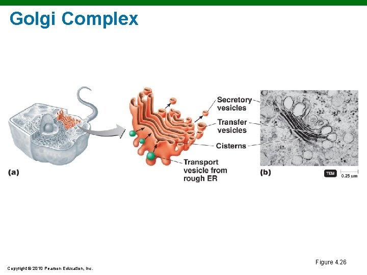
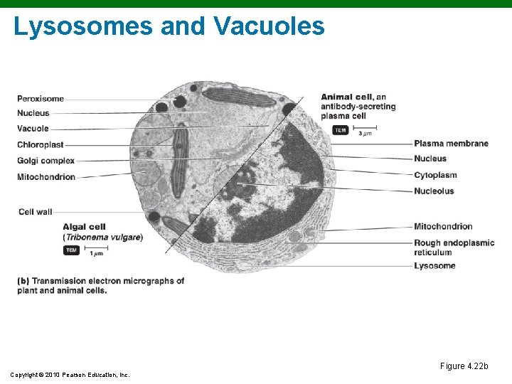
Organelles § § § Nucleus: Contains chromosomes ER: Transport network Golgi complex: Membrane formation and secretion Lysosome: Digestive enzymes Vacuole: Brings food into cells and provides support Copyright © 2010 Pearson Education, Inc.
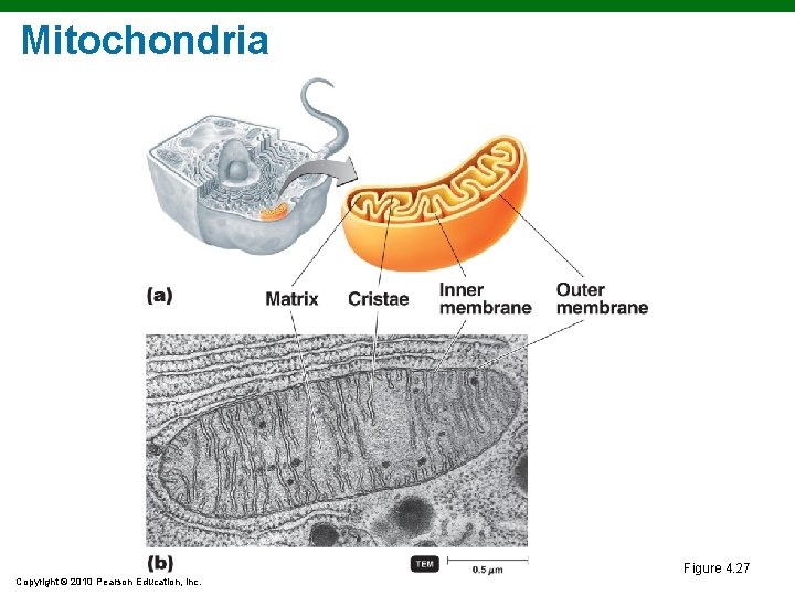
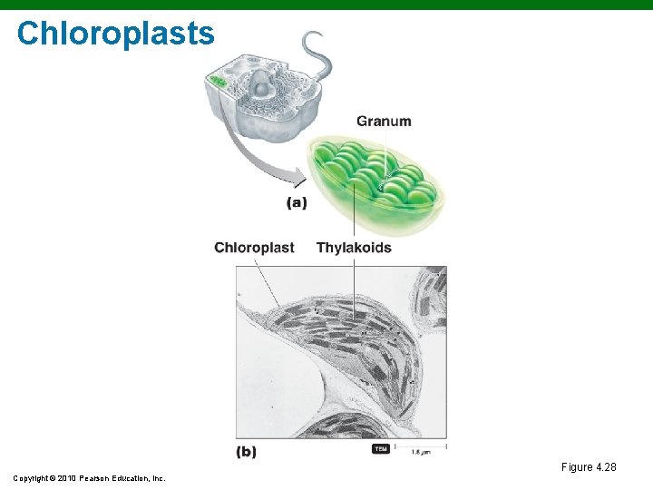
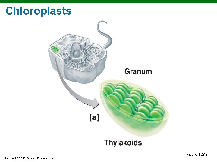
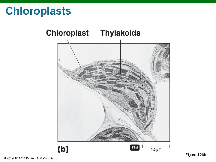
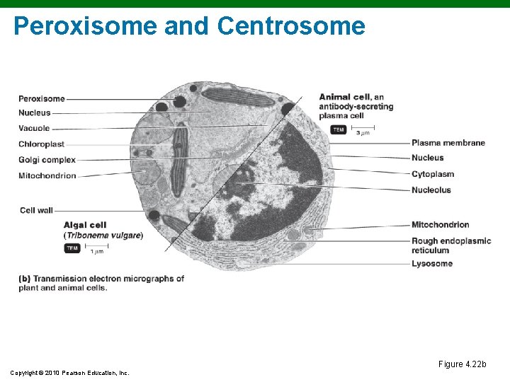
Organelles § § Mitochondrion: Cellular respiration Chloroplast: Photosynthesis Peroxisome: Oxidation of fatty acids; destroys H 2 O 2 Centrosome: Consists of protein fibers and centrioles Copyright © 2010 Pearson Education, Inc.
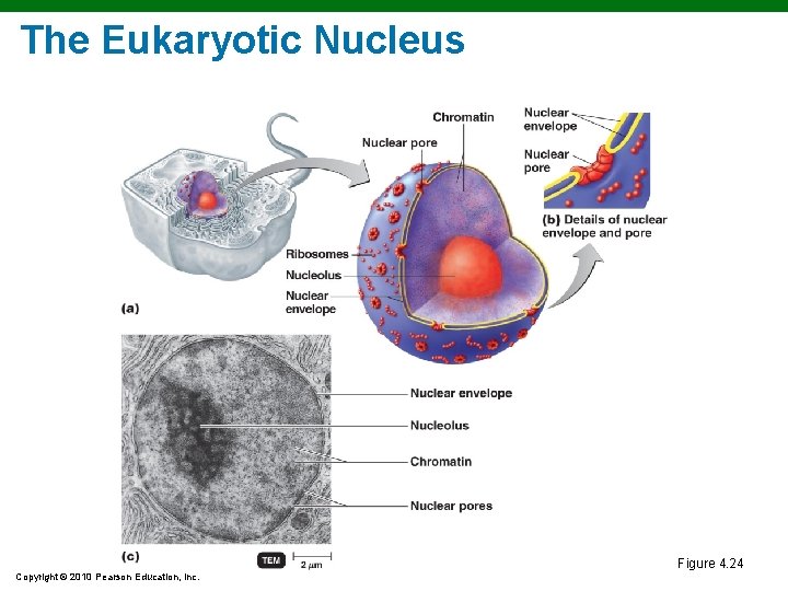
The Eukaryotic Nucleus Figure 4. 24 Copyright © 2010 Pearson Education, Inc.
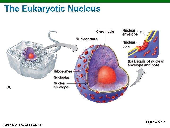
The Eukaryotic Nucleus Figure 4. 24 a–b Copyright © 2010 Pearson Education, Inc.
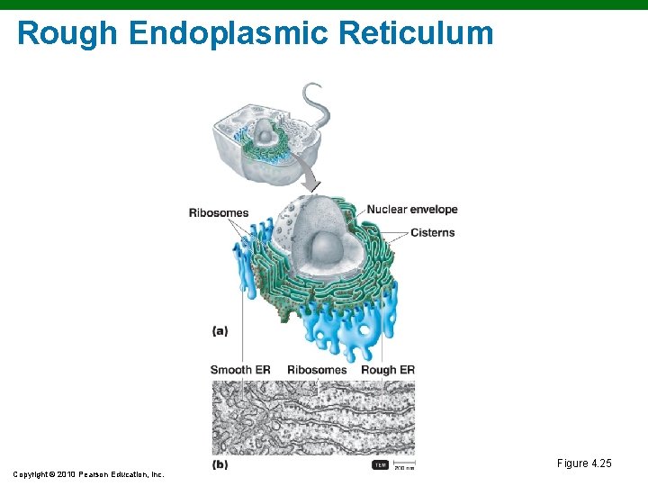
Rough Endoplasmic Reticulum Figure 4. 25 Copyright © 2010 Pearson Education, Inc.
Detailed Drawing of Endoplasmic Reticulum Figure 4. 25 a Copyright © 2010 Pearson Education, Inc.
Micrograph of Endoplasmic Reticulum Figure 4. 25 b Copyright © 2010 Pearson Education, Inc.
Golgi Complex Figure 4. 26 Copyright © 2010 Pearson Education, Inc.
Lysosomes and Vacuoles Figure 4. 22 b Copyright © 2010 Pearson Education, Inc.
Mitochondria Figure 4. 27 Copyright © 2010 Pearson Education, Inc.
Chloroplasts Figure 4. 28 Copyright © 2010 Pearson Education, Inc.
Chloroplasts Figure 4. 28 a Copyright © 2010 Pearson Education, Inc.
Chloroplasts Figure 4. 28 b Copyright © 2010 Pearson Education, Inc.
Peroxisome and Centrosome Figure 4. 22 b Copyright © 2010 Pearson Education, Inc.
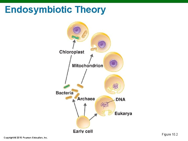
Endosymbiotic Theory Figure 10. 2 Copyright © 2010 Pearson Education, Inc.
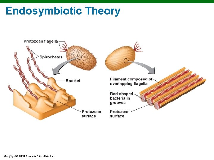
Endosymbiotic Theory Copyright © 2010 Pearson Education, Inc.