From sangarunsahotmail com This mean of cells The


 linear molecules (chromosomes) with histone proteins linear molecules coupled in RNA-synthesis")


 molecules")


")


 Structure - sheets of unit membrane with ribosomes on the outside")
 Function -transports chemicals between cells and within cells - provides a")
 and protein enzymes")
 - vesicles")
 - inner membrane")


 and protein Function")


- Slides: 58
From sangarun_sa@hotmail. com
This mean of cells: The cell is one of the most basic units of life There are millions of different types of cells. There are cells that are organisms onto themselves, such as microscopic amoeba and bacteria cells.
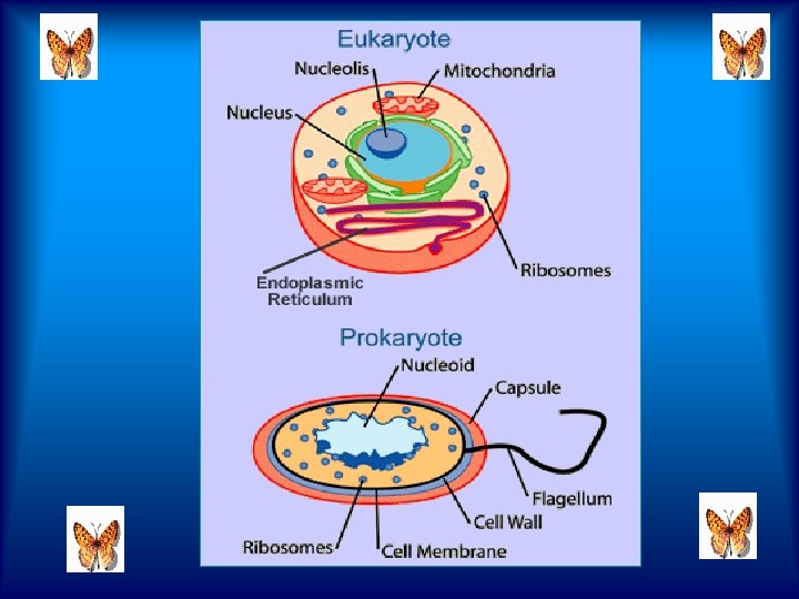
Anatomy of cells There are two types of cells: eukaryotic and prokaryotic. Prokaryotic cells are usually independent, while eukaryotic cells are often found in multicellular organisms.
And there are cells that only function when part of a larger organism, such as the cells that make up your body. The cell is the smallest unit of life in our bodies. In the body, there are brain cells, skin cells, liver cells, stomach cells, and the list goes on. All of these cells have unique functions and features.
Eukaryotic cells Prokaryotic cells
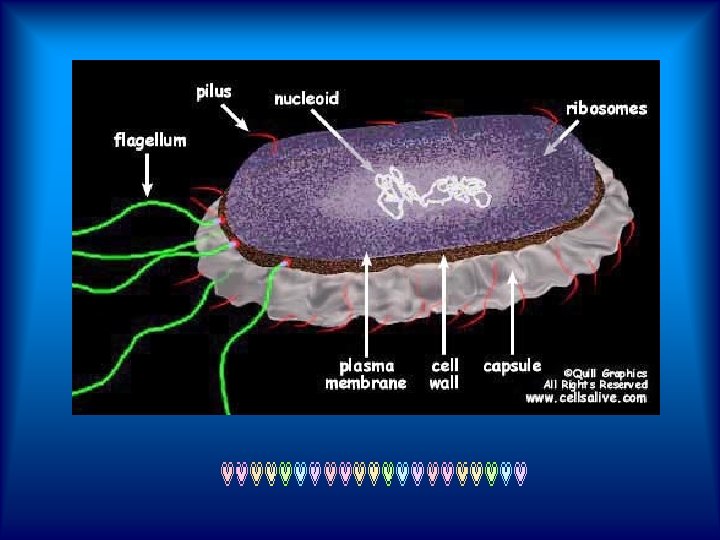
Prokaryotic cells The prokaryote cell is simpler, and therefore smaller, than a eukaryote cell, lacking a nucleus and most of the other organelles of eukaryotes. There are two kinds of prokaryotes: bacteria and archaea; these share a similar structure.
Procaryotic: These cells are simple in structure, with no recognizable organelles. They have an outer cell wall that gives them shape. Just under the rigid cell wall is the more fluid cell membrane. The cytoplasm enclosed within the cell membrane does not exhibit much structure when viewed by electron microscopy
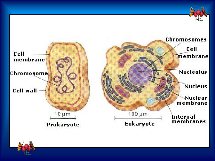
Living cells are divided into two types - procaryotic and eucaryotic (sometimes spelled prokaryotic and eukaryotic). This division is based on internal complexity. The following pages can provide graphic roadmaps to the organization of both of these cell types.
Eukaryotic cells are about 15 times wider than a typical prokaryote and can be as much as 1000 times greater in volume. The major difference between prokaryotes and eukaryotes is that eukaryotic cells contain membrane-bound compartments in which specific metabolic activities take place. Most important among these is a cell nucleus, a membrane-delineated compartment that houses the eukaryotic cell's DNA. This nucleus gives the eukaryote its name, which means "true nucleus. " Other differences include:
Eukaryotic Eucaryotic: The cells of protozoa, higher plants and animals are highly structured. These cells tend to be larger than the cells of bacteria, and have developed specialized packaging and transport mechanisms that may be necessary to support their larger size. Use the Interactive animation of plant and animal cells to learn about their respective organelles.
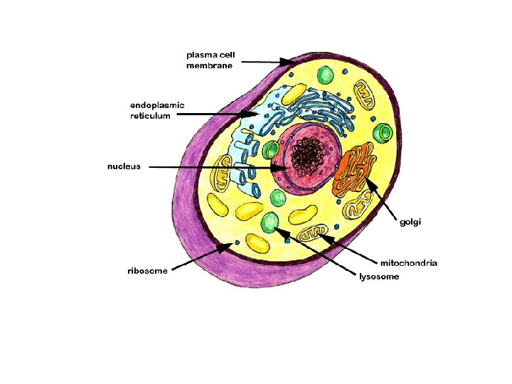
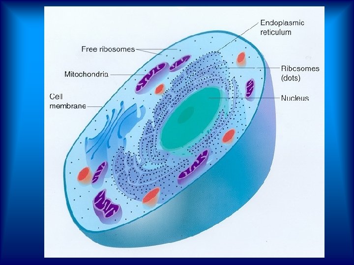
Eukaryotic Cell
Table 1: Comparison of features of prokaryotic and eukaryotic cells Prokaryotes Eukaryotes Typical organism bacteria, archaea protists , fungi, plants, animals Typical size ~ 1– 10 µm ~ 10– 100 µm (sperm cells, apart from the tail, are smaller) Type of nucleus nucleoid region; no real nucleus with double membrane
DNA circular (usually) linear molecules (chromosomes) with histone proteins linear molecules coupled in RNA-synthesis (chromosomes) cytoplasm inside the with histone nucleus protein synthesis in cytoplasm Ribosomes 0 S+30 S 60 5 S+40 S
unicellular
Amoeba Free-living amoeba protozoan groups that inhabit soils and natural waters are extremely diverse, not only in their structure but also in the manner in which they feed, reproduce, and move. The amoebas are a diverse group of free-living protozoa that probably evolved from a number of different primitive protozoan ancestors.
Euglena is a genus of unicellular protists, of the class Euglenoidea of the phylum Euglenozoa (also known as Euglenophyta). They are single-celled organisms. Currently, over 1, 000 species of Euglena have been described. There are many to be discovered. Marin et al. (2003) revised the genus to include several species without chloroplasts, formerly classified as Astasia and Khawkinea.
Cyanobacteria blue-green algae Cyanobacteria as bluegreen algaeis phylum of bacter are aquatic and photosynthetic, that is, they live in the water, and can manufacture their own food. Because they are bacteria, they are quite small and usually unicellular,
though they often grow in colonies large enough to see. They have the distinction of being the oldest known fossils, more than 3. 5 billion years old, in fact! It may surprise you then to know that the cyanobacteria are still around; they are one of the largest and most important groups of bacteria on earth.
Paramecium is a group of unicellular ciliate protozoa, which are commonly studied as a representative of the ciliate group, and range from about 50 to 350 μm in length. Simple cilia cover the body, which allow the cell to move with a synchronous motion (like a caterpillar).
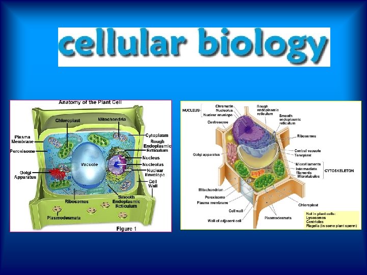
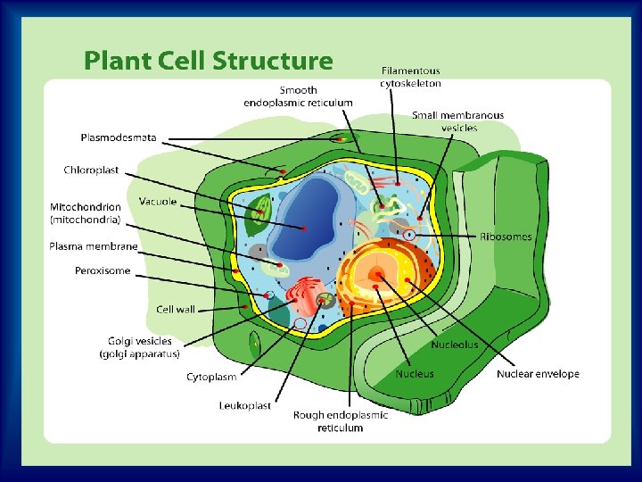
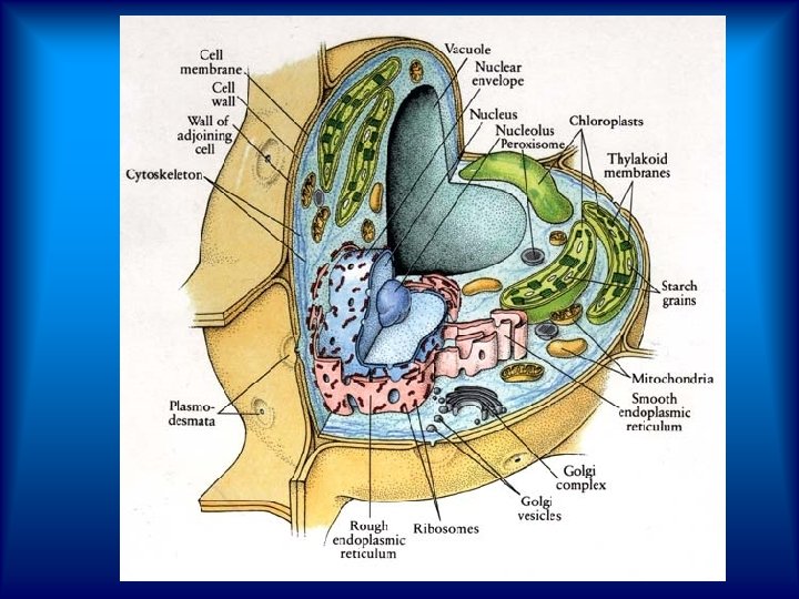
Plant Cell Structure
Animal Cell Structure
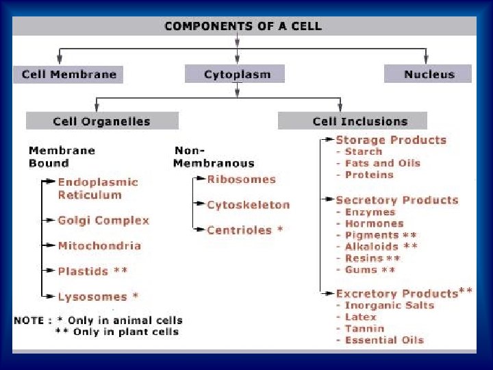
Unit Membrane Typical Structure - composed of protein and lipid (fat) molecules
Cell Membrane Structure - same as unit membrane. Function - acts as a boundary layer to contain the cytoplasm (fluid in cell) - interlocking surfaces bind cells together - selectively permeable to select chemicals that pass in and out of cells
Cell Wall Structure - a non-living secretion of the cell membrane, composed of cellulose - cellulose fibrils deposited in alternating layers for strength - contains pits (openings) that make it totally permeable
Function - provides protection from physical injury - together with vacuole, provides skeletal support
Chloroplast Structure - composed of a double layer of modified membrane (protein, chlorophyll, lipid) - inner membrane invaginates to form layers called "grana" (sing. , granum) where chlorophyll is concentrated
Centriole Structure - nine triplets of microtubules form one centriole - two centrioles form one centrosome Function - forms spindle fibres to separate chromosomes during cell division
Vacuole Structure - a single layer of unit membrane enclosing fluid in a sack Function - produces turgor pressure against cell wall for support - stores water and various chemicals - may store insoluble wastes
Endoplasmic Reticulum (ER) Structure - sheets of unit membrane with ribosomes on the outside - forms a tubular network throughout the cell
Endoplasmic Reticulum (ER) Function -transports chemicals between cells and within cells - provides a large surface area for the organization of chemical reactions and synthesis
Ribosome Structure - non-membraneous, spherical bodies composed of RNA (ribonucleic acid) and protein enzymes Function - site of protein synthesis
Golgi Apparatus Structure - stacks of flattened sacs of unit membrane (cisternae) - vesicles pinch off the edges Function - modifies chemicals to make them functional - secretes chemicals in tiny vesicles - stores chemicals - may produce endoplasmic reticulum
Mitochondrion Structure - composed of modified double unit membrane (protein, lipid) - inner membrane infolded to form cristae Function - site of cellular respiration ie. the release of chemical energy from food Glucose + Oxygen ------> Carbon Dioxide + Water + Energy (ATP)
Lysosome Structure - membrane bound bag containing hydrolytic enzymes - hydrolytic enzyme = (water split biological catalyst) i. e. using water to split chemical bonds
Lysosome Function - break large molecules into small molecules by inserting a molecule of water into the chemical bond
Nucleus nuclear envelope nucleolus chromatin nucleoplasm
Nucleus The nucleus consists of the nuclear envelope, nucleolus, chromatin, and nucleoplasm. Nuclear Envelope Structure - two unit membranes with a fluid-filled space - nuclear pores present - outer membrane may be continuous with endoplasmic reticulum
Nucleus Function - selectively permeable to control movement in or out - contains nuclear contents
Chromatin Structure - composed of long thin strands of DNA Function - contains instructions that control cell metabolism and heredity
Nucleolus Structure - non - membraneous matrix of RNA (ribonucleic acid) and protein Function - instructions in DNA are copied here - works with ribosomes in the synthesis of protein
Chromosomes -Usually in the form of chromatin - Contains genetic information - Composed of DNA - Thicken for cellular division - Set number per species (i. e. 23 pairs for human)
Write answers to the following: 1. Are euglena unicellular or multicellular? 2. What Kingdom do euglena belong to? What Phylum? 3. What organelle carries out photosynthesis? 4. On which end is the flagellum located? 5. Define autotrophic.
6. Define heterotrophic. 7. Describe the two ways in which the euglena get their nutrients. 8. What is the eyespot used for? 9. What is the function of the nucleus? 10. What is the function of the contractile vacuole? What would happen if the cell did not have this organelle.
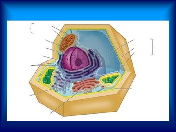
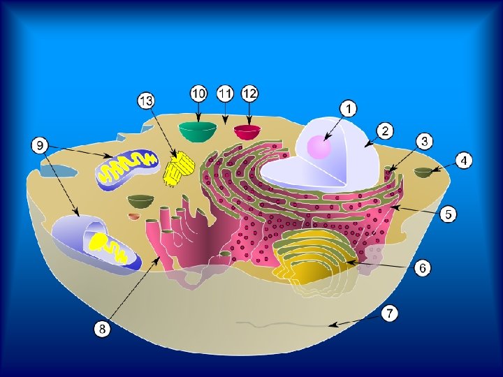
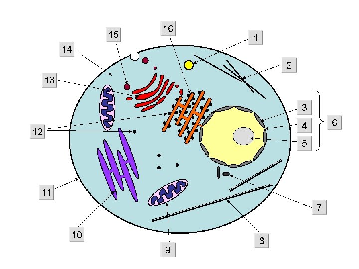
Cell Structure and Function Quiz Lable the diagram
Lable the diagram
Write answers to the following: 11. This structure is made of DNA . 12 Produces ATP 13. Creates turgor pressure . 14 New proteins are made on the 15. This organelle controls entry into the cell *************