Specific learning objectives Functional anatomy of eye Photoreceptor

• • Specific learning objectives: Functional anatomy of eye Photoreceptor mechanism Image-forming mechanism Visual pathway Visual Acuity, light and dark adaptations Color Vision

eye

• EYE has two major parts: • Optical system Helps to focus and form an image on the receptor cells-light rays fall. • Neural system. Transmits optical signal in the form of Aps along the optic nerve to visual cortex.

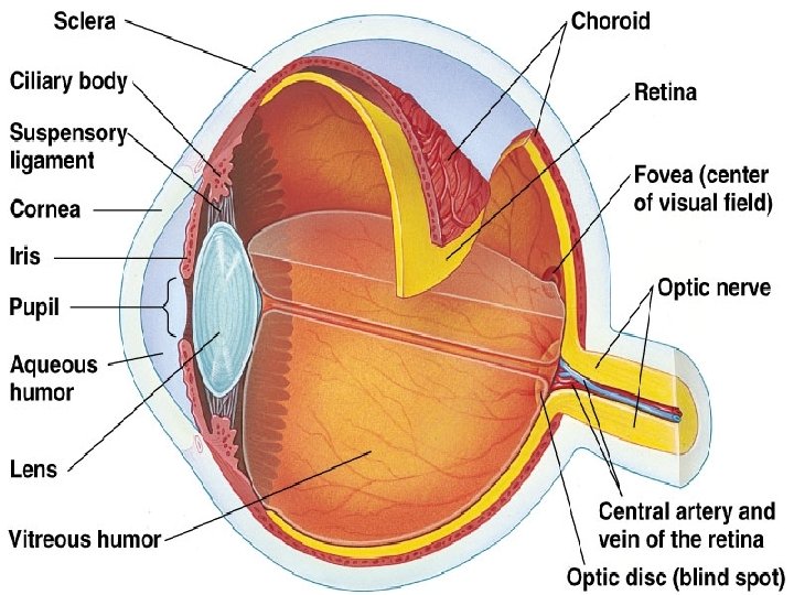
• Eye ball-hollow spherical organ • Optical system –Anterior part of the eye ball • Visual receptors –posterior surface-optic nerve arises and proceeds towards occipital cortex.

• • Interior of eyeball is divided into three spaces: Anterior chamber Posterior cavity

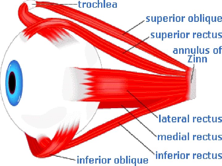
• Sclera • White avascular fibrous coat-composed of collagen fibers. • Shape to eyeball • Protective function • Extraocular muscles-regulates the eyeball movement. • Central portion of eye-transparent cornea

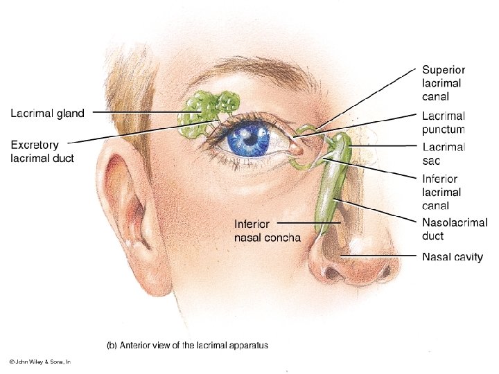
• Lacrimal gland • • • Lateral corner of eye. Secretes tear Keeps cornea moistened. Prevent infection Drained through naso-lacrimal duct.

Choroid-posterior 2/3 rd of eyeball Numerous blood vessels Nourishes structure of eyeball Front thickened portion-ciliary body-absorb extra amount of light. • Ciliary body • Attached to suspensory ligament at one end. • Other end-crystalline lens • •

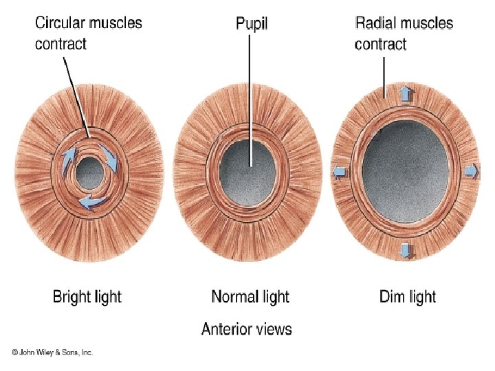
• Two types of smooth muscle: • Circular and longitudinal. • Accommodation for near vision. • • Iris Pigmented and opaque muscular structure. Colour to eye. Center-aperture-pupil-light enters the eye.

• • Two types of muscles: Sphincter and dilator pupillae Determines size of pupil. Functions Regulates intensity of light. Absorbs extra amount of light. Prevents entry of light through periphery of lens. Increases the depth of focus by constriction of pupil.

• Retina • Outer pigmented layer-attached to inner surface of choroid. • Inner layer-nerve cells and nerve fibersphotoreceptors.

• • Crystalline lens Circular biconvex Formation of image on retina. Has no blood supply.

• • All the nerve fibers-retina-optic nerve-brain Optic disc-optic nerve leaves eye. Blind spot Macula lutea-posterior pole of eye. Marks the location of fovea centralis. Absence of rods and densely packed cones. Greatest visual acuity.

• Line joining the anterior pole-posterior pole of the eyeball-optical axis • Line joining the fixation point to fovea centralis-visual axis.

Fluids in the eye: • Aqueous Humor-anterior cavity • Vitreous Humor-posterior cavity.
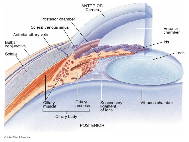

• Aqueous humor-protein –free clear fluid • Formed- Ciliary processes-posterior chamberpupil-anterior chamber. • Aqueous pressure-15 -18 mm Hg higher than intracranial pressure. • Helps to maintain shape of eye.

• Composition • High content of Na. Cl, vitamin C, lactic acid and hyaluronic acid.

Outflow of Aqueous Humor: Aqueous Humor is formed in ciliary processes Through the pupil In to the anterior chamber Anterior to the lens The angle b/w cornea and the iris Canal of Schlemm Extra ocular veins

• Functions • It provides nutrition to all avascular structures of eye. • It maintains intraocular pressure. • It maintains shape of the eye.

• Vitreous Humour • Interior between the lens and retina is filled with albumin and hyaluronic acid.

Functions of vitreous humor: • Prevent the wall of eyeball from collapsing. • It maintain the intraocular pressure and keep the intraocular structures in position. • Acts as a refractive medium.

Glucoma/Ocular Hypertension: Pressure increase to 60 -70 mm of Hg. Normal : 15 mm of Hg(12 -20 mm of Hg) Pressure above 25 -30 mm of Hg can cause blindness if maintained for long duration. Causes: • Blockage of canal of Schlemm • Excessive production of the fluid

2 types: Ø Primary: • Open angle: after 40 yrs • Closed angle: after 60 yrs. Ø Secondary : Cataracts, Trauma, Intraocular haemorrage.

Retina
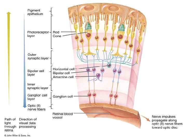

LAYERS OF RETINA: 1. 2. 3. 4. 5. 6. 7. 8. 9. 10. Pigmented epithelium Layers of rods and cones External limiting membrane Outer nuclear layer Outer synaptic layer Inner nuclear layer Inner synaptic layer ganglion cell layer Optic nerve Internal limiting membrane.

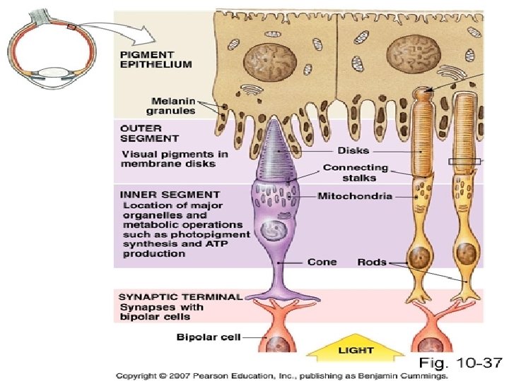
1. Outer pigmented epithelial layer. • • Rich in melanocytes. Prevents scattering of light. Phagocytosis. Storage of Vit A.

• Rods and cones-Photoreceptors • Each rod and cone is divided into outer segment, inner segment and a synaptic zone.

• Outer and inner segments form the layer of rods and cones. outer segment Modified cilia-piles of flattened discs. Discs - Rhodopsin and Iodopsin

• Rods-thin , rod like appearance • Old discs-shed and removed by pigmented epithelium. • New discs-inner edge of outer segment. • Synapse with several rod bipolar cells

• Cones • Conical outer segment • Renewal –diffused-multiple sites at outer segment.

• Inner segment: Rich in mitochondria In cones-thick, oval and is larger • Synaptic Zone. Synaptic vesicles –glutamate.

• External limiting membrane. Glial tissues. • Outer nuclear layer Nucleus of rods and cones • Outer synaptic layer synapse between ends of rods and cones with dendrites of bipolar cells and horizontal cell processes.

• Inner nuclear layer o Bipolar cells o Horizontal cells o Amacrine cells-synaptic contacts with dendrites of both ganglion and bipolar cells. • Inner synaptic layer Site of processing of visual image.

• Ganglion cell single layer of cell containing round cells • Optic nerve axons of ganglion cells • Internal limiting membrane separates retina from vitreous humour

• How light rays reach photoreceptors Light rays Ganglion cell Bipolar cell photoreceptors

• Functions of Rods • sensitive to light. • dim light vision or night vision or scotopic vision. • Low threshold

• Functions of cones. • High threshold. • bright light vision or daylight vision or photopic vision. • Cones are also responsible for acuity of vision and the color vision.

Rods : 1. 120 million. 2. Mainly in peripheral retina. 3. Slender , Elongated, Rod like. 4. More pigments/rod. 5. Only one type of pigment-Rhodopsin. 6. Cannot detect color. 7. Functions better in dim light. 8. Loss- Night blindness Cones: 1. 6 million 2. Mainly in the central retina. 3. Conical shape. 4. Less pigments/cone. 5. Three types: blue colour(cyanolabe) Green (chlorolabe) red (erythrolabe) 6. Can detect color. 7. Functions better in day light 8. Loss-functional blindness.

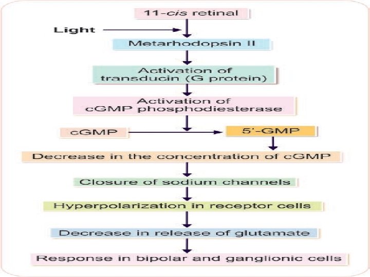
• • • Chemistry of Rhodopsin is a conjugated protein. Protein called opsin and a chromophore. Opsin -scotopsin. Chromophore - retinal. Retinal is the aldehyde of vitamin A or retinol. Retinal is present in the form of 11 -cis retinal known as retinine 1

Rhodopsin –Retinal visual cycle

RHODOPSIN LIGHT ENERGY BATHORODOPSIN LUMIRHODOPSIN METARHODOPSIN 1 SCOTOPSIN 11 -CIS RETINAL 11 -CIS RETINOL ISOMERASE METARHODOPSIN 2 ALL-TRANS RETINAL ALL-TRANS RETINOL (Vit A)

• Rhodopsin --metarhodopsin II Resynthesis of Rhodopsin • all-trans retinal - 11 -cis retinal ( enzyme retinal isomerase). • 11 -cis retinal immediately combines with scotopsin to form rhodopsin.

PHOTOTRANSDUCTION The process by which light energy is converted into receptor potential in visual receptor.

• In darkness-visual receptors -resting membrane potential is about – 40 m. V. • sodium ions leak back into the rod cellsreduce the electronegativity inside rod cell • Dark current.

Maintenance of dark current in outer segment of rod cell

Functions of retina: 1. Visual functions: • Light sense: Help to perceive light. • Form sense: Appreciate the shape. • Color sense: Perceive and recognize different color and different intensities of the same color

2. Reflexes: • Light reflex • Accommodation reflex. 3. Help to maintain the Tone , Posture and Equlilibrium.

IMAGE FORMING MECHANISMS

• Light rays-falls on eye-image formation on retina: • Refraction of light by cornea and lens • Ciliary muscle activity-accommodation of lens • Change in pupil size –iris muscles.

• Principles of optics • Refraction-Change in the direction of light rays • Lens-transparent glass-two spherical surfaces 3 types: Convex Concave Cylindrical

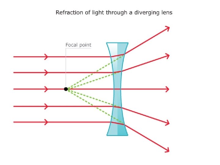
• Optical centre/nodal point-centre point of lens • Principal axis-line joining centre of two spherical part lens surfaces. • Principal focus vconvex lens-point on principal axis-light rays –converges v. Concave lens-point on principal axis-light rays –diverges
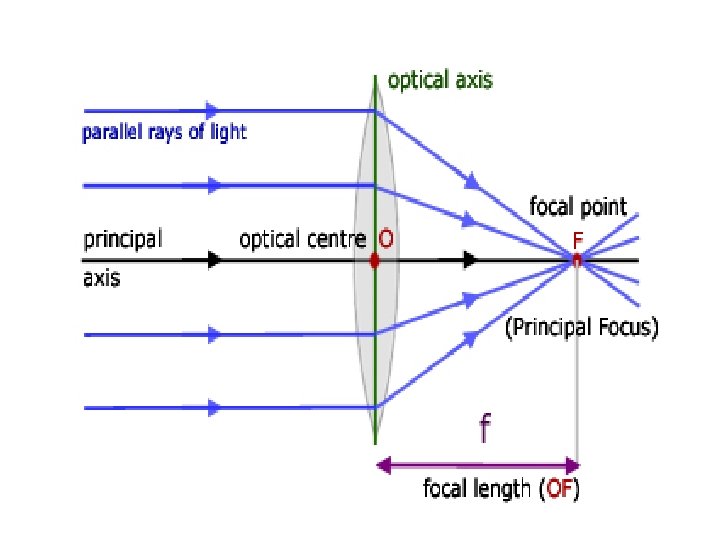

• Focal length-distance between optic centre and principal focus of the lens. • Light rays-distant object(>6 m)-parallel and object closer(<6 m)-diverging.

• Power • Reciprocal of focal length. • S. I-dioptre. • Convex lens-positive focal length and concave lens-negative.

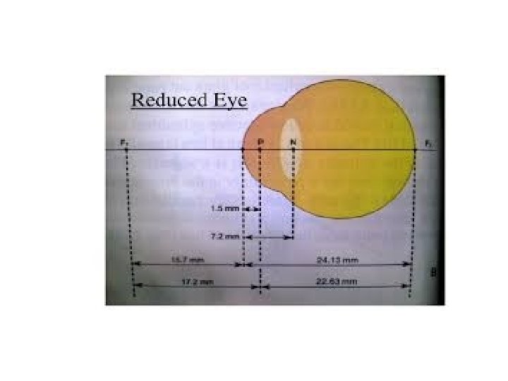
• Reduced /schematic Eye. • Differences in refractive indices of eye structure • Reduced eye –single spherical surface –single principal and nodal point. • Nodal point-7 mm anterior surface of cornea.

• Human eye-24 mm in length-focal length 17 mm. • Refractive power of reduced eye-59 D • Normal eye(59 D)-behaves as the reduced eye.

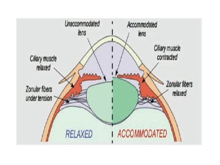
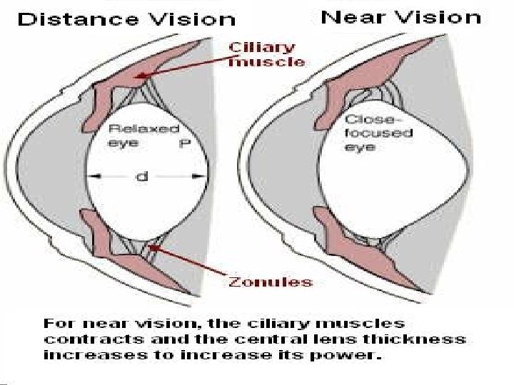
ACCOMMODATION: The ability of the eye ball by which the near objects are clearly focused on the retina. • • • Changing the curvature of lens. Ciliary muscle contracts Suspensory ligaments relax Tension on lens decreases. Lens becomes convex.

Near point: The nearest point to the eye at which an object can be brought in to clear focus by accommodation. Far point: The farthest point which can be brought to focus.

• Near vision • Nearest point to the eye, at which object seen clearly. • Accommodation is maximum.
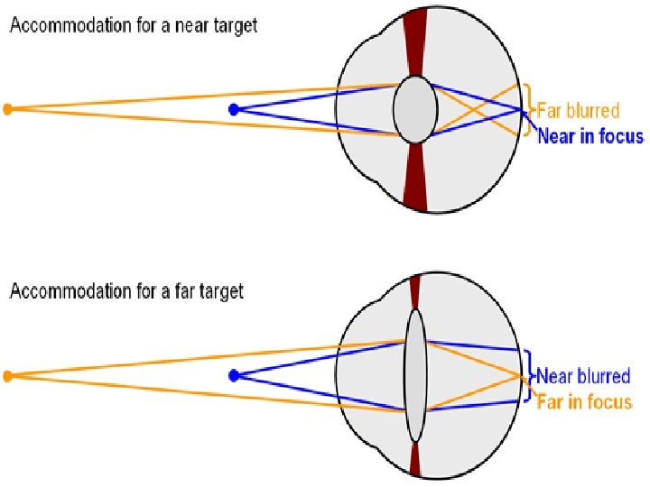

Changes associated with near vision: • Change in the anterior curvature of the lens. • Constriction of pupil. • Convergence of the eye ball.

Far vision: Near vision: • Ciliary muscles relax. • Ciliary muscles contract. • Lens is held under tension by the lens ligament. • Lens ligaments are relaxed. • Lens flattens. • Lens convex shape.

• Defects of image forming mechanisms

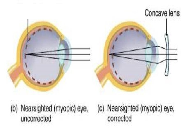
Emmetropia= Normal eye. Myopia/Near sightedness: Objects are focused infront of retina. Cause: • Too long an eye ball. • Stronger curvature of the eye. Seen in: • 5 % of newborns and infants. Correction : • Biconcave lens • Surgical correction. Refractive power +ive

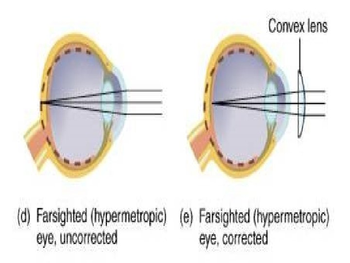
Hypermetropia/Farsightedness. Object is focused beyond the retina. Cause: • Too short an eye ball. • Weak curvature of the eye. Seen in: • New born and infants 80% • Rare in young adults. Correction : • Biconvex lens. Refractive power : -ive

Astigmatism: Failure to focus light at one point- blurring of the image. Cause: due to the faulty curvature of the cornea. Correction: • Cyclindrical lens
.")
Presbyopia: Lens looses its elastic nature and becomes relatively solid mass( Denaturation of proteins). • Accommodation power decreases. • Far point not affected. • Eye remains focused at a constant distance. Correction: Bifocal lens

Visual pathway
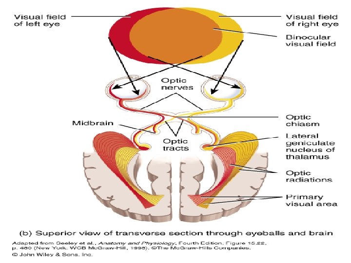

• Optic nerve: Complete blindness in one side. • Optic chiasma: Bi-temporal Hemianopia. • Temporal side nerve: Bi-nasal Hemianopia. • Optic tract, Lateral geniculate body, Optic radiations: Homonymous Hemianopia. • End of optic radiation(occipital cortex): Quadratic Hemianopia

Visual cortex: Area : 17 Primary Visual Area. Highest area for perception of visual sense. Visual association areas: Area 18 -Visuo Psychic Area. • Visual orientation, Depth perception. • Relay of information from visual cortex to other areas of brain. • Interpretation and integration in the light of past experience.

Lesion: Fails to recognize the nature of the object. Area 19 -Occipital eye field. Deviation and movement of the eye ball. Lesion: • visual hallucination • Conjugate deviation of eye to opposite side. Area 8 –Frontal eye field Function same as Area 19

Other connections: Optic chiasma-limbic system. Circadian rhythms and sexual cycles in birds and some animals. Occipital cortex- frontal eye field. Movement of the eye ball Occipital cortex-spinal cord. Posture, Equilibrium, Visuo spinal reflexes. Optic tract -3 rd cranial nucleus Light refluxes.

Visual acuity: The degree to which the details and contours of objects are perceived/Minimal separable distance b/w two objects to see them as two. Functions of cone.

Factors affecting visual acuity: 1. Optical factors: • • • Curvature of the Cornea and Lens. Elasticity of the eye. Conditions of ciliary muscles and lens ligaments. 2. Retinal factors: • • Functional status of retina. Highest in the Fovea and decreases towards the periphery.

3. Stimulus factors: • • • Size of the object. Distance from the eye. Color of the object. Shape, brightness, duration for which the object is viewed. Age. Test: • Distant vision: Snellens chart. (normal is 6/6) • Near vision Jaeger’s chart

Visual field: The area of the external world that can be seen when the gaze is fixed at a point. Limits: Medially: Bridge of the Nose. Superiorly: Roof of the orbit. Inferiorly: The cheeks. Field is not circular.

Monocular and binocular visual fields:

Advantages of Binocular vision: • Optical defect of one eye is corrected by the other. • Field of vision is increased. • Stereoscopic vision. Useful for the perception of depth. Tests: • Confrontation test. • Perimetry.

Normal aberrations of vision:

Spherical Aberration: Crystalline lens of the eye is not regularly formed Rays coming through the peripheral edges not focused on the retina. • Iris covers the peripheral parts of the lens. • The central part of the lens has higher refractive power which help in focusing all light rays at one point

Chromatic aberration: Different colored rays bend differently and so not focused exactly at one point. Light rays passing through the periphery of the lens is affected most- color fringes appear. Color fringes are ignored by the brain. Corrected by iris. Due to the difference in the refractive indices of the media

Reflexes: 1. Pupillary reflux a. Light direct indirect b. Accommodation. • Conjunctival reflux
 OPTIC CHIASMA OPTIC TRACT")
Light reflex: LIGHT RETINA OPTIC NERVE OPTIC TRACT (SAME SIDE) OPTIC CHIASMA OPTIC TRACT (OPP SIDE) CILIARY GANGLION SHORT CILIARY NERVES SPHINCTER PUPILLAE CONSTRICTION OF THE PUPIL

Pathway for accommodation:

Near object Retina Visual cortex. Frontal eye field 3 rd cranial nucleus Short ciliary nerves Constrictor Pupillae Pupillary constriction Ciliary muscles Anterior curvature of lens increases Medial rectus contraction Convergence of the eye ball

Adaptation: Getting accustomed or used to a new condition. Dark adaptation: the decrease in the visual threshold or increase in the sensitivity of the eye to light. Processes: • Removal of the after images from the retina. (Bright light) • Resynthesis of the bleached visual purple in the rods(Dim light)

2 responses: 1. Fast response. • First drop in the visual threshold is rapid but small in magnitude. • Due to adaptation of cones. • 4 -5 min. 2. Slow response. • Further drop in the visual threshold occurs slowly over a period of 25 min • Adaptation of rods • Resynthesis of Rhodopsin.
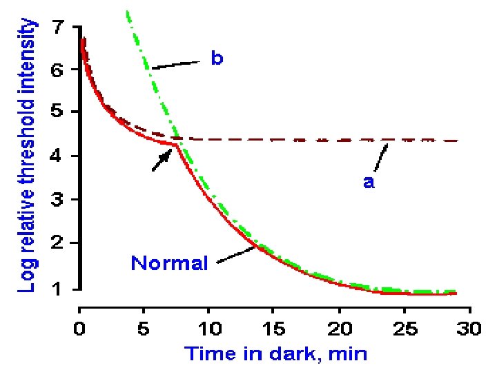

Changes occurring during dark adaptation: • Dilation of pupil. • Photoreceptor function, Cones-> Rods. • Resynthesis of Rhodopsin. Changes occurring during light adaptation. • Pupil constrict. • Photoreceptor function , Rods-> Cones. • Photo pigments are bleached and their concentration decrease.

Refraction: The bending of light rays at angulated interface. Refractive index. The ratio of velocity of light in the air by the velocity in the substance. Refractive power: =1 M/focal length =1 M/17 mm =1000 mm/17 mm =59 Diopters.

Anisometria: Difference in the refraction in 2 eyes. Aphakia: Crystalline lens is being removed.

Color vision

Achromatic vision: It is the sensation of white color and no color has been assigned to it. Chromatic vision: Spectral colors vision and extra spectral color vision. Primary colors: Red Green blue. Complimentary color: when two colors are mixed an appropriate amounts= white. After image: After one stops looking at a color he may continue to see it for a short time(+ive after image) Or he may see its complimentary color(-ive after image)
.")
Theories of color vision; 1. Thomas young and Von –Helmholtz's theory( Trichromatic color theory). Three different types of cones each containing a different photosensitive pigment and maximum sensitivity to one type of primary color.

2. Mullers doctrine of specific nerve energy theory: There are specific nerve fibers with specific ganglion cells responding to three primary colors.

Granites dominator and modulator theory. Two types of ganglion cells: • Dominators: they respond to the whole visual spectrum Detects intensity of the light but not the color. Y ganglion.

Modulators: respond maximum to a narrow wave length of light. Blue: 450 -470 nm(Peak=445 nm). Green: 520 -540 nm(Peak=535 nm). Red: 500 -600 nm(Peak=570 nm) they respond to the entire visual spectra to a varied degree. Hence they are responsible for color vision X ganglions

Hering ‘s opponent color theory • Extension of first theory. • 4 primary color: Red Blue Green Yellow • Photo chemical substances give one sensation on breakdown and other on resynthesis.

Three types of pigments: • Cyanopsin: blue color , max 445 nm, S-cone. • Iodopsin: green, max 535 nm, M-Cone. • Phorphyropsin : Red, Max-570 nm, L-Cone.

Color blindness: This is based on the young Helmholtz theory of color vision: 8%of males, 0. 4% females Red and green color blindness is X linked.

Types of color blindness:

Trichromats: weakness for one primary color. Protanomolous: Red color , 6%, sex linked Deuteranomolous: Green color, 6%, sex linked Tritanomolous: Blue color, rare, not sex linked.

Dichromats: 2 cone system present • Protanopia: Blindness to red , Phorphyropsin absent. • Deutranopia: Blindness to green, Iodopsin absent. • Tritanopia: Blindness to blue, Cyanopsin absent.

Monochromats: one cone system present They can see black , white and shades of grey.

Test for color vision. • Ishihara chart • Wool sorting test. • Edridge green lantern method
- Slides: 120