
Respiratory system Functional Anatomy Respiratory system Image from

- Slides: 16
Respiratory system Functional Anatomy Respiratory system Image from Encyclopaedia Britannica Inc
Aims of the session By the end of the session you should • Understand the structure of the pulmonary system. • Understand the function of the pulmonary system. • Understand the mechanism of breathing
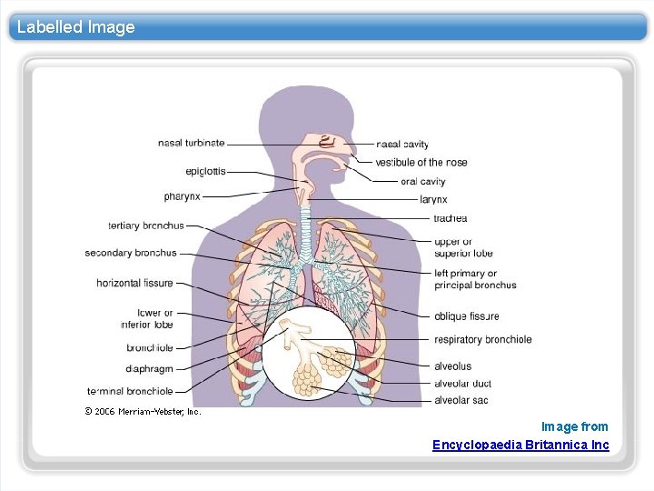
Labelled Image from Encyclopaedia Britannica Inc
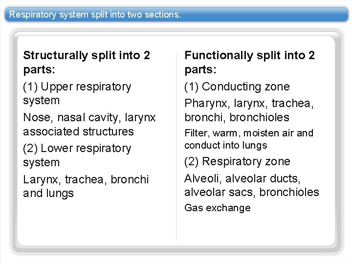
Respiratory system split into two sections. Structurally split into 2 parts: (1) Upper respiratory system Nose, nasal cavity, larynx associated structures (2) Lower respiratory system Larynx, trachea, bronchi and lungs Functionally split into 2 parts: (1) Conducting zone Pharynx, larynx, trachea, bronchioles Filter, warm, moisten air and conduct into lungs (2) Respiratory zone Alveoli, alveolar ducts, alveolar sacs, bronchioles Gas exchange
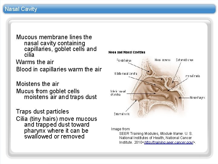
Nasal Cavity Mucous membrane lines the nasal cavity containing capillaries, goblet cells and cilia Warms the air Blood in capillaries warm the air Moistens the air Mucus from goblet cells moistens air and traps dust Traps dust particles Cilia (tiny hairs) move mucous and trapped dust toward pharynx where it can be swallowed or removed Image from SEER Training Modules, Module Name. U. S. National Institutes of Health, National Cancer Institute. 2010<http: //training. seer. cancer. gov/>.
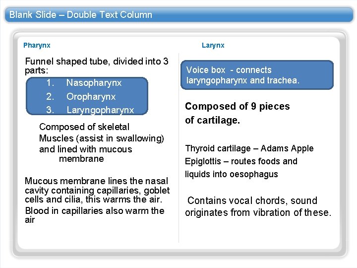
Blank Slide – Double Text Column Pharynx Funnel shaped tube, divided into 3 parts: 1. Nasopharynx 2. Oropharynx 3. Laryngopharynx Composed of skeletal Muscles (assist in swallowing) and lined with mucous membrane Mucous membrane lines the nasal cavity containing capillaries, goblet cells and cilia, this warms the air. Blood in capillaries also warm the air Larynx Voice box - connects laryngopharynx and trachea. Composed of 9 pieces of cartilage. Thyroid cartilage – Adams Apple Epiglottis – routes foods and liquids into oesophagus Contains vocal chords, sound originates from vibration of these.
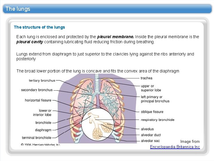
The lungs The structure of the lungs Each lung is enclosed and protected by the pleural membrane. Inside the pleural membrane is the pleural cavity containing lubricating fluid reducing friction during breathing. Lungs extend from diaphragm to just superior to the clavicles lying against the ribs anteriorly and posteriorly The broad lower portion of the lung is concave and fits the convex area of the diaphragm Image from Encyclopaedia Britannica Inc
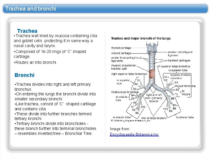
Trachea and bronchi Trachea • Trachea wall lined by mucosa containing cilia and goblet cells protecting it in same way a nasal cavity and larynx. • Composed of 16 -20 rings of ‘C’ shaped cartilage. • Routes air into bronchi. Bronchi • Trachea divides into right and left primary bronchus • On entering the lungs the bronchi divide into smaller secondary bronchi • Like trachea, consist of ‘C’ shaped cartilage and contains cilia • These divide into further branches termed tertiary bronchi • Tertiary bronchi divide into bronchioles these branch further into terminal bronchioles – resembles inverted tree – Bronchial Tree. Image from Encyclopaedia Britannica Inc
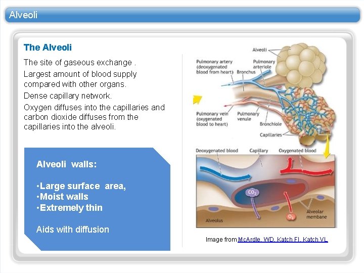
Alveoli The site of gaseous exchange. Largest amount of blood supply compared with other organs. Dense capillary network. Oxygen diffuses into the capillaries and carbon dioxide diffuses from the capillaries into the alveoli. Alveoli walls: • Large surface area, • Moist walls • Extremely thin Aids with diffusion Image from Mc. Ardle, WD, Katch FI, Katch VL
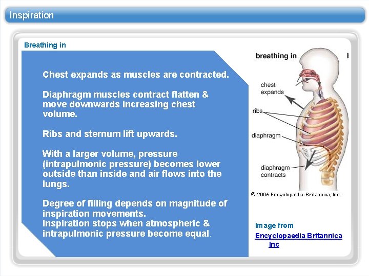
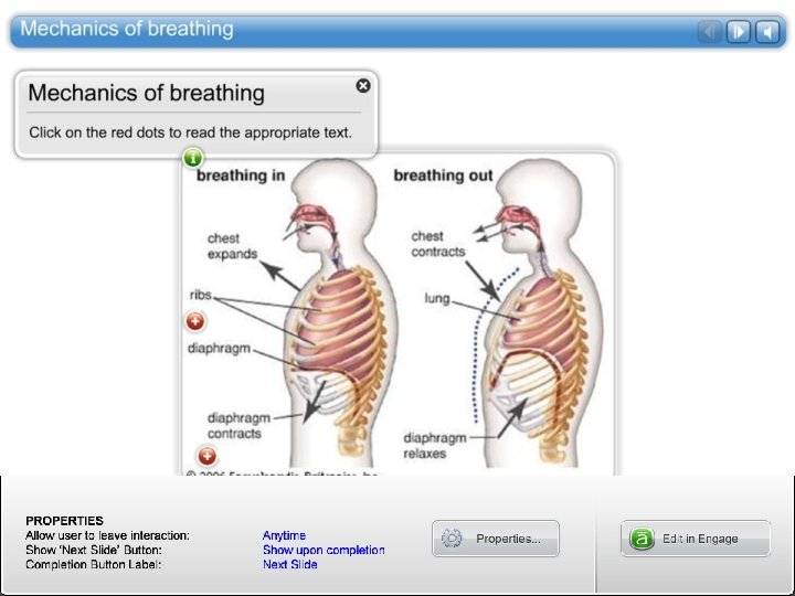
Inspiration Breathing in Chest expands as muscles are contracted. Diaphragm muscles contract flatten & move downwards increasing chest volume. Ribs and sternum lift upwards. With a larger volume, pressure (intrapulmonic pressure) becomes lower outside than inside and air flows into the lungs. Degree of filling depends on magnitude of inspiration movements. Inspiration stops when atmospheric & intrapulmonic pressure become equal. Image from Encyclopaedia Britannica Inc
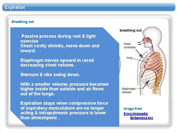
Expiration Breathing out Passive process during rest & light exercise Chest cavity shrinks, move down and inward. Diaphragm moves upward in recoil decreasing chest volume. Sternum & ribs swing down. With a smaller volume, pressure becomes higher inside than outside and air flows out of the lungs. Expiration stops when compressive force of expiratory musculature are no longer acting & intrapulmonic pressure is lower than atmoshperic. Image from Encyclopaedia Britannica Inc
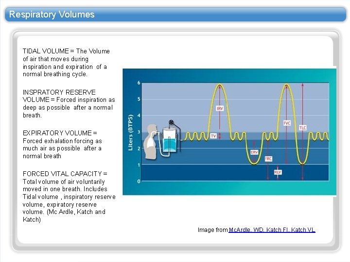
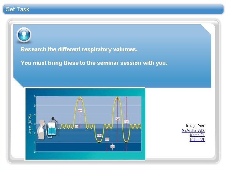
Respiratory Volumes TIDAL VOLUME = The Volume of air that moves during inspiration and expiration of a normal breathing cycle. INSPRATORY RESERVE VOLUME = Forced inspiration as deep as possible after a normal breath. EXPIRATORY VOLUME = Forced exhalation forcing as much air as possible after a normal breath FORCED VITAL CAPACITY = Total volume of air voluntarily moved in one breath. Includes Tidal volume , inspiratory reserve volume, expiratory reserve volume. (Mc Ardle, Katch and Katch) Image from Mc. Ardle, WD, Katch FI, Katch VL
Set Task Research the different respiratory volumes. You must bring these to the seminar session with you. Image from Mc. Ardle, WD, Katch FI, Katch VL
Summary In summary: The respiratory system is split into two sections. Air flows from a high pressure to a low pressure. Gaseous exchange occurs at the alveoli.