Anatomy of Shoulder Complex Dr Fadel Naim Orthopedic


 Of the accessory nerve n n Runs downward in the")


 Joint n n n A ball-and-socket type of synovial joint Relatively unstable")


- Slides: 101
Anatomy of Shoulder Complex Dr. Fadel Naim Orthopedic Surgeon Faculty of Medicine IUG-Gaza
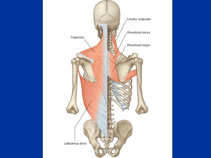
Trapezius n Origin – – – n Medial third superior nuchal line on the occipital bone External occipital protuberance Ligament nuchae Spine of 7 th cervical vertebra Spinous processes and supraspinous ligaments to T 12 INSERTION – Upper fibers to lateral third of posterior border of clavicle – Middle fibers to medial acromion and superior lip of spine of scapula – Lower fibers to medial end of spine of scapula n Action – Laterally rotates, elevates and retracts scapula. – If scapula is fixed, extends and laterally flexes neck n Nerve – Motor fibers form spinal accessory nerve (Cranial nerve XI) – Sensory fibers spinal nerves C 3 and C 4
Levator Scapulae n Origin – Posterior tubercles of transverse processes of C 1 -4 n INSERTION – Upper part of medial border of scapula opposite the supraspinous fossa n Action – Raises medial border of scapula – In conjunction with middle fibers of trapezius and rhomboids, pulls the scapula medially and upward n Nerve – Anterior primary rami of C 3 and C 4 and dorsal scapular nerve (C 5)
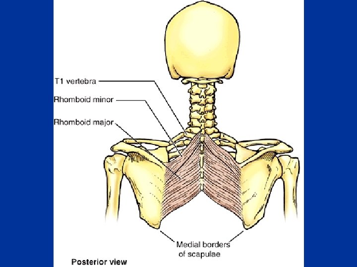
Rhomboid Minor n Origin – Lower ligamentum nuchea – Spines of C 7 and T 1 n INSERTION – Small area of posteromedial border of scapula at level of spine, below levator scapulae n Action – Elevates the medial border of the scapula and pulls it medially n Nerve – Dorsal scapular nerve (C 5)
Rhomboid Major n Origin – Spines of T 2 -T 5 and supraspinous ligaments n INSERTION – Lower half of posteromedial border of scapula, opposite the infraspinous fossa n Action – Elevates the medial border of the scapula and pulls it medially n Nerve – Dorsal scapular nerve (C 5) (from root )
Deltoid n ORIGIN – Anterior fibers: Lateral third of clavicle – Middle fibers: Acromion – Posterior fibers: spine of scapula to deltoid tubercle n INSERTION – Deltoid tuberosity – Middle of lateral surface of humerus n ACTION – Abducts arm – anterior fibers flex and medial rotate – posterior fibers extend and lateral rotate n NERVE – Axillary nerve (C 5, 6) (from posterior cord) Forms the rounded contour of the shoulder
Supraspinatus n Origin – Medial three quarters of supraspinous fossa of scapula – Upper surface of spine n INSERTION – Superior facet on greater tuberosity of humerus – Capsule of shoulder joint n Action – Abducts arm – Stabilizes shoulder joint n Nerve – Suprascapular nerve
Infraspinatus n Origin – Medial three quarters of infraspinous fossa of scapula n INSERTION – Middle facet of greater tuberosity of humerus – Capsule of shoulder joint n Action – Laterally rotates arm – Stabilizes shoulder joint n Nerve – Suprascapular nerve
Teres Minor n Origin – Middle third lateral border of scapula above teres major n INSERTION – Inferior facet of greater tuberosity of humerus (below infraspinatus) – Capsule of shoulder joint n Action – Laterally rotates arm – Stabilizes shoulder joint n Nerve – Axillary nerve
Pectoralis Minor n n Origin 3, 4, 5 ribs INSERTION – Medial and upper surface of coracoid process of scapula n Action – Elevates ribs if scapula fixed – Pulls shoulder downward and forward n Nerve – Medial pectoral nerve (from medial cord of brachial plexus)
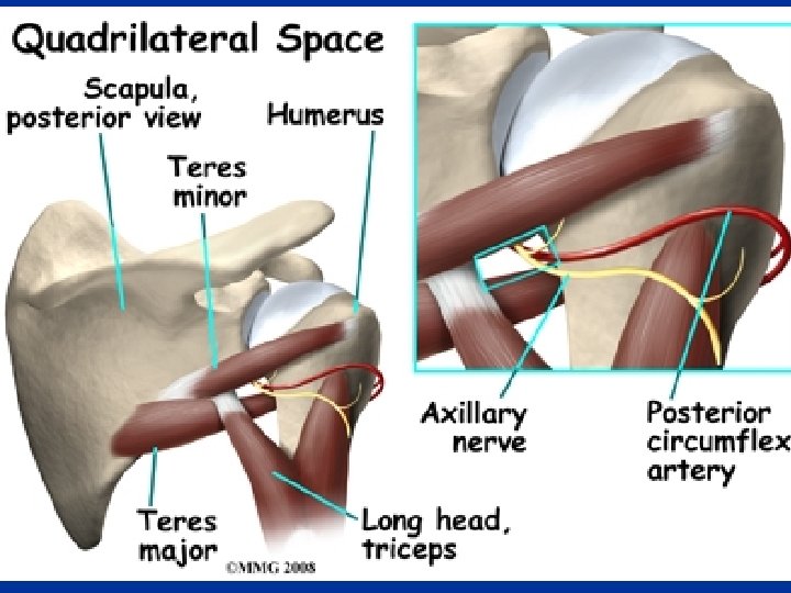
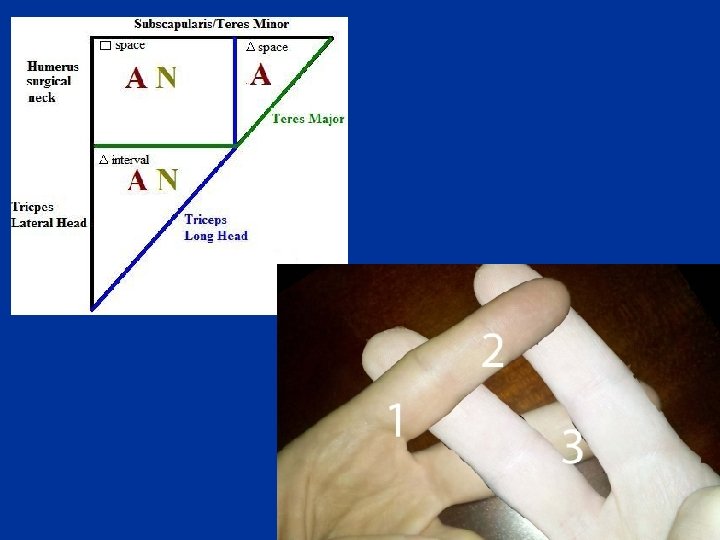
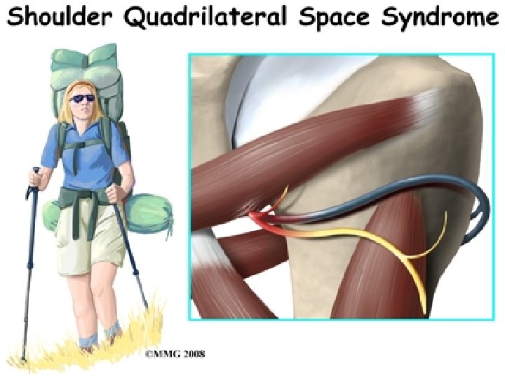
Quadrangular space Triangular interval
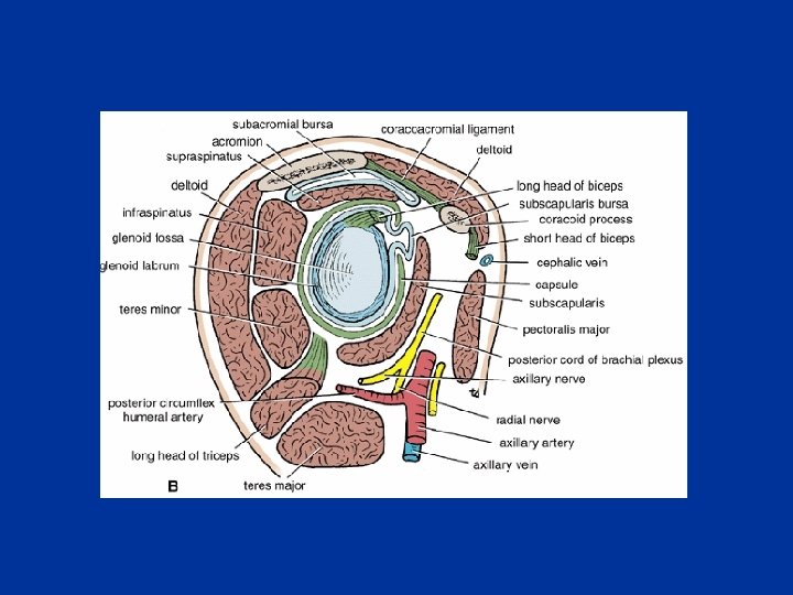
Quadrangular space n It is bounded: – Superiorly by: • Subscapularis • Teres minor – Inferiorly by: • Teres major – Medially by: • Long head of triceps – Laterally by: • Humerus The axillary nerve and the posterior circumflex humeral vessels pass backward through this space
Triangular space n n an area of communication between the axilla and the posterior scapular region formed by: – the medial margin of the long head of triceps brachii – the superior margin of teres major – the inferior margin of teres minor The circumflex scapular artery and vein pass through this gap
Triangular interval n formed by: – the lateral margin of the long head of triceps brachii; – the shaft of the humerus; – the inferior margin of teres major The radial nerve, the profunda brachii artery and associated veins pass through it
Quadrangular space syndrome n n n Hypertrophy of the quadrangular space muscles or fibrosis of the muscle edges may impinge on the axillary nerve. Uncommonly, this produces weakness of the deltoid muscle. Typically it produces atrophy of the teres minor muscle – which may affect the control that the rotator cuff muscles exert upon shoulder movement
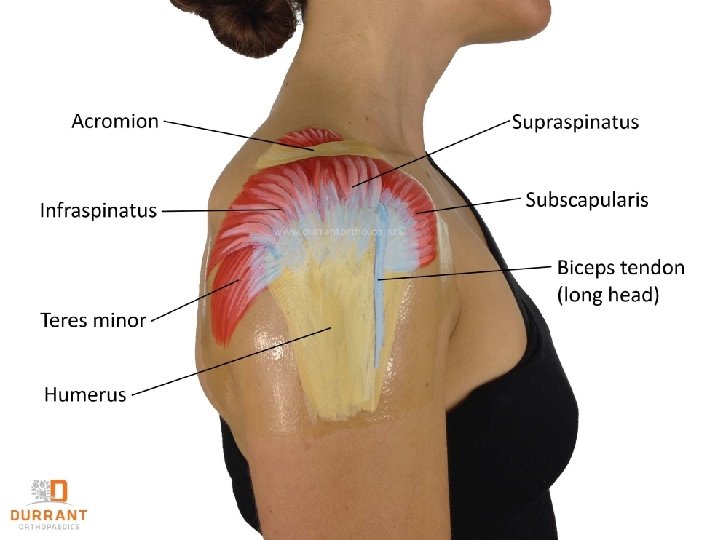
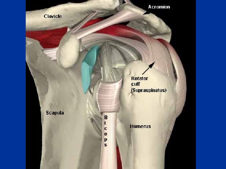
Rotator Cuff Muscles n Four of the scapulohumeral muscles – – n n n Supraspinatus Infraspinatus Teres minor Subscapularis Referred to as SITS muscles Called rotator cuff muscles because they form a musculotendinous cuff around the glenohumeral joint {Dynamic stabilizers (“cuff”)} All except the supraspinatus are rotators of the humerus
n The tendons of the muscle blend with the fibrous capsule of the glenohumeral joint to form a musculotendinous rotator cuff, which reinforces the capsule on three sides: – Anteriorly – Superiorly – Posteriorly n The cuff is weakest anteroinferiorly, making shoulder dislocation most common in this direction.
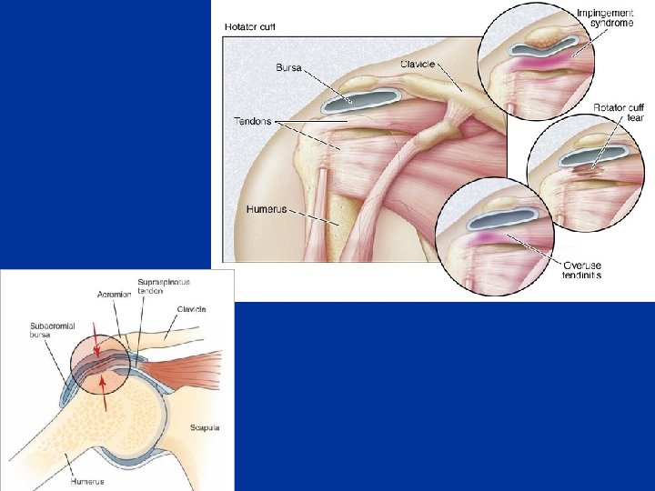
n Injury to rotator cuff can occur in any of the following manners: – Acutely, following trauma; – As a result of chronic impingement and overuse (excessive abduction); – Compromised blood supply. n The most frequently injured tendon is the supraspinatus, probably because it is relatively avascular n Additionally, the supraspinatus tendon is subject to significant trauma as it is compressed between the acromion and the humeral head during abduction
Impingement
Types Of Acromion n Three distinct types of acromion can be seen on the angled outlet Y view: – The type I acromion, which is flat, is the "normal" acromion. – The type II acromion is more curved and downward dipping, – the type III acromion is hooked and downward dipping, obstructing the outlet for the supraspinatus tendon. n Cadaveric studies have shown an increased incidence of rotator cuff tears in persons with type II and type III acromions
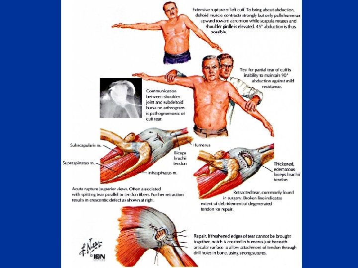
Rotator Cuff Injuries n Injury or disease may damage the musculotendinous rotator cuff, producing instability of the glenohumeral joint n Trauma may tear or rupture one or more of the tendons of the muscles forming the rotator cuff n Acute tears may occur when the arm is violently pushed into abduction n The patient reports a sharp pain in the anterosuperior part of the shoulder n Rotator cuff injuries are also common in persons with throwing activities n Rotator cuff tears also follow dislocation of the shoulder
n n n Degenerative tendonitis of the rotator cuff is common, especially in old people. To test for this disease, the person is asked to lower the fully abducted limb slowly and smoothly. From an approximately 90 ° abduction, abduction the limb will suddenly drop to the side in an uncontrolled manner if the rotator cuff (especially the supraspinatus part) is diseased and torn The injury often results from an indirect force to the abducted arm, such as a fall in a person older than 45 years Acute tears are uncommon in young persons.
This injury causes tenderness around the greater tubercle of the humerus and pain during 45 ° of passive abduction. n X-rays may be normal, but can reveal: n – Narrowing of the distance between the acromion and humeral head (impingement) – Erosion of the inferior acromion – Sclerosis of the greater tuberosity
Subacromial Bursitis 130° 50° n n The tendon of the supraspinatus is separated from the coracoacromial ligament, acromion, and deltoid by the subacromial bursa. Painful arc syndrome – When this bursa is inflamed abduction of the arm is extremely painful during the arc of 50 to 130 °. n n n The pain may radiate as far distally as the hand. Acute pain is also felt lateral to the acromion. A painful arc signifies an impingement of a painful structure during the movement of the humerus in relation to the roof of the shoulder
n The commonnest pathological situations causing painful arc are: – 1. Tendinitis of Supraspinatus – 2. Tendinitis of Infraspinatus – 3. Tendinitis of Subscapularis – 4. Tendinitis of the long head of the Biceps – 5. Subacromial Bursitis – 6. AC Sprain n Less frequent pathologies associated with a painful arc are: • metastases in the head of the humerus • metastases in the acromion • Instability of the shoulder
Glenohumeral motions
Glenohumeral motions
Glenohumeral motions
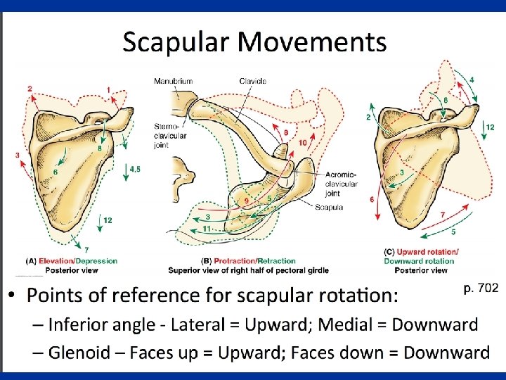
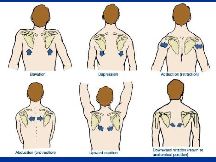
Scapular motions
Scapular motions Protraction – Retraction
Scapular Motions Rotational Elevation Of Glenoid Cavity Rotational Depression Of Glenoid Cavity
4 -43
Muscles Involved In Shoulder Movements Movement Prime movers Secondary movers Glenohumeral Flexion Anterior deltoid Coracobrachialis Pectoralis major (clavicular head) Extension Latissimus dorsi Teres major Posterior deltoid Teres minor Triceps Abduction Deltoid (mid) Supraspinatus Anterior/posterior deltoid Serratus anterior
Muscles Involved In Shoulder Movements Movement Prime movers Secondary movers Glenohumeral Adduction Pectoralis major Teres major Latissimus dorsi External rotation Infraspinatus Posterior deltoid Teres minor Internal rotation Subscapularis Pectoralis major Latissimus dorsi Teres major Anterior deltoid
Muscles Involved In Shoulder Movements Movement Prime movers Secondary movers Scapular Retraction Rhomboid major/minor Trapezius Protraction Serratus anterior Upward rotation Trapezius (upper and lower) Serratus anterior (upper and lower) Pectoralis minor
Muscles Involved In Shoulder Movements Movement Prime movers Secondary movers Scapular Downward rotation Rhomboids (major/minor) Pectoralis minor Elevation Trapezius Levator scapulae Rhomboids Depression Latissimus dorsi Pectoralis minor Latissimus dorsi
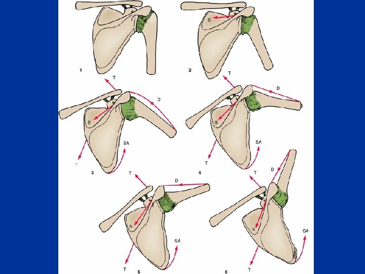
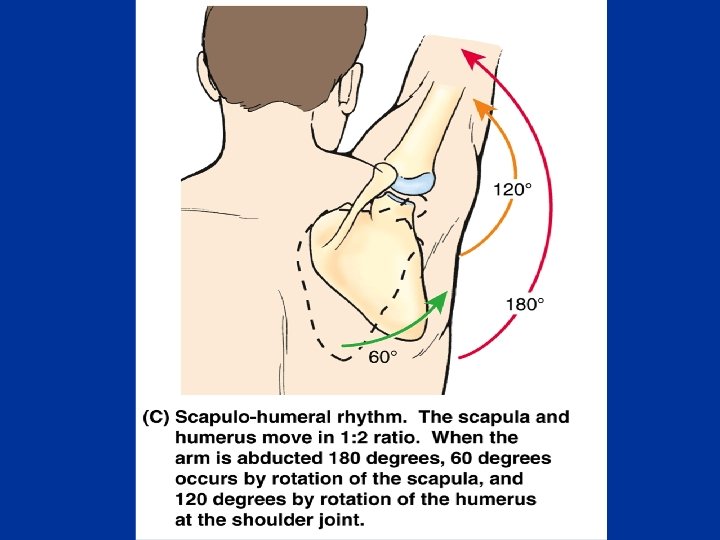
n n The supraspinatus initiates abduction and the deltoid takes over once the humerus is abducted past 15 degrees During full abduction of the arm we have to laterally rotate our humerus to move the greater tuberosity out of the way. n Even with this maneuver, space is too limited to allow for the range of motion that is seen in a normal individual. n The remainder of the range of motion is made possible by scapular rotation. n Once we have abducted the arm past 20 degrees or 30 degrees, for every 3 degrees of abduction at the glenohumeral joint, 1 degree occurs at the scapulothoracic surface and only 2 degrees occurs at the glenohumeral joint.
Spinal Part (ramus externus) Of the accessory nerve n n Runs downward in the posterior triangle of the neck on levator scapulae muscle Accompanied by branches from anterior rami of the 3 rd and 4 th cervical nerves Runs beneath the anterior border of the trapezius muscle It supplies the trapezius muscle
The suprascapular nerve n n The suprascapular nerve arises from the trunk formed by the union of the 5 th and 6 th cervical nerves. It innervates the suprasinatus and infraspinatus muscles. It enters the supraspintous fossa through the suprascapular notch, notch below, the superior transverse scapular lig. It then passes beneath the supraspinatous, and curves around the lateral border of the spine of the scapula to the infraspintous fossa
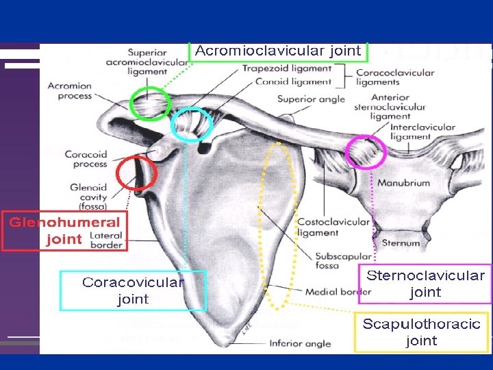
§ The pectoral girdle involves the: § SC joint § AC joint § Glenohumeral joint § § Generally, these joints move at the same time Functional defects in any of the joints impair movements of the pectoral girdle.
Sternoclavicular Joint n n n The only articulation between the upper limb and the axial skeleton A saddle type of synovial joint functions as a ball and socket joint The sternal end of the clavicle articulates with the manubrium of the sternum and the 1 st costal cartilage Can be palpated because the sternal end of the clavicle lies superior to the manubrium of the sternum The articular surfaces are covered with fibrocartilage
n Articular capsule: – The fibrous part of the articular capsule surrounds the SC joint, including: • the epiphysis at the sternal end of the clavicle • the periphery of the articular disc – attached to the margins of the articular surfaces n A synovial membrane: – lines the fibrous part of the articular capsule and both surfaces of the articular disc n Nerve supply: – The Supraclavicular nerve and nerve to Subclavius n Blood Supply: – The SC joint is supplied by internal thoracic and suprascapular arteries
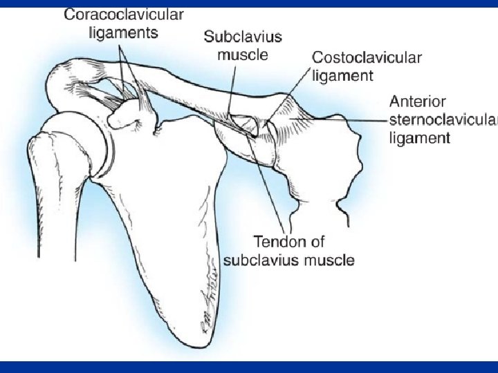
Ligaments of the SC Joint Anterior and posterior SC ligaments: n n The interclavicular ligament n – – Strengthens the capsule superiorly It extends from the sternal end of one clavicle and passes to the sternal end of the other clavicle. Attached to the superior border of the manubrium of the sternum. – n Reinforce the capsule anteriorly and posteriorly. The costoclavicular ligament: n Anchors the inferior surface of the sternal end of the clavicle to the 1 st rib and its costal cartilage, limiting elevation of the pectoral girdle
articular disc Articular disc n n Divides the SC joint into two compartments Attached to: – – – n Anterior and posterior SC ligaments The interclavicular ligament. The interior capsule Superiorly to clavicle Inferiorly to 1 st costal cartilage The articular disc serves as a shock absorber of forces transmitted along the clavicle from the upper limb
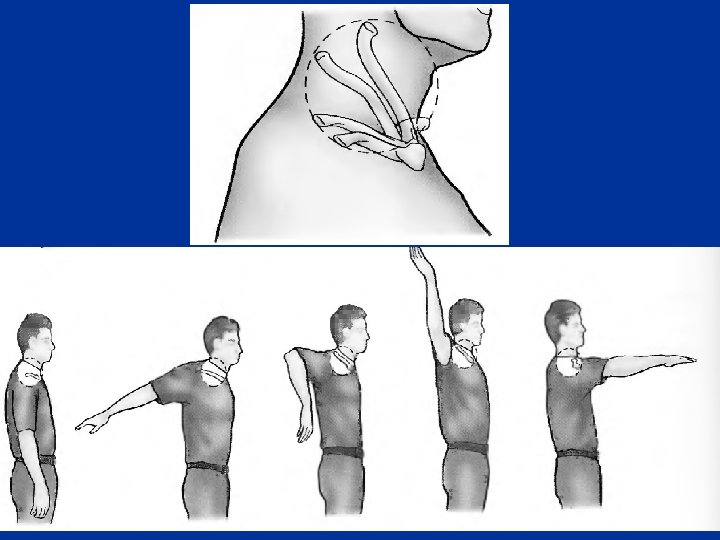
Movements of the SC Joint n n n Although the SC joint is extremely strong, it is significantly mobile to allow movements of the pectoral girdle and upper limb Mobility of the clavicle at the sternoclavicular joint is essential for the freedom of movement of the upper limb During full elevation of the limb, the clavicle is raised to approximately a 60 ° angle The SC joint moves up to 25 to 30 ° along its long axis in several direction: – anteriorly – Posteriorly – Inferiorly Forward and backward in the medial compartment Elevation and depression in the lateral compartment
Dislocation of the SC Joint n n The rarity of dislocation of the SC joint attests to its strength The force of a blow is usually transmitted along the long axis of the clavicle. The clavicle may break near the junction of its middle and lateral thirds, but it is uncommon for the SC joint to dislocate. Most dislocations of the SC joint in persons younger than 25 years result from fractures through the epiphyseal plate because the epiphysis at the sternal end of the clavicle does not dose until 23 to 25 year
n n Anterior dislocations of the SCJ may occur from an indirect mechanism such as a blow to the anterior shoulder. The force of blow causes rotation of the shoulder backwards and transmits the stress to the SCJ
n Posterior SCJ dislocation may occur secondary to direct trauma to the anteromedial aspect of the clavicle that drives it backward n Often serious associated injuries require treatment that take treatment precedence over the dislocation, for example: • • Tracheal rupture or erosion Pneumo-thorax Laceration of the superior vena cava Occlusion of the subclavian artery and/or vein
Acromioclavicular Joint n n n The AC joint is a plane type of synovial joint It is located 2 to 3 cm from the point of the shoulder formed by the lateral part of the acromion The acromial end of the clavicle articulates with the acromion The articular surfaces, covered with fibrocartilage, are separated by an incomplete wedge-shaped articular disc Blood Supply of the Acromioclavicular Joint – suprascapular and thoracoacromial arteries n Nerve Supply of the AC Joint – Supraclavicular, lateral pectoral, and axillary nerves supply the AC joint
Articular Capsule of the AC Joint The sleeve-like, relatively loose fibrous capsule is attached to the margins of the articular surfaces n A synovial membrane lines the fibrous capsule. n Relatively weak, strengthened superiorly by fibers of the trapezius n
Ligaments of the AC Joint n n The integrity of the joint is maintained by extrinsic ligaments, distant from the joint itself. The superior and inferior AC ligament: – a fibrous band extending from the acromion to the clavicle strengthens the AC joint superiorly
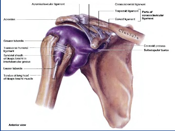
The coracoclavicular ligament: n – – a strong pair of bands that unites the coracoid process of the scapula to the clavicle, clavicle anchoring the clavicle to the coracoid process. The coracoclavicular ligament consists of two ligaments which are often separated by a bursa. : The conoid ligament 1. – – – The vertical conoid ligament is an inverted triangle (cone), The apex inferiorly is attached to the root of the coracoid process in front of the scapular notch. Its wide attachment is to the conoid tubercle on the inferior surface of the clavicle Trapezoid ligament 2. – – Horizontal ligament attached to the superior surface of the coracoid process extends laterally to the trapezoid line on the inferior surface of the clavicle.
The coracoclavicular ligament ü The strength of the AC joint depends on the strong coracoclavicular ligament ü provides the means by which the scapula and free limb are (passively) suspended from the clavicular strut üA greater part of the body weight is transmitted through it ü The rotatory movements of the scapula occur at this important ligament ü As long as the coracoclavicular ligament is intact, the acromion cannot be driven inferior to the clavicle. ü The ligament, however, does permit protraction and retraction of the acromion
Movements of the AC Joint n n The acromion rotates on the acromial end of the clavicle These movements are associated with motion at the scapulothoracic joint No muscles connect the articulating bones to move the AC joint The thoraco-appendicular muscles that attach to and move the scapula cause the acromion to move on the clavicle.
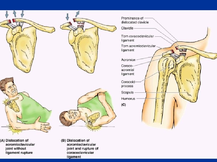
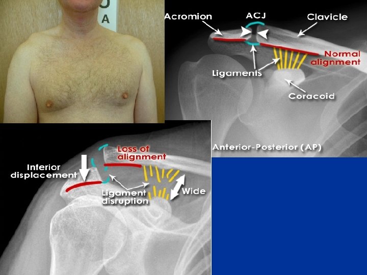
Dislocation of the AC Joint "shoulder separation" n n n The AC joint itself is weak and easily injured by a direct blow. In contact sports it is not uncommon for dislocation of the AC joint to result from a hard fall on the shoulder or from a fall on the outstretched upper limb. The AC injury, often called a "shoulder separation" When the coracoclavicular ligament tears, the shoulder separates from the clavicle because of the weight of the upper limb. Rupture of the coracoclavicular ligament allows the fibrous capsule of the joint to also be torn so that the acromion can pass inferior to the acromial end of the clavicle Dislocation of the AC joint makes the acromion more prominent, prominent and the clavicle may move superior to this process
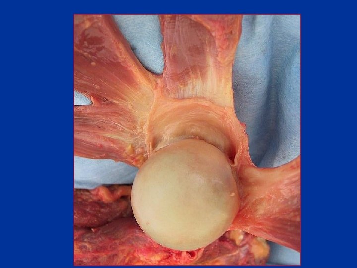
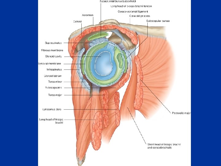
Glenohumeral (Shoulder) Joint n n n A ball-and-socket type of synovial joint Relatively unstable The large, round humeral head articulates with the relatively shallow glenoid cavity of the scapula The glenoid is deepened slightly but effectively by the ringlike glenoid labrum The glenoid cavity accepts little more than a third of the humeral head, head which is held in the cavity by the tonus of the rotator cuff muscles Both articular surfaces are covered with hyaline cartilage.
Glenoid labrum A fibrocartilaginous ring that attaches to the rim of the glenoid fossa n Encompasses the proximal portion of the head of the humerus. n If it is damaged then the joint becomes very unstable and is prone to dislocation n
Articular Capsule of the Glenohumeral Joint n The loose fibrous capsule surrounds the glenohumeral joint n Attached medially to the margin of the glenoid cavity and laterally to the anatomical neck of the humerus n Superiorly, the articular capsule encroaches on the root of the coracoid process so that the fibrous capsule encloses the proximal attachment of the long head of the biceps brachii (supraglenoid tubercle of scapula ) within the joint.
n The articular capsule of the glenohumeral joint has two apertures: apertures – The opening between the tubercles of the humerus is for passage of the tendon of the long head of the biceps brachii – The other opening situated anteriorly, inferior to the coracoid process, process allows communication between the subscapular bursa and the synovial cavity of the joint.
n The inferior part of the articular capsule is the only part not reinforced by the rotator cuff muscles Ø Its weakest area. n Here the capsule is particularly lax and lies in folds when the arm is adducted n It becomes taut when the arm is abducted. n The synovial membrane lines the fibrous capsule and reflects from it onto the glenoid labrum and neck of the humerus, humerus as far as the articular margin of the head n The synovial membrane also forms a tubular sheath for the tendon of the long head of the biceps brachii, brachii where it passes into the joint cavity and lies in the intertubercular groove, groove extending as far as the surgical neck of the humerus
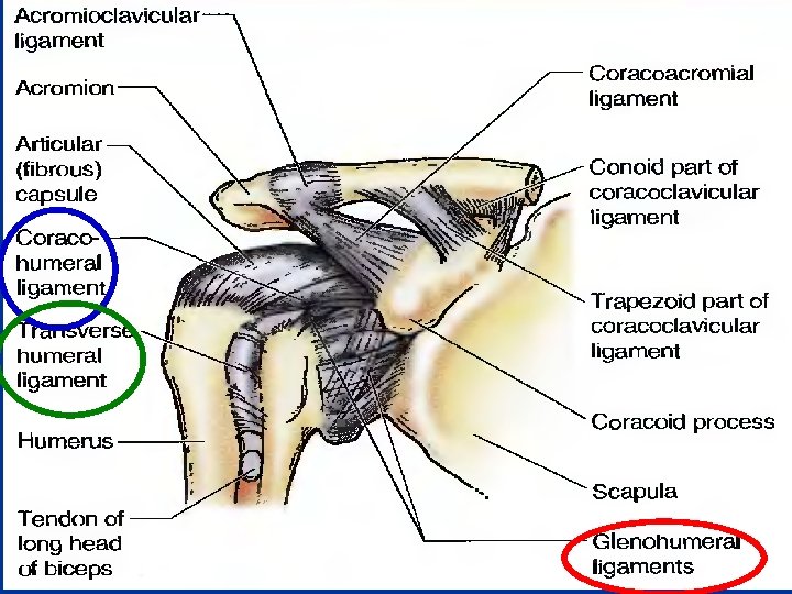
Ligaments of the Glenohumeral Joint n The glenohumeral ligaments: ligaments – – – strengthen the anterior aspect of the articular capsule three fibrous bands: evident only on the internal aspect of the capsule radiate laterally and inferiorly • from the glenoid labrum at the supraglenoid tubercle of the scapula • blend distally with the fibrous capsule as it attaches to the anatomical neck of the humerus.
Ligaments of the Glenohumeral Joint n The coracohumeral ligament – a strong, broad band strengthens the capsule superiorly – passes from the base of the coracoid process to the anterior aspect of the greater tubercle of the humerus
Ligaments of the Glenohumeral Joint n The transverse humeral ligament – a broad fibrous band that runs obliquely from the greater to the lesser tubercle of the humerus, bridging over the intertubercular groove. – The ligament converts the groove into a canal that holds the synovial sheath and tendon of the biceps brachii in place during movements of the glenohumeral joint
The Coracoacromial Arch n An extrinsic, protective osseoligamentous structure formed by: – The smooth inferior aspect of the acromion – The coracoid process of the scapula – The coracoacromial ligament n Overlies the head of the humerus, preventing its superior displacement n So strong that a forceful superior thrust of the humerus will not fracture it n The supraspinatus muscle passes under this arch n Movement of the supraspinatus tendon, is facilitated as it passes under the arch by the subacromial bursa, which lies between the arch and the tendon and tubercle
Bursae Around the Glenohumeral Joint n n Several bursae containing synovial fluid Bursae are located where – Tendons rub against bone, ligaments or other tendons – Where skin moves over a bony prominence. n Some of them communicate with the joint cavity (the subscapular bursa) Øopening a bursa may mean entering the cavity of the shoulder joint.
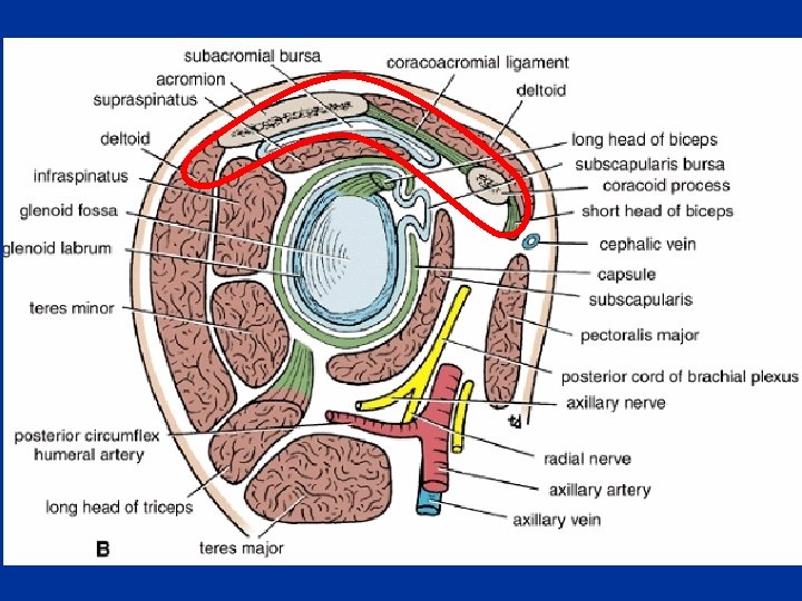
Subacromlal Bursa n Large bursa lies between the deltoid, the supraspinatus tendon, and the fibrous capsule of the glenohumeral joint. n It does not communicate with the glenohumeral joint cavity unless there has been a full-thickness rotator cuff tear n The subacromial bursa is located inferior to the acromion and coracoacromial ligament, between them and the supraspinatus n This bursa facilitates movement of the supraspinatus tendon under the coracoacromial arch and of the deltoid over the fibrous capsule of the shoulder joint and the greater tubercle of the humerus.
Subdeltoid Bursa n n This bursa lies between the deltoid muscle and the fibrous joint capsule over the head of the humerus. It does not communicate with the glenohumeral joint cavity, but may communicate with the subacromial bursa.
n n In patients who have injured their shoulder or who have supraspinatus tendinopathy, the bursa may become inflamed, making movements of the glenohumeral joint painful. These inflammatory changes may be treated by injection of a corticosteroid and local anesthetic agent
Subscapular Bursa n n Located between the neck of the scapula and the subscapularis muscle Often communicates with the glenohumeral joint cavity through an opening in the fibrous joint capsule
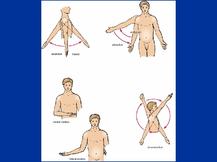
Movements of the Glenohumeral Joint n The glenohumeral joint has more freedom of movement than any other joint in the body. n This freedom results from: 1. 2. n The glenohumeral joint allows movements around three axes and permits: 1. 2. 3. 4. n the laxity of its articular capsule the large size of the humeral head compared with the small size of the glenoid cavity. Flexion-extension Abduction-adduction Rotation (medial and lateral) of the humerus Circumduction is an orderly sequence of flexion, abduction, extension, and adduction or the reverse.
n Blood Supply of the Glenohumeral Joint – The glenohumeral joint is supplied by the anterior and posterior circumflex humeral arteries and branches of the suprascapular artery. n Innervation of the Glenohumeral Joint – The suprascapular, axillary, and lateral pectoral nerves supply the glenohumeral joint.
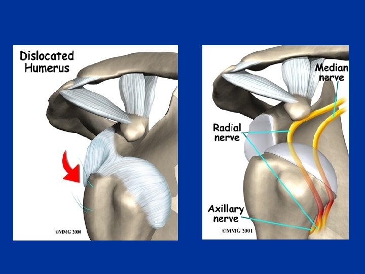
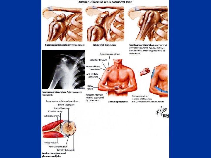
Dislocation of the Glenohumeral Joint n Because of its freedom of movement and instability, the glenohumeral joint is commonly dislocated by direct or indirect injury. n Most dislocations of the humeral head occur in the downward (inferior) direction 1. 2. the presence of the coracoacromial arch the support of the rotator cuff n Anterior or posterior dislocations n Posterior dislocations account for only 2 -3% of shoulder dislocations
n n n Anterior dislocation of the glenohumeral joint occurs most often in young adults, particularly athletes. It is usually caused by excessive extension and lateral rotation of the humerus. The head of the humerus is driven inferoanteriorly, and the fibrous capsule and glenoid labrum may be stripped from the anterior aspect of the glenoid cavity in the process.