The Gluteal Region Dr Fadel Naim Orthopedic Surgeon


 B. intergluteal cleft C. buttock D.")


.")


- Slides: 81
The Gluteal Region Dr. Fadel Naim Orthopedic Surgeon Faculty of Medicine IUG
l. The lower limbs specialized for locomotion l. The primary function of the lower limbs: l. Support the weight of the body l. Provide a stable foundation in: l. Standing l. Walking l. Running l. Similar in structure in many respects to the upper limbs l. Have less freedom of movement l. The upper limb is united to the trunk by only a small joint, (the sternoclavicular joint) l. The two hip bones articulate: Posteriorly with the trunk at the strong sacroiliac joints Anteriorly with each other at the symphysis pubis. § § Ø The lower limbs are more stable
Organization Of The Lower Limb l l The lower limbs are divided into different regions and compartments The regions: § § § l The gluteal region The thigh The knee The leg The ankle The foot The thigh and the leg are compartmentalized l Each compartment with own muscles § § Perform group functions Own distinct nerve and blood supply
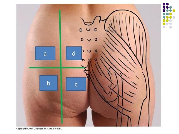
The Gluteal Region l The gluteal region bounded l l l superiorly by the iliac crest inferiorly by the fold of the buttock. The region is largely made up of the gluteal muscles and a thick layer of superficial fascia
A. Level of Iliac crest (L 4, ) B. intergluteal cleft C. buttock D. gluteal fold E. thigh F. Gluteal sulcus
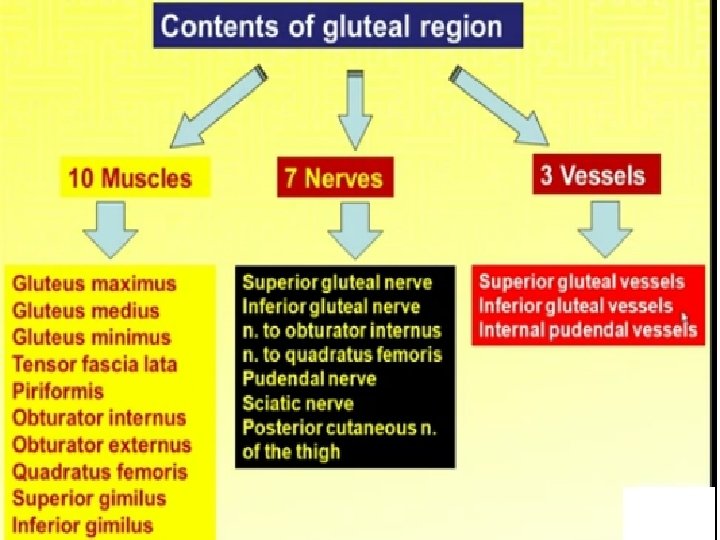
l The gluteal region contains: l Bones l Ligaments l Muscles l Vessels l Nerves
Cutaneous Nerve supply: l l l Upper lateral quadrant: Lateral branches of iliohypogastric (L 1) and T 12 Upper medial quadrant: Posterior rami of L 1, 2, 3 & S 1, 2, 3 Lower lateral quadrant: branches from lateral cutaneous nerve of thigh (L 2, 3) Lower medial quadrant: branches from posterior cutaneous nerve of thigh (S 1, 2, 3) Skin in the floor of the intergluteal cleft: branches from lower sacral and coccygeal nerves Dermatomes
Fascia Of The Buttock l l The superficial fascia is thick and impregnated with large quantities of fat. l It contributes to the prominence of the buttock. The deep fascia is continuous below with the deep fascia of the thigh (fascia lata). l l l It is attached to the iliac crest. In the gluteal region, it splits to enclose the gluteus maximus muscle It continues as a single layer that covers the outer surface of the gluteus medius
The Iliotibial Tract • On the lateral surface of the thigh, thickened to form a strong, wide band • From the tubercle of the iliac crest and below to the lateral condyle of the tibia • Forms a sheath for the tensor fasciae latae muscle • Receives the greater part of the insertion of the gluteus maximus
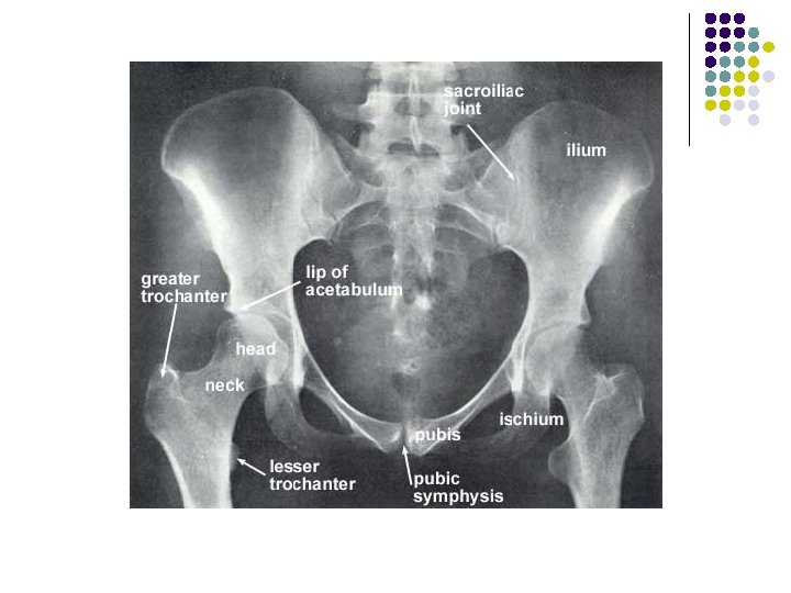
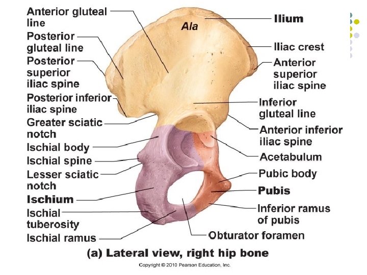
Bones Of The Gluteal Region Hip bone 1. 2. 3. l l Meet one another at the acetabulum Articulate with the sacrum at the sacroiliac joints l l The ilium The ischium The pubis Form the anterolateral walls of the pelvis Articulate with one another anteriorly at the symphysis pubis.
The Ilium l The upper flattened part of the hip bone l The iliac crest l l l l Can be felt through the skin along its entire length Anterior superior iliac spine Anterior inferior iliac spine Posterior superior iliac spine posterior inferior iliac spine. The iliac tubercle lies about 5 cm behind the anterior superior spine. Greater sciatic notch
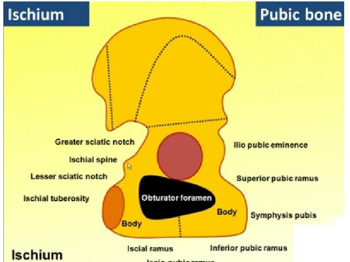
The ischium l l L shaped The body l l The ramus l l A lower thinner part, The ischial spine l l An upper thicker part Projects from the posterior border of the ischium and intervenes between the greater and lesser sciatic notches. l Converted into greater and lesser sciatic foramina by the presence of the sacrospinous and sacrotuberous ligaments The ischial tuberosity l Forms the posterior aspect of the lower part of the body of the bone.
The pubis l Divided into: l l l A body A superior ramus An inferior ramus l The bodies of the two pubic bones articulate with each other in the midline anteriorly at the symphysis pubis l The superior ramus joins the ilium and ischium at the acetabulum l The inferior ramus joins the ischial ramus below the obturator foramen. l The obturator foramen in life is filled in by the obturator membrane l The pubic crest forms the upper border of the body of the pubis, and it ends laterally as the pubic tubercle
The Acetabulum l l l On the outer surface of the hip bone is a deep depression, called the acetabulum Articulates with the head of the femur to form the hip joint The inferior margin of the acetabulum is deficient and is marked by the acetabular notch The articular surface of the acetabulum is limited to a horseshoe shaped area and is covered with hyaline cartilage. The floor of the acetabulum is non-articular and is called the acetabular fossa
Femur l l Articulates above with the acetabulum and below with the tibia and the patella The upper end of the femur has l l A head A neck Greater and lesser trochanters The head forms about two thirds of a sphere l Articulates with the acetabulum of the hip bone to form the hip joint
Femur Fovea capitis l l A small depression in the center of the head for the attachment of the ligament of head. Part of the blood supply to the head of the femur from the obturator artery is conveyed along this ligament and enters the bone at the fovea
The Neck of the Femur l l l Connects the head to the shaft Pass downward, backward, and laterally Makes an angle about 1250 with the long axis of the shaft. § l (Slightly less in the female) The size of this angle can be altered by disease
The Greater And Lesser Trochanters l l l Large eminence situated at the junction of the neck and the shaft Connecting the two trochanters are the intertrochanteric line anteriorly, where the iliofemoral ligament is attached Prominent intertrochanteric crest posteriorly, on which is the quadrate tubercle
Linea Aspera l l The shaft of the femur is smooth and rounded on its anterior surface but posteriorly has a ridge, the linea aspera Attachment of muscles and intermuscular septa. The margins of the linea aspera diverge above and below. On the posterior surface of the shaft below the greater trochanter is the gluteal tuberosity for the attachment of the gluteus maximus muscle
l l l The medial margin continues below as the medial supracondylar ridge to the adductor tubercle on the medial condyle The lateral margin becomes continuous below with the lateral supracondylar ridge. The shaft becomes broader toward its distal end and forms a flat, triangular area on its posterior surface called the popliteal surface
l l l The lower end of the femur has lateral and medial condyles, separated posteriorly by the intercondylar notch. The anterior surfaces of the condyles are joined by an articular surface for the patella. The two condyles take part in the formation of the knee joint. Above the condyles are the medial and lateral epicondyles The adductor tubercle is continuous with the medial epicondyle.
Arthritis Of The Hip Joint l The head of the femur can be palpated on the anterior aspect of the thigh just inferior to the inguinal ligament and just lateral to the pulsating femoral artery. l Tenderness over the head of the femur usually indicates the presence of arthritis of the hip joint.
Blood Supply To. The Femoral Head l l l l In the young, the epiphysis of the head is supplied by a small branch of the obturator artery, which passes to the head along the ligament of the femoral head. The upper part of the neck of the femur receives a profuse blood supply from the medial femoral circumflex artery. These branches pierce the capsule and ascend the neck deep to the synovial membrane. As long as the epiphyseal cartilage remains, no communication In adult anastomosis is present Fractures of the femoral neck interfere with or completely interrupt the blood supply from the root of the femoral neck to the femoral head. The blood flow along the small artery may be insufficient to sustain the viability of the femoral head ischemic necrosis gradually takes place.
Coxa Valga And Coxa Vara l l The neck of the femur is inclined at an angle with the shaft; the angle is about 1600 in the young child and about 1250 in the adult. An increase in this angle is referred to as coxa valga l l l A decrease in this angle is referred to as coxa vara, l l l ( congenital dislocation of the hip) In this condition, adduction of the hip joint is limited. ( fractures of the neck of the femur and in slipping of the femoral epiphysis) In this condition, abduction of the hip joint is limited. Shenton's line is a useful means of assessing the angle of the femoral neck on a radiograph of the hip region
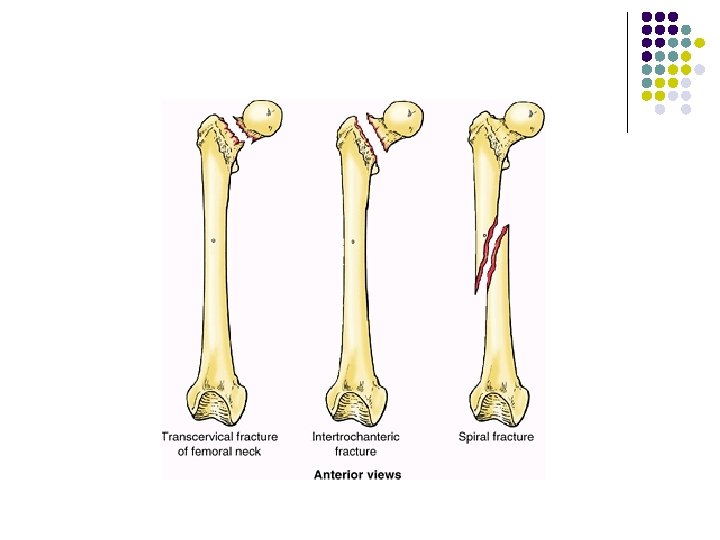
Subcapital Fracture l l Fractures of the neck of the femur are common The subcapital fracture l l l occurs in the elderly (common in women after menopause ) usually produced by a minor trip or stumble. Avascular necrosis of the head is a common complication. If the fragments are not impacted, considerable displacement occurs. The strong muscles of the thigh including the rectus femoris, the adductor muscles, and the hamstring muscles, pull the distal fragment upward, so that the leg is shortened The gluteus maximus, the piriformis, the obturator internus, the gemelli, and the quadratus femoris rotate the distal fragment laterally, as seen by the toes pointing laterally.
Trochanteric fractures l l In the young and middle aged as a result of direct trauma. Extracapsular Both fragments have a profuse blood supply. If not impacted, the pull of the strong muscles will produce shortening and lateral rotation of the leg
Fractures of the shaft of the femur l l usually occur in young and healthy persons. In fractures of the upper third of the shaft of the femur l the proximal fragment is: l l flexed by the iliopsoas abducted by the gluteus medius and minimus laterally rotated by the gluteus maximus, the piriformis, the obturator internus, the gemelli, and the quadratus femoris The lower fragment is: l l l adducted by the adductor muscles pulled upward by the hamstrings and quadriceps laterally rotated by the adductors and the weight of the foot
Fractures Of The Middle Third Of The Shaft Of The Femur, l The distal fragment is pulled upward by the hamstrings and the quadriceps resulting in considerable shortening. l The distal fragment is also rotated backward by the pull of the two heads of the gastrocnemius
Fractures Of The Distal Third Of The Shaft l The distal fragment is smaller and is rotated backward by the gastrocnemius muscle to a greater degree l May exert pressure on the popliteal artery and interfere with the blood flow through the leg and foot l Considerable traction on the distal fragment is usually required to overcome the powerful muscles and restore the limb to its correct length before manipulation and operative therapy to bring the proximal and distal fragments into correct alignment.
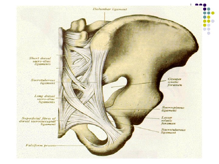
Ligaments Of The Gluteal Region l The two important ligaments in the gluteal region : l Sacrotuberous and sacrospinous ligaments. l The function of these ligaments is to stabilize the sacrum and prevent its rotation at the sacroiliac joint by the weight of the vertebral column.
Ligaments Of The Gluteal Region l Sacrotuberous ligament l l connects the back of the sacrum to the ischial tuberosity Sacrospinous ligament l connects the back of the sacrum to the spine of the ischium
l SI Ligaments: l Sacrotuberous Ligament: l Arises from ischial tuberosity to blend in with inferior fibers of posterior SI ligaments Sacrotuberous Ligament Ischial Tuberosity
l SI Ligaments: l Sacrospinous Ligament: l Originates from the ischial spine and attaches to the coccyx Sacrospinous Ligament
Foramina Of The Gluteal Region l The two important foramina in the gluteal region are l l The greater sciatic foramen The lesser sciatic foramen
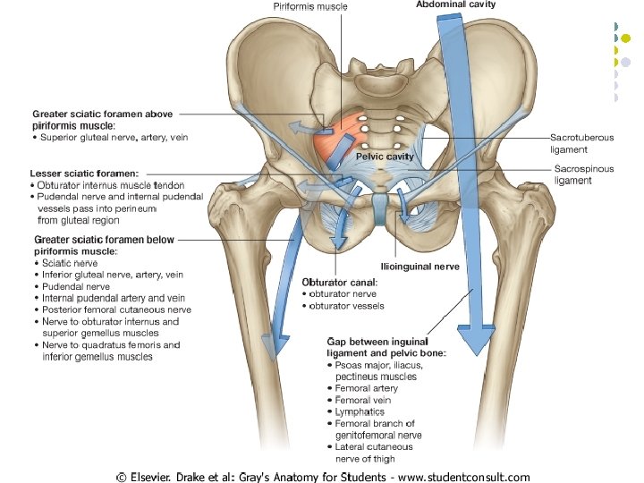
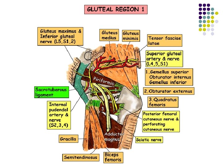
Greater Sciatic Foramen l l l Formed by the greater sciatic notch of the hip bone and the sacrotuberous and sacrospinous ligaments. It provides an exit from the pelvis into the gluteal region. The following structures exit the foramen 1. Piriformis 2. Sciatic nerve 3. Posterior cutaneous nerve of the thigh 4. Superior and inferior gluteal nerves 5. Nerves to the obturator internus and quadratus femoris 6. Pudendal nerve 7. Superior and inferior gluteal arteries and veins 8. Internal pudendal artery and vein
Structures passing through the greater sciatic foramen Above the piriformis: Superior gluteal vessels & nerve Piriformis: an important landmark Below the piriformis: Inferior gluteal vessels & nerve Sciatic nerve Posterior cutaneous nerve of thigh Pudendalnerve & Internal pudendal vessels Nerve to obturator internus Nerve to quadratus femoris
Lesser Sciatic Foramen l l Formed by the lesser sciatic notch of the hip bone and the sacrotuberous ligaments. Entrance into the perineum from the gluteal region. Enables nerves and blood vessels that have left the pelvis through the greater sciatic foramen above the pelvic floor to enter the perineum below the pelvic floor. The following structures pass through the foramen 1. Tendon of obturator internus muscle. 2. Nerve to obturator internus. 3. Pudendal nerve. 4. Internal pudendal artery and vein.
Structures passing through the lesser sciatic foramen Entering: Pudendal nerve & Internal pudendal vessels Exiting: Tendon of obturator internus Nerve to obturator internus
Gluteal Muscles l The gluteal muscles share a common compartment but are organized into two layers, superficial and deep: l The superficial layer consists of: 1. 2. The three large glutei (maximus, medius, and minimus) The tensor of the fascia lata. l l All have proximal attachments to the posterolateral (external) surface and margins of the ala of the ilium Mainly extensors, abductors, and rotators of the thigh.
Gluteal Muscles l The deep layer consists of: l Smaller muscles covered by the inferior half of the gluteus maximus 1. Piriformis 2. Obturator internus 3. Gemelli 4. Quadratus femoris § § All have distal attachments on or adjacent to the intertrochanteric crest of the femur. Lateral rotators of the thigh Stabilize the hip joint Working with the strong ligaments of the hip joint to steady the femoral head in the acetabulum.
Muscles of the Gluteal Region • • • Gluteus maximus Gluteus medius Gluteus minimus Tensor fascia lata Piriformis Superior Gemellus Inferior Gemellus Obturator internus Quadratus femoris
Gluteus Maximus l l l The largest muscle in the body. Superficial in the gluteal region largely responsible for the prominence of the buttock. l Origin: l l Insertion: l l the outer surface of the ilium the posterior surface of the sacrum and coccyx the sacrotuberous ligament The fibers pass downward and laterally Most are inserted into the iliotibial tract Some of the deeper fibers are inserted into the gluteal tuberosity of the femur. Nerve supply: l Inferior gluteal nerve.
Gluteus Maximus l • Action: l l l It extends and laterally rotates the hip joint Through the iliotibial tract it helps maintain the knee joint in extension. It is most commonly used as an extensor of the trunk on the thigh • The chief antigravity muscle of the hip. • It is used in standing up from a sitting position, running & climbing up stairs. • In each case extension of the hip moves the trunk upwards. • The muscle must be extremely powerful to raise the weight of the body against gravity. • This is called "forced extension".
Gluteus Maximus And Bursitis l Three bursae 1. Between the tendon of insertion and the greater trochanter, 2. Between the tendon of insertion and the vastus lateralis Overlying the ischial tuberosity. Bursitis, or inflammation of a bursa, can be caused by acute or chronic trauma. An inflamed bursa becomes distended with excessive amounts of fluid and can be extremely painful. The bursae associated with the gluteus maximus are prone to inflammation. 3. l l l
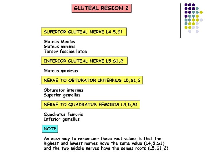
Gluteus Medius l l The gluteus medius is a thick, fan-shaped muscle Its posterior part is covered by the gluteus maximus l origin: l l Insertion: l l l The fibers pass downward and laterally Attached to the lateral surface of the greater trochanter. Nerve supply: l l From the outer surface of the ilium. Superior gluteal nerve. Action: l l Acting with the gluteus minimus Powerfully abducts the thigh at the hip joint. Most important action takes place in walking or running The anterior fibers also medially rotate thigh.
Gluteus Minimus l The gluteus minimus is fan shaped and lies deep to the gluteus medius. l Insertion: l l l Nerve supply: l l The fibers pass downward and laterally Attached to the lateral surface of the greater trochanter. Superior gluteal nerve. Action: l l l Acting with the gluteus medius Powerfully abducts the thigh at the hip joint. The anterior fibers also medially rotate thigh.
Gluteal Region Muscles
l Action of abductors of thigh when walking l l The three muscles contract and steady the pelvis on the lower limb While walking the pelvis is held in position and does not tilt downward on the unsupported
Injury to the Superior Gluteal Nerve l l The gluteus medius and minimus muscles may be paralyzed when poliomyelitis involves the lower lumbar and sacral segments of the spinal cord. They are supplied by the superior gluteal nerve (L 4 and 5 and SI). Paralysis of these muscles seriously interferes with the ability of the patient to tilt the pelvis when walking. This observation is referred to clinically as a positive Trendelenburg test
Tensor Fasciae Latae l Origin: l l Insertion: l l l The fibers run downward and backward inserted into the iliotibial tract. Nerve supply: l l From the outer edge of the iliac crest between the anterior superior iliac spine and the iliac tubercle Superior gluteal nerve. Action: l l l It exerts traction on the iliotibial tract assists the gluteus maximus muscle in maintaining the knee in the extended position. As long as the iliotibial tract remains in front of the axis of flexion of the knee, it assists in keeping the knee extended. l In standing upright, the upward pull of the iliotibial tract is the most important factor in keeping the knee extended l The quadriceps muscles may be relaxed.
Piriformis l l l The piriformis muscle lies partly within the pelvis at its origin. It emerges through the greater sciatic foramen to enter the gluteal region. Its position in the gluteal region serves to separate the superior gluteal vessels and nerves from the inferior gluteal vessels and nerves
Piriformis l Origin: l l Insertion: l l l The fibers pass downward and laterally through the greater sciatic foramen Attached to the upper border of the greater trochanter. Nerve supply: l l From the anterior surface of the 2 nd, 3 rd, and 4 th sacral vertebrae within the pelvis. Anterior rami of the first and 2 nd sacral nerves. Action: l Lateral rotator of the thigh at the hip joint.
Piriformis Syndrome l l l Caused by an entrapment of the sciatic nerve as it exits the Greater Sciatic notch in the gluteal region. Piriformis syndrome is also known as "wallet sciatica" or "fat wallet syndrome, " as the condition can be caused or aggravated by sitting with a large wallet in the rear pocket. This particular syndrome mimics sciatica, and that being the case, it is often misdiagnosed as sciatica.
Piriformis Syndrome l There are two normal variations for the exit of the sciatic nerve in this region. l The first places the sciatic nerve inferior to the Piriformis muscle and superior the gemellus muscle. l Entrapment in this area is likely due to a myospasm or contracture of either of these two muscles.
Piriformis Syndrome l The second common site of entrapment is when the sciatic nerve actually pierces the piriformis muscle itself. This can occur in about 1% to 10% of all humans. l In this case myospasm and or contraction of the piriformis muscle itself § can lead to: § pain along the back of the thigh to the knee § loss of sensation or numbness and tingling in the sole of the foot.
Triceps coxae 1. 2. 3. l l Obturator Internus Gemelli Superior Gemelli inferior These small muscles form a tricipital muscle (triceps coxae) located between the piriformis and quadratus femoris. The common tendon of the triceps runs horizontally to the greater trochanter of the femur.
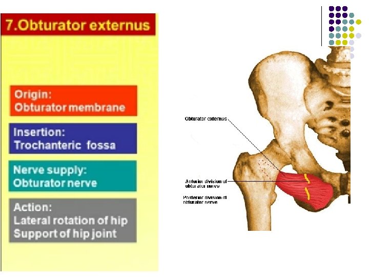
Obturator Internus l l l A fan-shaped muscle that lies partly within the pelvis at its origin. It emerges through the lesser sciatic foramen to enter the gluteal region. Origin: l l Insertion: l l l The tendon passes out of the pelvis through the lesser sciatic foramen and is joined by the superior and inferior gemelli. The common tendon is inserted into the upper border of the greater trochanter. Nerve supply: l l From the pelvic surface of the obturator membrane and the surrounding bones Nerve to the obturator internus from the sacral plexus. Action: l Lateral rotator of the thigh at the hip joint.
Gemellus Superior l l The gemellus superior is a small muscle. Origin: l l Insertion: l l With the tendon of the obturator internus Nerve supply: l l Spine of the ischium. Nerve to the obturator internus from the sacral plexus. Action: l Lateral rotator of the thigh at the hip joint.
Gemellus Inferior l l The gemellus inferior is a small muscle. Origin: l l Insertion: l l With the tendon of obturator internus Nerve supply: l l Upper margin of the ischial tuberosity. Nerve to the quadratus femoris from the sacral plexus. Action: l Lateral rotator of the thigh at the hip joint.
SIX LATERAL ROTATORS Piriformis. l Obturator internus. l Gemelli (superior and l inferior ). Obturator externus. l Quadratus femoris. l
Sciatic Nerve l l The nerve appears below the piriformis muscle and curves downward and laterally, Lying successively on: l The root of the ischial spine l The superior gemellus l The obturator internus l The inferior gemellus l The quadratus femoris Reach the back of the adductor magnus muscle It is related posteriorly to the posterior cutaneous nerve of the thigh and the gluteus maximus.
Muscle Origin Insertion Action Nerve supply Gluteus maximus Outer surface of ilium, sacrum, coccyx, sacrotuberous ligament Iliotibial tract, gluteal tuberosity of femur Extends and laterally rotates thigh at hip; through iliotibial tract it extends knee joint Inferior gluteal nerve Gluteus medius Outer surface of ilium Greater trochanter of femur Abducts thigh at Superior gluteal hip; tilts pelvis nerve when walking Gluteus minimus Outer surface of ilium Greater trochanter of femur Abducts thigh at Superior gluteal hip; anterior nerve fibers medially rotate thigh Tensor fasciae latae Iliac crest Iliotibial tract Assists gluteus maximus in extending the knee joint Superior gluteal nerve
Muscle Origin Insertion Action Nerve supply Piriformis Anterior surface of sacrum Greater trochanter of femur Lateral rotator of thigh Sacral nerve S 1 and S 2 Superior gemellus Spine of ischium Greater trochanter of femur Lateral rotator of thigh Sacral plexus Obturator internus Inner surface of obturator membrane Greater trochanter of femur Lateral rotator of thigh Sacral plexus Inferior gemellus Ischial tuberosity Greater trochanter of femur Lateral rotator of thigh Sacral plexus
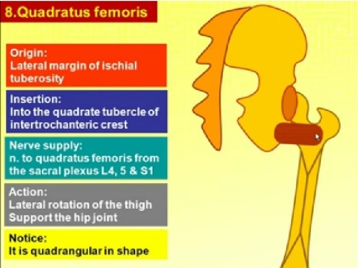
Muscle Origin Insertion Action Nerve supply Obturator externus Outer surface of obturator membrane Greater trochanger of femur Lateral rotator of thigh Obturator nerve Quadratus femoris Ischial tuberosity intertrochanteric crest of femur Lateral rotator of thigh Sacral plexus
ARTERIES OF THE GLUTEAL REGION Superior Gluteal Artery l l l A branch from the internal iliac artery Enters the gluteal region through the upper part of the greater sciatic foramen above the piriformis It divides into branches that are distributed throughout the gluteal region.
Inferior Gluteal Artery l l l A branch from the internal iliac artery Enters the gluteal region through the lower part of the greater sciatic foramen, below the piriformis It divides into numerous branches that are distributed throughout the gluteal region.
The Trochanteric Anastomosis l l l The trochanteric anastomosis provides the main blood supply to the head of the femur. The nutrient arteries pass along the femoral neck beneath the capsule The following arteries take part in the anastomosis: 1. 2. 3. 4. The superior gluteal artery The inferior gluteal artery The medial femoral circumflex artery The lateral femoral circumflex artery.
Arterial supply to Femoral head • Medial & lateral femoral circumflex arteries • Superior and inferior gluteal arteries • Post. obturator artery via artery of femoral ligament TROCHANTERIC ANASTOMOSIS Posterior view
The Cruciate Anastomosis l l l At the level of the lesser trochanter of the femur Together with the trochanteric anastomosis, provides a connection between the internal iliac and the femoral arteries. The following arteries take part in the anastomosis: 1. 2. 3. 4. The inferior gluteal artery The medial femoral circumflex artery The lateral femoral circumflex artery The first perforating artery, a branch of the profunda artery.