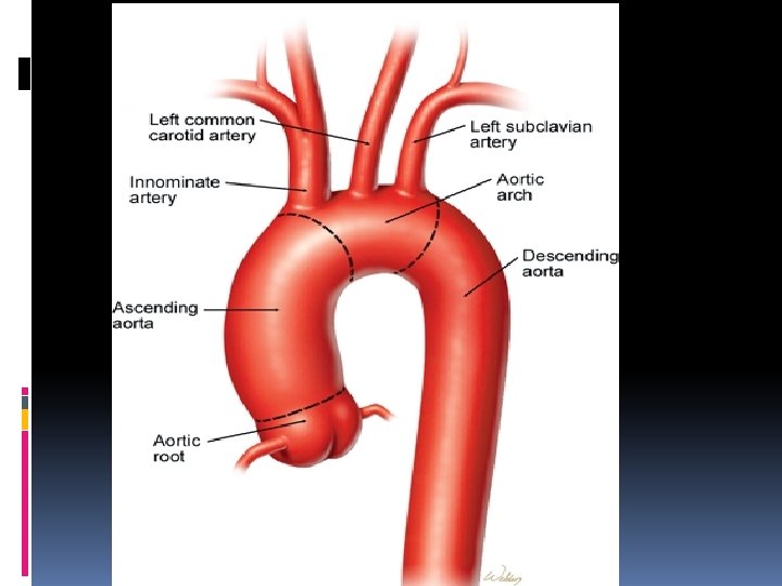
Anatomy Of Aorta Aortic Valve Echocardiographic point of


 to maintain the leaflet distended")


 Age Dilatation Aortic wrapping")


- Slides: 51
Anatomy Of Aorta & Aortic Valve “Echocardiographic point of view” Rasoul Azarfarin MD, FACC Professor of Anesthesiology Fellowship of Cardiac Anesthesia Rajaie Cardiovascular Medical & Research Center
“nessuno effetto in natura e sanza ragione; intendi la ragione e non ti bisogna sperienza““ “nothing in nature is without reason; understand the reason and you don’t need experience”
“perche il buso della arteria aorto e trianghulare” “why the orifice of the aortic artery is triangular” Royal Library, Windsor 19117 v. Leonardo’s sketch of a tricuspid valve inserted in a circle in its open and closed configuration. (The Royal Collection © 2004, Her Majesty Queen Elisabeth II)
Anatomical design Two fixed rings: The skeleton of the root
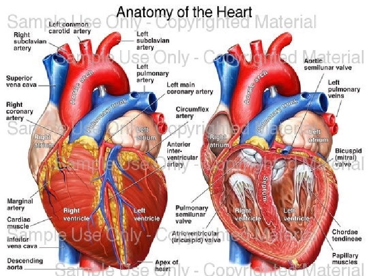
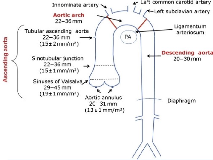
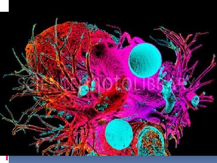
Aortic root The aortic root is a dynamic unit that allows easy opening and closing and shares stress for the valve leaflets ST junction Sinuses of Valsalva Annulus Leaflets
Echocardiographic view ST junction The sinotubular junction is larger than the annulus with a diameter ratio of 1. 3 in a normal adult human heart Valsalva sinuses Aortic annulus
Echocardiographic anatomy valve size average leaflet length average sinus height average coaptation height 21 (n=4) 21. 3 mm (20. 6 -21. 8) 21. 4 mm (20. 9 -22. 0) 11. 2 mm (10. 8 -12. 6) 22 (n=2) 22. 2 mm (21. 8 -22. 5) 22. 3 mm (22. 1 -22. 6) 11. 7 mm (11. 4 -12. 0) 23 (n=6) 23. 4 mm (22. 6 -24. 0) 23. 2 mm (22. 7 -23. 8) 12. 3 mm (11. 6 -12. 8) 25 (n=2) 25. 3 mm (25. 2 -25. 5) 25. 1 mm (25. 0 -25. 3) 12. 9 mm (12. 7 -13. 1) V. Vijay et al. EACTS/ESTS Joint Meeting 2000
Geometry From structure to function…
“per la qual cosa langolo piu ottuso e piu forte chellangolo retto del to” “the more obtuse angle is stronger than the right angle of the square”
“i pannicul delli 4 usscoli son piu deboli cheli 3 usscioli perchecolli loro angholi son piu remoti dalla basa del triangolo loro che quel de 3 ussciolj” “the membranes of four valve-cusps are weaker than those of three valve-cusps because their central angles are more remote from the bases of their triangles than those of the three valve-cusps ”
Folding and unfolding of the free edge of the leaflets is necessary for opening and closure of the aortic valve tricuspid valve bicuspid valve degree of folding Robicsek F et al. Ann Thorac Surg 2004; 77: 177 -185
Functional geometry The progressive increase in aortic diameter maintain the leaflets flat through the whole sequence of leaflet opening
Composition of aortic root Annulus collagen only at the nadir of leaflets attachment Sinuses elastic tissue ST junction elastic component but important collagen support Interleaflet triangles divide the sinuses and are exposed to ventricular pressure
“lla sagace natura provide dj durissima resisstentia nella infima baseza delcerchio dellinpeto” “the wisdom of nature provides a very hard resistance at the lowest base of the circle of impetus”
“il sangue che col suo inpeto percote esso vsscjolo non potedo sfondarlo seguita ilsuo moto allo insu e allargha edjstende le grinze” the blood which with his impetus percusses this valve--cusp, not being able to rupture it, continues its upward motion, enlarges and distends the “ folds of the cusp”
Root deformation during the cardiac cycle: Isovolemic contraction - Expansion at the commissures “Pull--and--release” mechanism Aorta Root LV
Root deformation during the cardiac cycle: Ejection - Contraction at the annulus - Expansion at the commissures Aorta Root is morecylindrical to favor ejection Root LV
Root deformation during the cardiac cycle: Diastole - Re-expansion at the annulus - Contraction at the commissures Aorta Recoil to restore the static equilibrium Root LV
Mechanism of opening: sequence of leaflet opening Stellate orifice Small triangle Triangle Circular orifice
The paradoxes of the aortic valve The valve opens before the presence of forward flow Ejection continues even after the aortic pressure exceedes ventricular pressure The aortic valve already starts closing during ejection
Answer: From stellate orifice to small triangle Increase in ventricular pressure through the interleaflet triangle causes an increase of diameter at the commissures before the valve opens
From small triangle to triangle Sinuses expansion (increased 16%) to maintain the leaflet distended and flat
The paradoxes of the aortic valve The valve opens before the presence of forward flow Ejection continues even after the aortic pressure exceedes ventricular pressure The aortic valve already starts closing during ejection
Answer: From triangle to circular orifice Due to an increase in velocity the blood enter the aorta because of motion’s inertia more than pressuregradient” “Noble phenomenon”
Mechanism of leaflets closure: the role of the sinuses
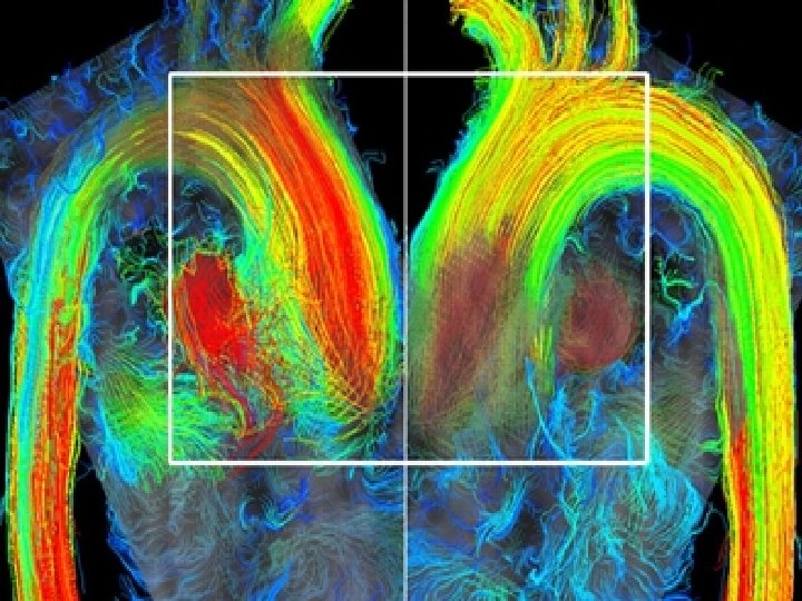
Old technique “by hand” Royal Library, Windsor 19117 v. New technique “phase contrast MRI”
“apresi per il moto incidete e sserrasi col motorefresso” “it is opened by the incident motion and closed by the reflected motion” “…jnpari tempo qual sara la proporzione delle varie largezze dessa canna” “will be proportional to the different sizes of the pipe” “…e poi si rivolta insu conmoto refresso e ritorna alla porta del suo primo introito” “…and then curls up and goes back to the entrance door”
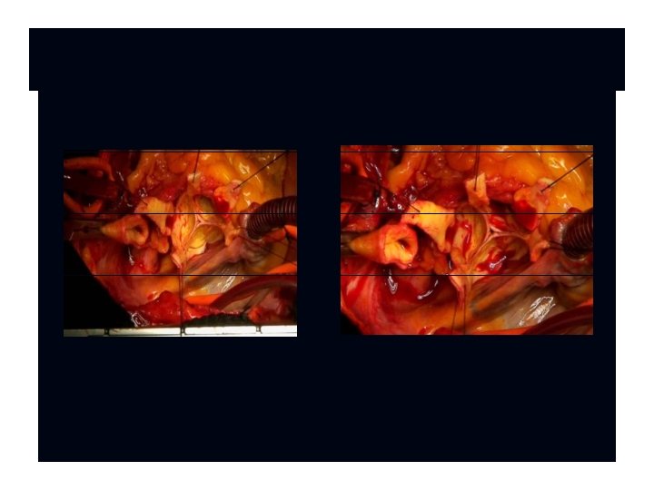
In the absence of Valsalva sinuses
In the presence of Valsalva sinuses
The paradoxes of the aortic valve The valve opens before the presence of forward flow Ejection continues even after the aortic pressure exceedes ventricular pressure The aortic valve already starts closing during ejection
Answer: a-b = rapid valve opening; b-c = slow systolic closure; c-d = rapid valve closing; RVOT = rapid valve opening time; RVCT = rapid valve closing time; ET = ejection time; D 1 = maximal leaflet displacement; SCD = slow closing displacement; D 2 = leaflet displacement before rapid valve closing Schematic drawing of an M-mode tracing describing the measured aortic valve opening and closing features
Concept of the functional unit sinus-cusp Shock absorbing Stress sharing
Stress and aortic valve At 100 mm. Hg the aortic valve withstand a pressure of: 1, 3 Kg vertically 0, 6 Kg horizzontally (200 g for each commissure) Elasticity is an important variable specially at sinus level
Stress contours in the aortic root when the flexible sinuses are present versus when the sinuses are absent Robicsek F. et al. Ann Thorac Surg 2002; 73: 1346 -1354
Five hundred frames/sec cinematography showing the leaflet surface during valve opening Robicsek F. et al. Ann Thorac Surg 2002; 73: 1346 -1354
Stiffening of the aortic wall at the level of the sinuses leads to loss: the physiologic pull--and--release function of the aortic root stress overload on the aortic leaflets (eventually cusp fibrosis and calcification, and in some cases hemodynamically significant aortic valve disease)
Cause of impairment of physiologic function Degenerative (increased aortic stiffness) Age Dilatation Aortic wrapping (? ) Surgery (root replacement) Absence of sinuses Non compliant sinuses
For a long-lasting aortic leaflet function Maintain or reconstruct the sinuses Natural shape of the sinuses Normal proportion of the components Large leaflet coaptation Elasticity of the wall