The Thorax 1 Axial vs Appendicular Skeleton Axial


 Manubrium Jugular (sternal) notch Articulation with rib NO")
 5 False ribs-")


 1 st rib T 1 Vertebrae")


 The lower border")


- Slides: 33
The Thorax 1
Axial vs. Appendicular Skeleton Axial Skeleton Skull = Cranium + Facial bones Vertebrae Ribs Sternum Appendicular Skeleton Bones of upper/lower limbs Limb Girdles
Thoracic Cage Is osteocartilaginous cage Formation: Sternum and costal cartilages Anteriorly 12 Thoracic vertebrae and 11 IV discs posteriorly Ribs laterally Forms protective cage around heart, lungs, and other organs Designed primarily to change in the intrathoracic pressure during respiration
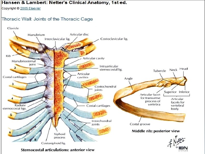
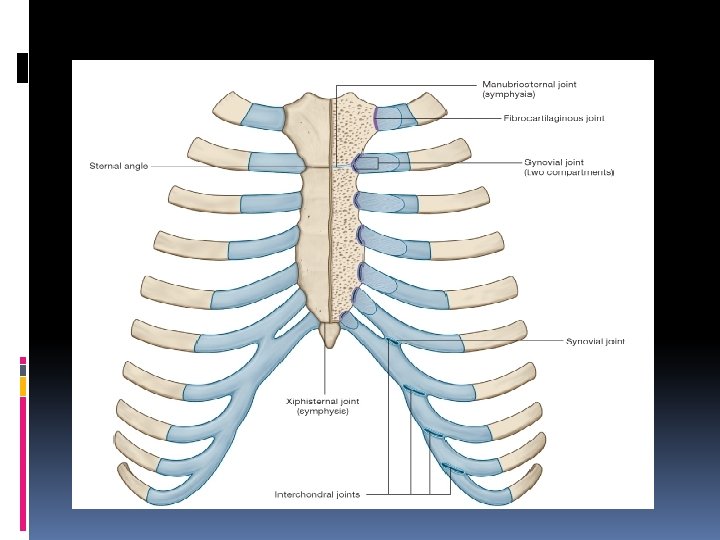
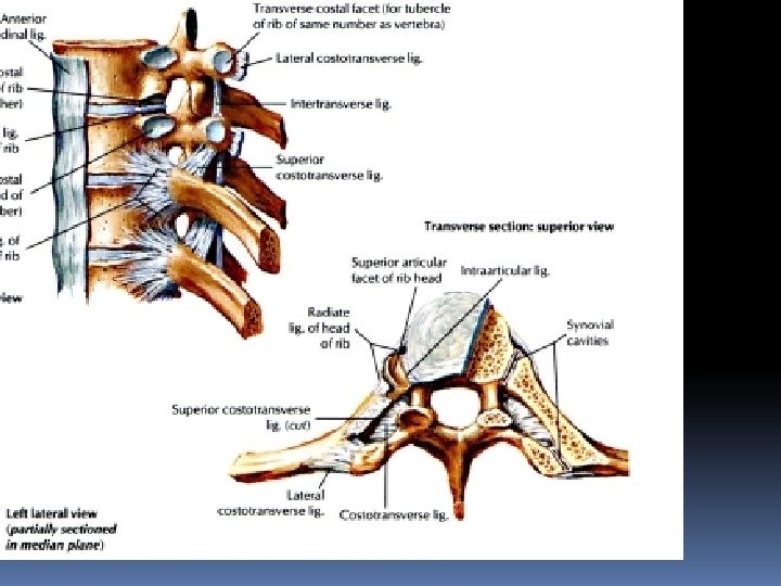
Synovial joint 1 ry cartilagenous 2 nd cartliagenous joint Synovial Fibrous
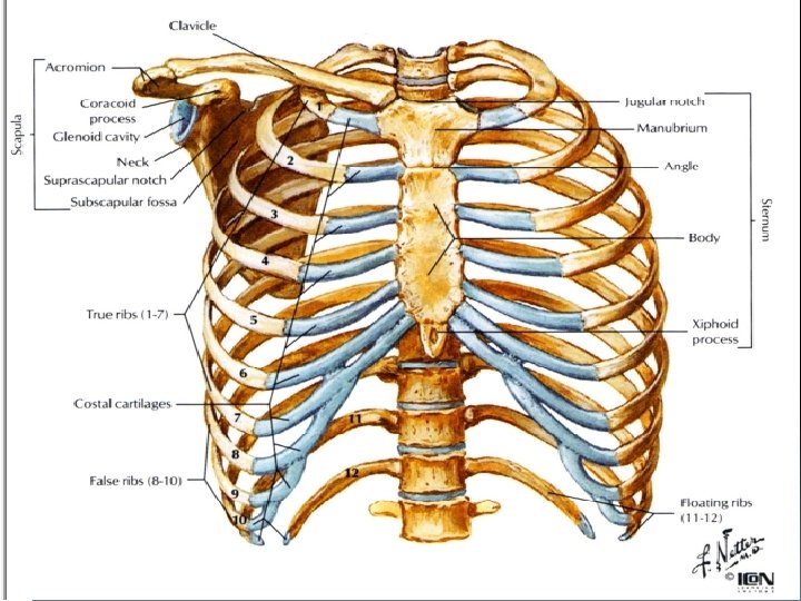
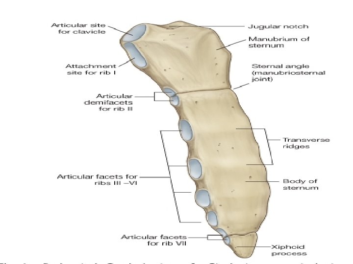
The Sternum (Composed of fused sternebrae) Manubrium Jugular (sternal) notch Articulation with rib NO 1 & 2 (upper part) Clavicular Articular facets Sternal Angle – 2 nd rib Body Articulates with ribs 2 -7 Xiphosternal joint Xiphoid process Cartilage-calcifies through time Partial attachment of many muscles
Ribs Usually, 12 pairs 7 True ribs-direct attachment to sternum (Vertebrosternal) 5 False ribs- indirect or no attachment to sternum (Vertebrochondral) Floating ribs-make up the last 2 of the 5 False ribs, no ventral attachment (Vertebral) The costal cartilage is formed by the 7 th, 8 th, 9 th and 10 th rib. Longest rib is 7 th Typical Ribs No 2 -9 (some books consider 2 Atypical) Atypical Ribs No 1, 10, 11, 12
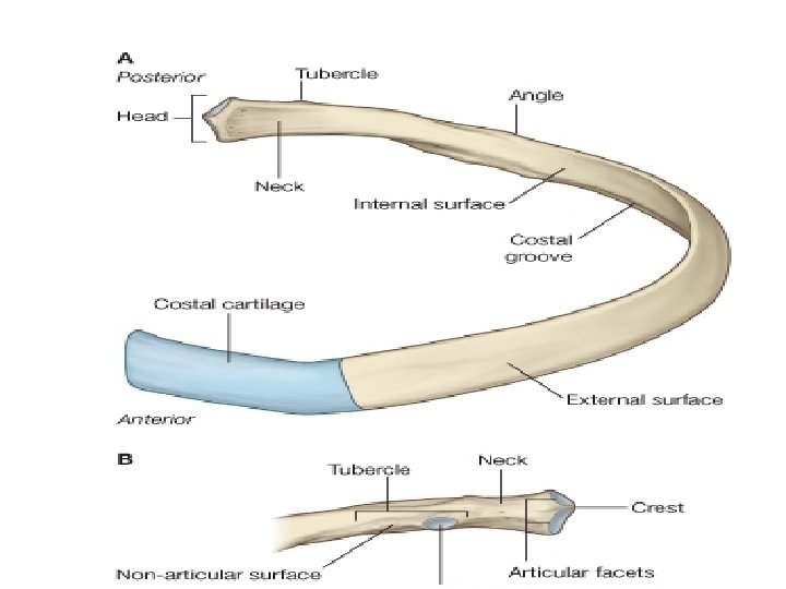
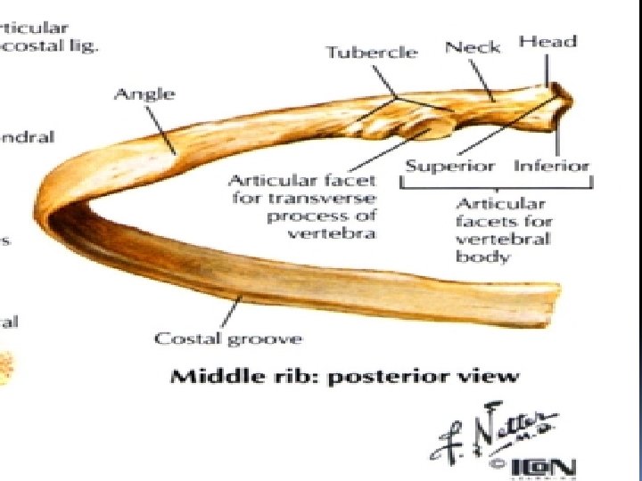
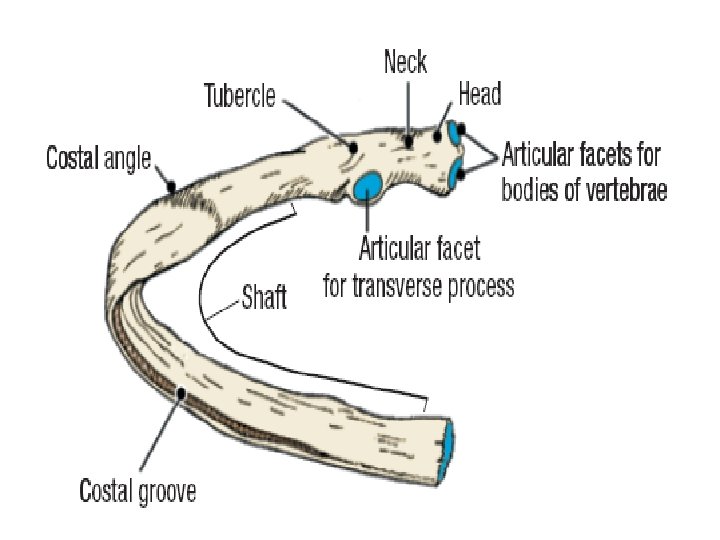
What should be found in typical rib? Typical Ribs 1. Head with 2 articular facets articulates with corresponding vertebra and vertebra above 2. Neck 3. Tubercle formed of medial smooth part articulate with the transverse process of the corresponding vertebra and lateral rough part 4. Angle 5. Shaft has 2 surfaces And 2 borders 1. Subcostal Groove
What are the Atypical ribs and their numbers? Ø No 1 - short, flat (superio-inferior), wide, Supports Subclavian vessels Ø No 1, 10 -12 articulate with only with the corrosponding vertebra Ø 11, 12 don’t articulate with transverse processes, or anteriorly at all Ø NB 2 nd has no twist, small non articular tubercle, tubercle in the outer surface
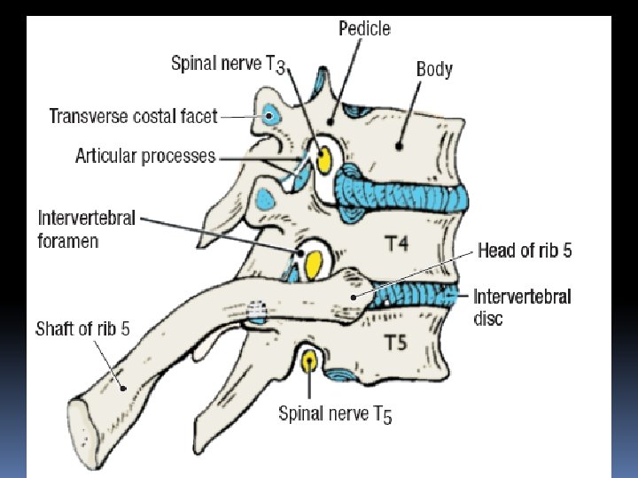
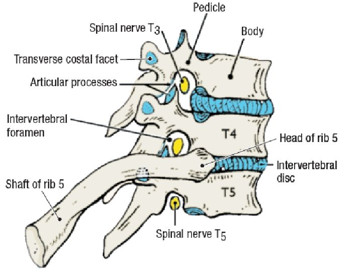
Typical rib articulation Posterior Attachment to Thoracic Vertebrae �Head of Rib 2 costal facets �Superior costal facet �Inferior costal facet of vertebra above it �Intervertebral disc �Tubercle of Rib Transverse Costal Facet �e. g. Rib No 5 articulates with Superior Costal Facet and Transverse Costal Facet of T 5 & Inferior Costal Facet of T 4 Anterior Attachment to Sternum Via costal cartilage
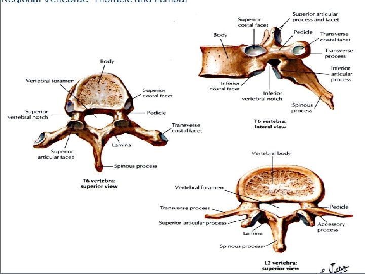
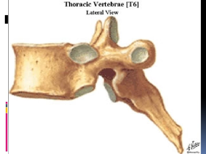
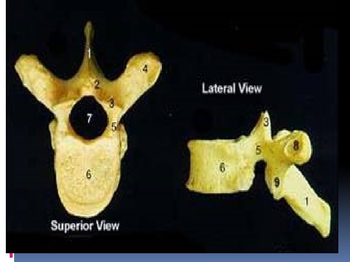
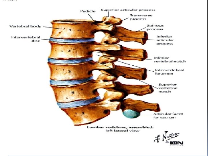
Thoacic vertebrae Transverse Costal Facets S/I Costal facets on vertebral body Spinous Processes long, point inferiorly Superior Articular Facets face Posteriorly Inferior Articular Facets face Anteriorly Vertebral Foramen is Circular Body is Heart-shaped Atypical 1, 9, 10, 11, and 12
Thoracic Cage Inlet composed of: Manubrium (upper border) 1 st rib T 1 Vertebrae Directed downward 30 -45° Seperated from neck partially via suprapleural membrane Outlet composed of: xiphisternum costal margin of 1 oth rib+ 10, 11 and 12 ribs T 12
Thoracic Cage Shape: truncated cone Transverse section kidney shape T>AP below 2 yr circular In infants ribs horizontal that’s why abdominal breathing Adult ribs oblique thoracic+abdominal
Landmarks Jugular notch corresponds with The 2 th thoracic vertebra in male, the 3 th thoracic vertebra in female Xiphisternal junction upper border of T 9 Scapula overlies ribs 2 -7 10 th rib is the lowest at thoracic outlet at level of L 3 T 3 spine same level with root of scapula spine
Sternal angle connects 2 nd costal cartilage laterally, corresponds with a) The lower border of 4 th thoracic vertebra (T 4/T 5) b) Mark the plane separates superior and inferior mediastinum c) The bifurcation of trachea in the adult d) The beginning and end of aortic arch e) The esophagus is crossed by the left main bronchus f) Thoracic duct reach the left side of T 4 g) The cardiac plexus situated at same level h) Pulmonary trunk divides just below the level i) Marks upper limit of heart base
Diaphragm Shape and position: dome-shaped between thorax and abdomen, consists of a peripheral muscular part and a central tendon Origin Sternal part: xiphoid process by 2 slips Costal part: lower six and costal cartilages Lumbar part: arises by two crura from upper 2 -3 lumbar vertebrae Arcuate part: from median , medial , and lateral
Insertion: central tendon formed of 3 leafs RT, LT and central Weak areas: triangular spaces without muscular tissue 1. Lumbocostal triangle: between costal and lumbar parts. 2. Sternocostal triangle: between costal and sternal parts.
Openings in the diaphragm Aortic hiatus-lies anterior to the body of the 12 th thoracic vertebra between the crura. It transmits the aorta, thoracic duct Esophageal hiatus -for esophagus and vagus nerves. Lt gatric oesophageal branches at level of T 10. Vena cava foramen -for inferior vena cava and RT phrenic nerve , through central tendon at T 8 level
Nerve and blood supply Phernic N motor & sensory to the central part. Pericardiophernic & Phernic arteries supply the central part. Peripheral parts supplied by lower 5 intercostal neurovascular bundles.
Action: Contraction: the dome moving downward, increases the volume of thoracic cavity which results in inspiration, at the same time the intraabdominal pressure is increased assists in defecation, vomiting or child birth. Relaxation: the dome returns to the former position, reduces the volume to the thoracic cavity, resulting in expiration.