LOCOMOTION AND TRANSPORT CONCEPT MAP OF CHAPTER 2


")


- Slides: 61
LOCOMOTION AND TRANSPORT
CONCEPT MAP OF CHAPTER 2 LOCOMOTION & SUPPORT IN HUMANS & ANIMALS TYPES OF SKELETON Muscles, ligaments & tendon SUPPORT IN PLANTS LOCOMOTION AQUATIC PLANTS TERRESTRIAL PLANTS Hydrostatic skeleton Birds Exoskeleton Fish Grasshopper Endoskeleton Axial skeleton Appendicular skeleton Earthworms
LEARNING OUTCOMES To explain the necessity for support and locomotion in humans and animals, To describe problems that could be faced by humans and animals in support and locomotion, To explain how problems in support and locomotion are overcome in humans and animals, To name the bones that make up the axial skeleton and appendicular skeleton of the human body
The Necessity for Support & Locomotion in Humans & Animals WHY DO HUMANS & ANIMALS NEED SUPPORT? 1. To find the food 2. To find partner for mating 3. To protect/escape from their predator 4. To shelter from bad environment
The Necessity for Support & Locomotion in Humans & Animals Without support, animals & humans would not be able to maintain their body shape their body collapse under the weight of their own tissues. Support are provided by some form of skeleton. Hydrostatic skeleton Exoskeleton endoskeleton
Support are provided by some form of skeleton: 1. Hydrostatic skeleton 2. Exoskeleton 3. Endoskeleton
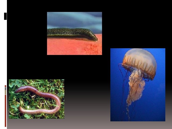
HYDROSTATIC SKELETON A fluid-filled internal body cavity in which the fluid is held under pressure. This also can be usedd to maintan the body shape & provide support for internal organ The cavity is surrounded by muscles arranged in layers. The body shape of the animal changes as these muscles contract & relax.
The animals are soft & flexible, the hydrostatic fluid does protect body parts by acting as a shock absorber. Examples : earthworm, jellyfish, leech & caterpillar.
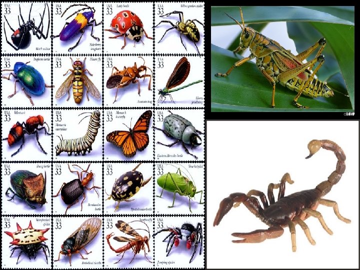
EXOSKELETON A rigid outer covering usually made up of protein, chitins &/@ calcium salt. Insects – the cuticle (covered with wax to prevent water loss from the body), cover the body’s surface. The exoskeleton is jointed / hinged = certain points of the skeleton are flexible & can bend enabling the movement.
Exoskeleton restrict the growth of animals the exoskeleton must be shed from time to time in order for the animal to grow. (ecdysis) Also found in the shells of molluscs & the bony plates of tortoises. Examples : insects, crabs, lobsters, tortoise
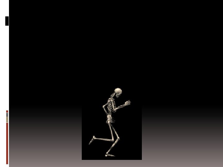
ENDOSKELETON Found in the bodies of all vertebrates including fish, amphibians & birds. Consist of hard skeleton of bones & cartilage found inside the body – made up of calcium & phosphate. Works with the muscular system to perform movement & locomotion. It support the body & protects the organs.
The Necessity for Support & Locomotion in Humans & Animals The functions of skeleton : Provide shape & support Enables movement (locomotion) Protects internal organs Stores calcium & phosphate ions Produces blood cells A firm base for the attachment of muscles
The Necessity for Support & Locomotion in Humans & Animals Problems that could be faced by humans and animals in support and locomotion, gravitational force, friction & resistance when moving around Aspect need to be considered when describing the locomotion of an animal : Stability – when it moves, it is temporarily unstable, but its stability will be restored when it stops. Support – must have enough support from its body’s skeleton Propulsion – must be propelled in order to move
HOW TO OVERCOME THE PROBLEMS? RESISTANCE & FRICTION – by streamlining their bodies. GRAVITATIONAL FORCE – most animals have their own supporting structures (fins – fishes, wings – birds & strong limbs – tetrapods & humans) provide the propulsive force to overcome the problem
The skeletal system together with its muscles are designed specially to overcome the problems associated with support & locomotion of humans & animals. To initiate locomotion, the force required is generated by contraction of muscles, whereas the movement is transmitted by the skeleton.
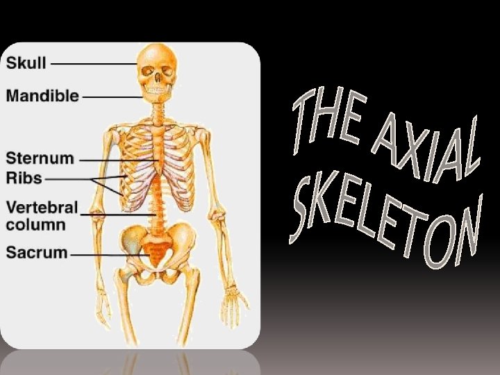
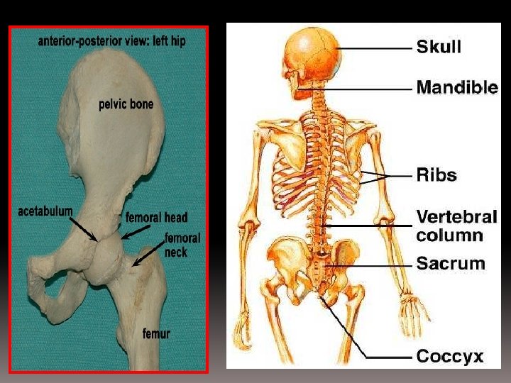
THE HUMAN SKELETAL SYSTEM The adult human skeleton consist of 206 bones. Divided into two main parts : the axial skeleton & the appendicular skeleton. The axial skeleton : made up of the bones that form the vertical axis of the body. It supports & protects the organs of the head, neck & trunk. (skull, vertebral column, rib cage)
The appendicular skeleton : made up of the bones that are attached to the axial skeleton. Include bones of the limbs, the pectoral girdle & the pelvic girdle.
FUNCTION OF THE SKELETON PROTECTION – the skull protects the brain, the vertebral column protects the spinal cord & the rib cage protects internal organs such as the heart. SUPPORT – act as a framework to support the soft body parts, to maintain the upright position & to keep the body stable.
FUNCTION OF THE SKELETON MOVEMENT – bones interact with the skeletal muscle. BLOOD CELL FORMATION – most of the blood cells are formed in the bone marrow of the long bones. MINERAL STORAGE – bones act as a reservoir for calcium & phosphorus.
SKELETAL SYSTEM HUMAN SKELETON AXIAL SKELETON APPENDICULAR SKELETON SKULL VERTEBRAL COLUMN RIB CAGE CERVICAL THORACIC LUMBAR SACRUM COCCYX PECTORAL GIRDLE PELVIC GIRDLE FORELIMB BONES HINDLIMB BONES
THE SKULL Made up of 8 cranial bones & 14 facial bones including the upper jaw & the lower jaw. The facial bones also provide support & protect the entrances to the respiratory system.
SKULL PARTS 8 Cranial bones FUNCTIONS n Are fused to form immovable joints called sutures. n Protect the brain & the sensory organs. Eye sockets n to protect the eye ball. Nasal bones n to support nose tissues Ear holes n to protect inner part of ears Maxilla (upper jaw) n to support upper teeth Mandible (lower jaw) n to support lower teeth, to enable eating & talking.
VERTEBRAL COLUMN Known as the spine/ backbone. Extends from the base of the skull to the pelvic girdle. Made up of 33 vertebrae separated from each other by discs of cartilage (intervertebral discs) which absorb shocks & serve as flex point. This S-shaped column supports & balances the body in a vertical plane & protects the spinal cord, supports the skull & provides a base for the attachment of muscles to the back.
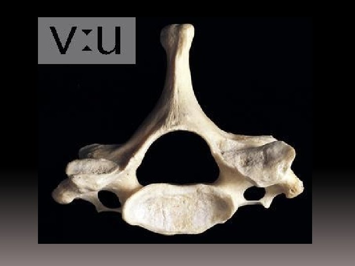
VERTEBRAL COLUMN The vertebrae differ in structure & function in different regions of the vertebral column. A vertebra typically consists of a main body (the centrum), a neural arch & transverse processes.
VERTEBRAL COLUMN
VERTEBRAL COLUMN STRUCTURE FUNCTION NEURAL SPINE Provides surface for attachment of ligaments & muscles. TRANSVERSE PROCESS Provides surface for attachment of ligaments & muscles. NEURAL ARCH/ VERTEBRAL FORAMEN CENTRUM Protects the spinal cord. NEURAL CANAL Provides the passage of nerves from the spinal cord. ARTICULATING SURFACE Provides surface which articulates with the next vertebra. Provides support & absorbs shocks.
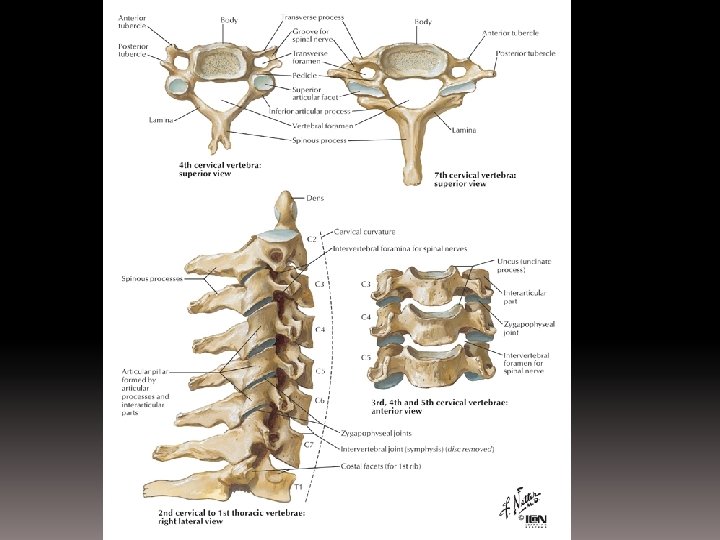
TYPES OF VERTEBRAE TYPE OF VERTEBRAE Cervical Vertebra POSITION NUMBER OF VERTEBRAE Below the skull 7 MAIN CHARACTER n n n n 1 st one – atlas vertebra 2 nd – axis vertebrae Large neural canal/vertebral foramen Short neural spine Flat centrum Short transverse processes Has a pair of vertebrarterial canals
CERVICAL VERTEBRA
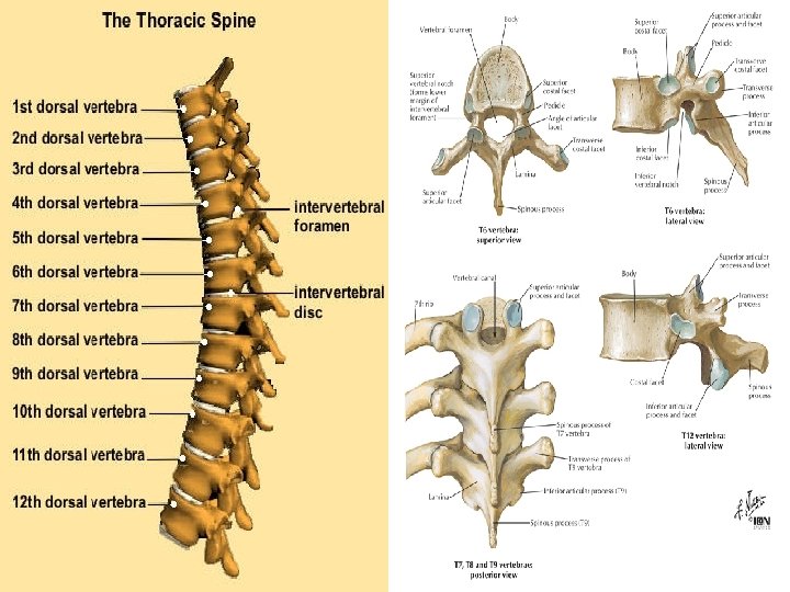
THORACIC VERTEBRA TYPE OF POSITION VERTEBRAE Thoracic Vertebra Thorax NUMBER OF VERTEBRAE 12 MAIN CHARACTER n Neural canal/ vertebral foramen is smaller than cervical vertebra’s n Long neural spine/ spinous processes (for attachment of back muscle) Thick & big centrum n n Short transverse processes
THORACIC VERTEBRA (12)
LUMBAR VERTEBRA TYPE OF VERTEBRAE Lumbar Vertebra POSITION NUMBER OF VERTEBRAE Waist 5 MAIN CHARACTER small neural canal/ vertebral foramen n n short neural spine thick & big centrum n n long transverse processes for muscle attachment
LUMBAR VERTEBRA
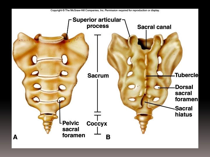
SACRUM & COCCYX TYPE OF VERTEBRAE Sacrum POSITION NUMBER OF VERTEBRAE Pelvic region 5 fused MAIN CHARACTER n Vertebrae fused to each other n Has four pairs of openings n. Triangular Coccyx Caudal region 4 fused shape Bones fused to each other forming a triangular shape which tapers at one end n
SACRUM & COCCYX
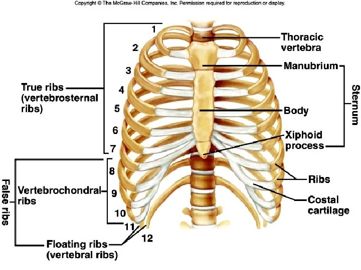
THE STERNUM & RIBS The rib cage consists of 12 pairs of ribs with the thoracic vertebrae at the back portion of the body & join to the sternum in the front portion. Movement of the rib cage are brought about by intercostal muscles between the ribs.
THE STERNUM & RIBS The sternum & ribs enclose & protect the internal organs (the lungs & heart) & play an important role in breathing.
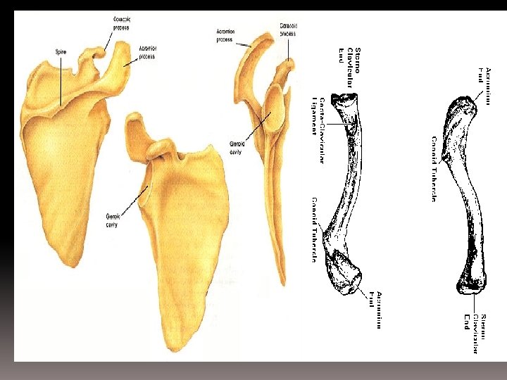
PECTORAL GIRDLE Consist of scapula & clavicle. Links the upper limbs bones to the axial skeleton. The scapula : bound by muscles to the back of the thorax. It is a flat, triangular bone which provides a surface for the attachment of muscles.
n The clavicle : a rodshaped bone placed horizontally above the scapula. n It links the scapula to the sternum. It limits the movements of the scapula.
PELVIC GIRDLE Consists of 6 fused bones – support the weight of the body from the vertebral column. Also protect the internal organs – urinary bladder & reproductive organs. Made up of two halves, each consists of 3 bones (ilium, pubis & ischium)
PELVIC GIRDLE The pelvic girdle is attached to the sacrum of the vertebral column. The asetabulum / socket for femur articulates with each side of the pelvic girdle at the hip joint.
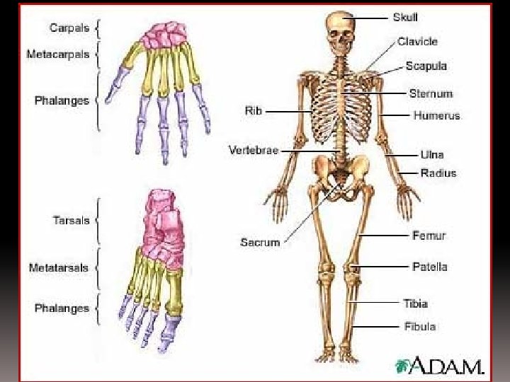
FORELIMB BONES Consists of humerus, radius & ulna. The Humerus : The long bones of the upper arm Rounded head end fits into an open socket of the scapula forming a ball-and-socket joint (allow movement in all planes)
The posterior end of the humerus forms a hinge joint with the ulnaradius bones, allowing movement in one plane only. The radius & ulna : The bones on the forearm in which the ulna is longer than the radius. It has a notch at its upper end which articulates the humerus at the elbow.
The carpals : The bones that form the wrist. Consists of 8 small bones The metacarpals : The rod-shaped ones that form the palm. The phalanges : The bones that form the fingers.
HINDLIMB BONES Consists of femur, tibia & fibula. The femur : The longest, strongest & heaviest bone in the body. Support the tight. Play an important role in maintaining the body’s upright position & in locomotion as it is attached to massive muscles. The head of femur fits into the pelvic girdle to form a joint. Other end, articulates with the tibia & fibula at the knee.
The tibia & fibula : The bones of the lower leg. Support the shank. Tibia larger than fibula & is the weight-bearing bone of the leg. Fibula is a long & thin bone – not bear any load more important for attachment of muscles than for support. Articulate with the tarsals of the ankle.
The tarsals : The 7 bones that form the ankle. The metatarsals : The 5 rod-shaped bones that form the foot. The phalanges : The bones that form the toes. The patella/kneecap : A small rounded, movable bone. Protect the knee joint.