Appendicular Skeleton Division of the skeleton consisting of

 Girdle")


 - a depression inferior to the acromion where")

 Humerus in the")


 DELTOID TUBEROSITY: a roughened V-shaped")


 n Longer")


")

 n n 8 carpals Held together by ligaments with four bones in")
 row are the: n")
 n n 5 metacarpals Each consists of a proximal base, an intermediate")
 n n n 14 in each hand Thumb has two (proximal and")


 Girdle n Functions: n n Support for vertebral column Protect pelvic organs")


 has 2, the rest")


 n. A point of contact between bones, between cartilage and bone")


 Depression")


- Slides: 93
Appendicular Skeleton Division of the skeleton consisting of the pectoral girdle, upper limbs, pelvic girdle and lower limbs.
Pectoral (Shoulder) Girdle
Pectoral Girdle n Attaches the bones of the upper limbs to the axial skeleton
Clavicle n n Also known as the collarbone Long, slender Sshaped bone that lies horizontally above the first rib (Transmits mechanical force from the upper limb to the trunk)
Scapula n Also known as the shoulder blade n Large, flat triangular bone on the posterior part of the thorax
n n n SPINE: A sharp ridge that runs diagonally across the back portion of the scapula body BODY – Main flat area of the scapula ACROMION: The lateral end of the spine. Where the scapula articulates with the clavicle
n n GLENOID CAVITY (glenoid fossa) - a depression inferior to the acromion where the head of the humerus sits CORACOID PROCESS – Projection anterior to the acromion for muscle attachment
Upper Limb
Upper Limb n n Consists of 30 bones (all paired up) Humerus in the arm Ulna and radius in the forearm 8 carpals, 5 metacarpals, and 14 phalanges in the hand
Humerus n n Longest and largest bone of the upper limb Articulates with the scapula at the shoulder and both the ulna and radius at the elbow
Humerus Bone Surface Markings n ANATOMICAL NECK: constricted portion distal to the head – site of the epiphyseal plate
n n BODY: Main portion of the bone (diaphysis) DELTOID TUBEROSITY: a roughened V-shaped area where the deltoid muscle attaches
n n CAPITULUM – small rounded process at the distal end that articulates with the head of the radius. RADIAL FOSSA - a depression that receives the head of the radius when the forearm is bent.
n n n TROCHLEA - a spoolshaped surface that articulates with the ulna. CORONOID FOSSA – a depression that receives part of the ulna when the forearm is bent. OLECRANON FOSSA a depression on the back of the bone that receives the ulna when the forearm is straightened.
Ulna n Located on the medial side of the forearm (pinky side) n Longer than the radius
Ulna Bone Surface Markings n n n The olecranon forms the prominence of the elbow on the proximal end. The coronoid process projection on the proximal, helps to hold the trochlea Trochlear Notch – depression formed by the olecranon and coronoid process
n The radial notch is a depression for the head of the radius. n A styloid process is a pointy projection at the distal end.
Radius n Located on the lateral side of the forearm (thumb side)
Radius Bone Surface Markings n Radial tuberosity a raised, roughened area that is where the biceps brachii muscle attaches to the bone n Styloid Process – pointy projection on the distal end
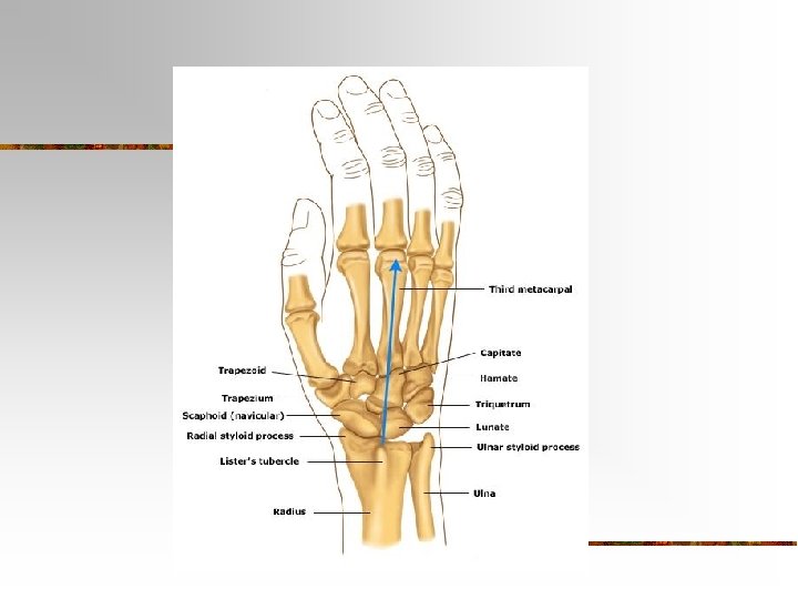
Carpus (Wrist) n n 8 carpals Held together by ligaments with four bones in each row Named for their shapes Short bones
n The carpals in the proximal (closest to the radius/ulna) row are the: n Scaphoid, Lunate, Triquetrum, and Pisiform n The carpals in the distal row are the: n Trapezium, Trapezoid, Capitate, and Hamate
Metacarpus (Palm) n n 5 metacarpals Each consists of a proximal base, an intermediate body, and a distal head Numbered I-V starting with the thumb Long bones
Phalanges (Fingers) n n n 14 in each hand Thumb has two (proximal and distal) In each of the other four digits, there are three (proximal, middle, and distal)
Disorders of the Upper Limb
Carpal Tunnel Syndrome n n Narrowing of the carpal tunnel causes compression of the median nerve The nerve compression causes pain, numbness, tingling, and hand muscle weakness
Rotator Cuff Injury n n Tears or inflammation of ligaments and tendons of the shoulder near the humerus Results in pain and loss of shoulder mobility
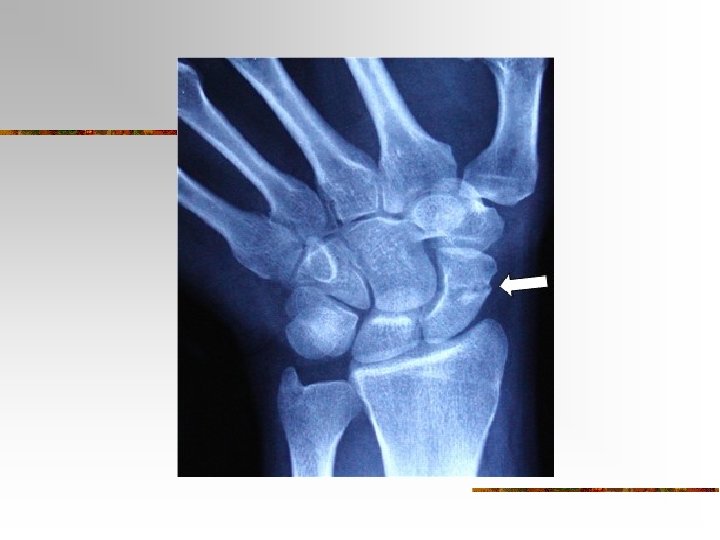
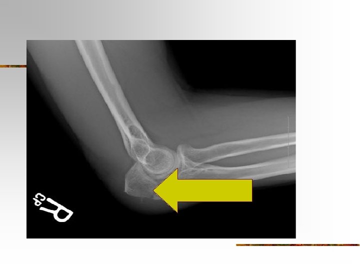
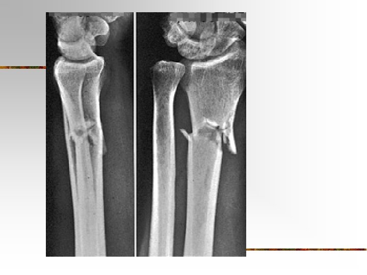
Fractures
Checkpoint Questions n n n Which bones make up a pectoral girdle? What is the function of the pectoral girdle? With which part of the scapula does the humerus articulate? What part of the ulna is called the “elbow”? What part of which bones are commonly called the “knuckles”? What bones form the upper limb, from proximal to distal?
The Pelvic Girdle and Lower Limb
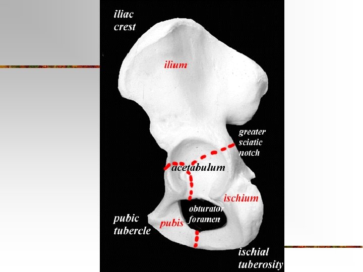
Pelvic (hip) Girdle n Functions: n n Support for vertebral column Protect pelvic organs Attach lower limbs Coxal Bones: Hip bones n 3 parts: Pubis, Ilium and Ischium
Articulations n n n Sacroiliac Joint – posterior articulation of the pelvic girdle Pubic Symphysis – anterior articulation of the pelvic girdle Acetabulum – attachment point of the femur n socket of the ball and socket joint
Coxal Bones n Pubis – anterior portion n n Ilium – superior portion n n Joined by pubic symphysis Iliac Crest – ridge at the top of the ilium Ischium – inferior portion n n Acetabulum – socket for the head of the femur Obturator Foramen – hole formed by the ischium and pubis
Pelvis n n n Combination of the sacrum, coccyx, and the 2 hip bones Greater (false) – Top portion that is not fully enclosed by bone Lesser (true) – Bottom portion the is completely surrounded by bone
Pelvis - continued n n n n Ilium Ischium Pubis Pubic Symphysis Sacrum Coccyx Pelvic Brim Pubic Arch
X-Ray of Pelvis
Comparison of Male and Female Pelvis Female Point of Comparison Male General Structure Light and Thin Heavy and Thick False Pelvis Shallow Deep Pelvic Inlet Larger and Oval Smaller and Heart Shaped Acetabulum Small, faces anteriorly Large and faces laterally Obturator Foramen Oval Round Pubic Arch Greater than 90 o Less than 90 o
Comparison of Male and Female Pelvis
Pelvimetry n Measurement of the size of the inlet and outlet of the birth canal.
Pelvic Girdle Checkpoint n n What is the name of the hip bone? What are the 3 parts of the hip bone? n n Can you identify them on a diagram? ? ? List 3 functions of the pelvic girdle What is the name of the “socket” where the head of the femur sits? List 3 differences between the male and female pelvis. Why are these present?
LOWER LIMB n n Includes the thigh, leg, ankle, foot and toes 30 bones in each n n n n Femur Patella Tibia Fibula Tarsals Metatarsals Phalanges
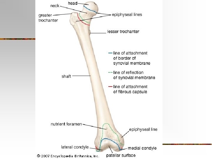
Femur Thigh Bone n Longest, strongest heaviest bone in the body n n Diaphysis has a medial bend to bring knees closer to the midline of the body
Femur continued n n n Body - diaphysis Head – “ball” of ball and socket joint Neck – common site of fractures Greater and Lesser Trochanters – used for muscle attachment Lateral and Medial Condyles – articulation with the tibia Patellar Surface
Patella n n n Sesamoid bone Develops in the tendon of the quadriceps femoris muscle Increases the leverage of the tendon and maintains the position of the tendon
Patellofemoral stress syndrome n n AKA “Runner’s Knee” Patella does not glide up and down between the femoral condyles but rather laterally causing pain.
Tibia n Shin Bone n Larger bones of 2 lower leg
Tibia Continued n n n Lateral and Medial Condyles – articulate with the femur Tibial Tuberosity – roughened area on anterior/proximal portion of the tibia for muscle attachment Medial Malleolus – bump on the inside of the ankle
Shin Splints n Soreness or pain along the tibia due to inflammation of the periosteum caused by the repeated tugging of the attached muscles and tendons. Often the result of walking or running up and down hills.
Fibula n n n Parallel and lateral to the tibia Articulates with the tibia and the talus (ankle bone) Lateral malleolus – bump on the outside of the ankle
The Foot
Tarsals n 7 Ankle Bones n n Posterior: Talus and Calcaneus Anterior: cuboid, navicular and 3 cuneiform
Metatarsals n n 5 bones make up the instep of the foot Numbered I-V starting medially
Phalanges n n 14 in each foot Hallux (Big Toe) has 2, the rest of the toes have 3
Bones of the Foot n n n A - Talus B – Navicular C - Cuneiform D - Cuneiform E – Distal Phalanx F – Middle Phalanx G – Proximal Phalanx H - Metatarsal I - Cuneiform J - Cuboid K - Calcaneous
Arches of the Foot n n Transverse and Longitudinal Arches enable the foot to support the weight of the body
LOWER LIMB CHECKPOINT n n n What are the bones of the lower limb? Describe the hip joint Describe the knee joint Which is the medial bone of the lower leg? What type of bone is the patella?
Joint (AKA Articulation) n. A point of contact between bones, between cartilage and bone or between teeth and bone
More Definitions n n n Arthrology – The study of joints Kinesiology – study of the movement of the human body Rheumatology – the field of medicine devoted to joint diseases and related conditions
Classification of Joints n Functional Classification – related to the degree of movement it permits n Structural Classification – classified by the presence or absence of space between bones and the type of connective tissue that binds them together
Functional Classifications n n n Synarthrosis – immovable joint Amphiarthrosis – slightly moveable joint Diarthrosis – Freely movable joint
Structural Classifications n n n Fibrous Joints Cartilaginous Joints Synovial Joints
Fibrous Joints n n Bones are held together by fibrous connective tissue that is rich in collagen fibers No synovial cavity
Fibrous joints n n Permits little or no movement Suture – connect bones of the skull Syndesmosis – the distance between the 2 bones is greater than in a suture. Ex – between tibia and fibula Gomphosis – a cone shaped peg (tooth) fits into a cavity (socket)
Cartilaginous Joints n n The bones are held together by cartilage No synovial cavity
Cartilaginous Joints n n n Allows little or no movement Synchondrosis – the connecting material is hyaline cartilage. Ex - epiphyseal plate Symphysis – ends of the bones are covered with articular cartilage but the cones are connected by a broad flat disc of fibrocartilage. Ex – pubic symphysis
Synovial Joints n Bones have a synovial cavity and are united by a dense irregular connective tissue and accessory ligaments
Synovial joints n Unique Characteristics: n n n Synovial cavity Articular cartilage Articular capsule Fibrous capsule – outer layer (can form ligaments) n Synovial membrane – areolar connective tissue with elastic fibers n n Synovial Fluid – secreted by the synovial membrane. It lubricates the joint, supplies nutrients and removes metabolic waste.
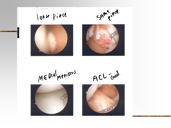
Synovial joints n n Accessory Ligaments – ligaments that are both inside and outside the articular capsule (ex – collateral ligaments of the knee) Menisci – pads of fibrocartilage that lie between articular surfaces of the bones and attach to the fibrous capsule n n Allows 2 bones of different shapes to fit more tightly Bursa – saclike structures between skin and bone or between tendons and bones situated to reduce friction
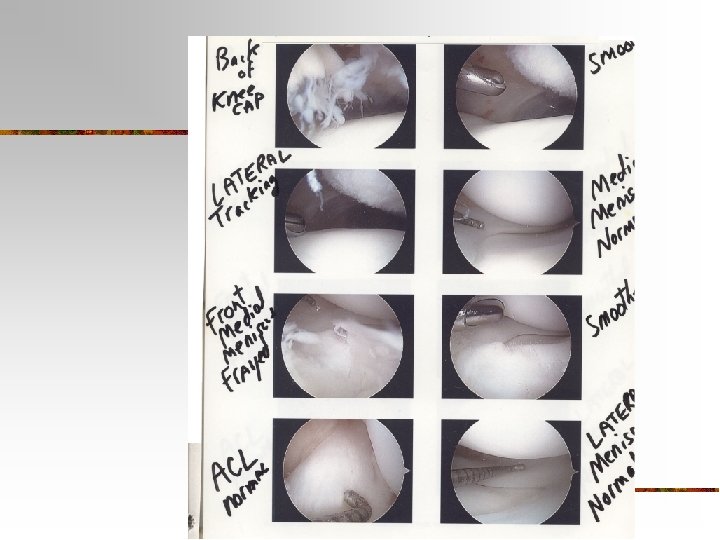
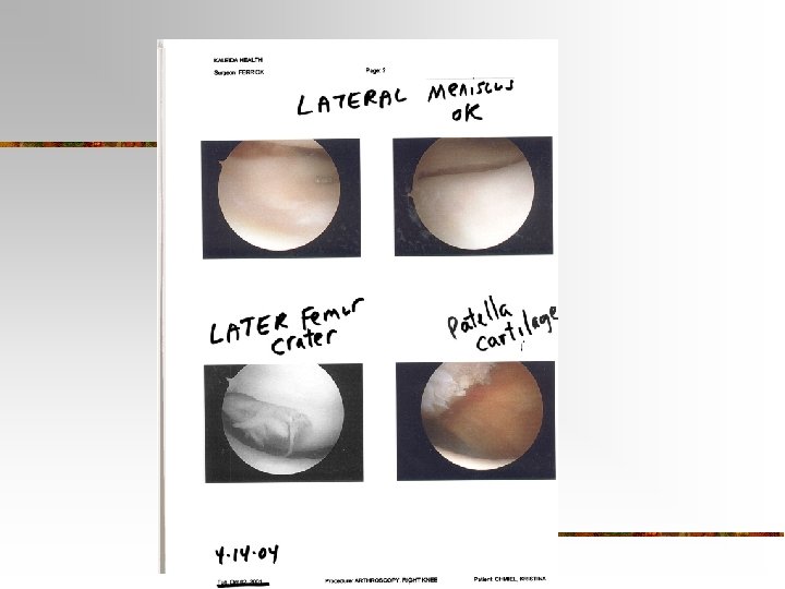
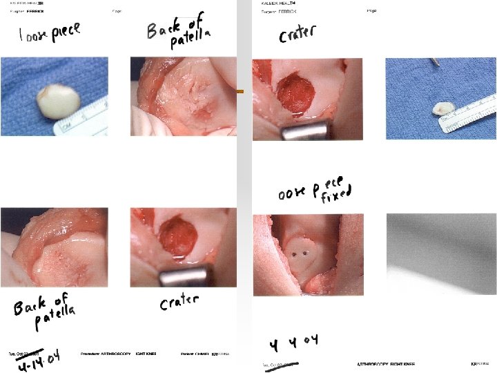
Torn Meniscus n The tearing of the cartilage in the knee is common among athletes. Damaged cartilage can wear away the joint and eventually develop into arthritis. The surgical repair of the knee may be assisted by arthroscopy (a small lighted tool is inserted into the knee for visualization)
Types of Synovial Joints n n n Planar – intercarpal, intertarsal, sternoclavicular Hinge – knee, elbow, ankle, fingers Pivot – atlanoaxial, radioulnar Condyloid – wrist and metacarpals Saddle – wrist and thumb Ball and Socket – shoulder, hips
SEE ALSO PAGE 163 IN THE TEXT BOOK FOR VERY GOOD ILLUSTRATIONS OF THE SYNOVIAL JOINTS
Types of Movements at Synovial Joints
Gliding n n n A simple movement in which relatively flat bone surfaces move back-and-forth and side-to-side relative to one another. Limited in range by the articular capsule and ligaments of the joint Occurs in Planar Joints (carpals, tarsals, sternoclavicular)
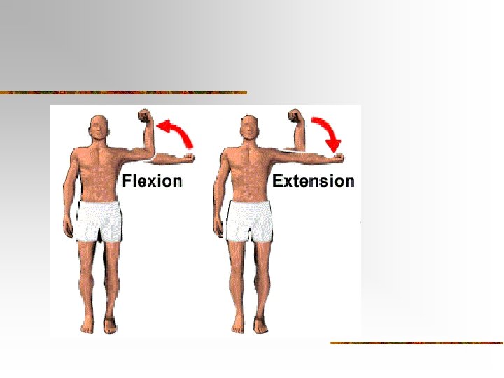
Angular Movements n There is an increase or decrease in the angle between articulating bones n Flexion, Extensions, Abduction, Adduction
Flexion/Extension n n Flexion - A decrease in the angle between articulating bones Extension – An increase in the angle between articulating bones Found in hinge, pivot, condyloid, saddle and ball-and-socket joints HYPEREXTENSION – extension beyond anatomical position
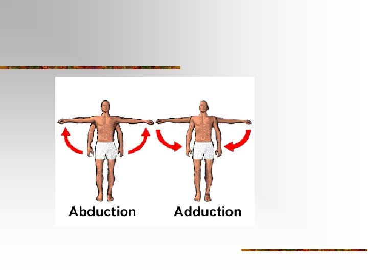
Abduction/Adduction n Abduction – movement away from the midline of the body Adduction – movement toward the midline of the body Found at condyloid, saddle and ball and socket joints
Circumduction n n Movement of the distal end of a body part in a circle Found at ball-and-socket joints
Rotation n n A bone revolves around its own longitudinal axis Found at pivot and ball and socket joints
Special Movements n n Occur only at certain joints Include elevation, depression, protraction, retraction, inversion, eversion, dorsiflection, plantar flexion, supination and pronation
n n Elevation - upward movement of a body part (closing your mouth) Depression – downward movement of a body part (opening your mouth) Protraction – movement of a body part forward (mandible or clavicles) Retraction – returning a protracted part to anatomical position
n n Inversion – movement of the soles medially so they face each other Eversion – movement of the soles laterally so they are away from eachother Dorsiflexion – Bending the feet upward (like standing on your heels) Plantar Flexion – Bending the feet down (like standing on your toes)
n n Supination – turning the palm upward Pronation – turning the palm downward