Echocardiographic Evaluation of Prosthetic Valves Part I Echo

 I. Mechanical II. Biological/Tissue III. Appearance of Normally")

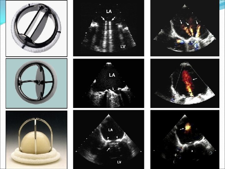
 Mechanical Stented Ball and cage (Starr-Edwards) Single tilting disc (Medtronic -Hall)")


")


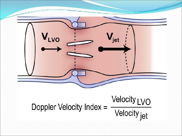
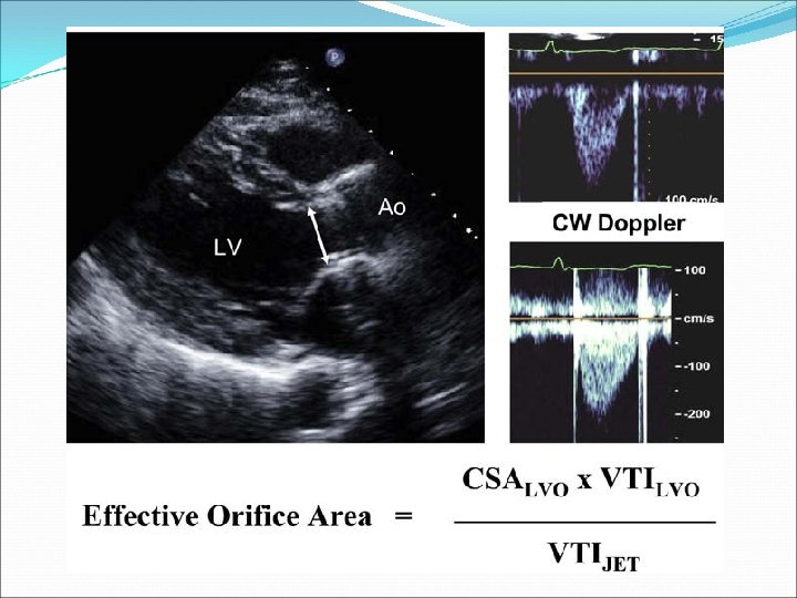
 Dimensionless ratio of the proximal velocity in the LVO tract")


 EOA Pr. AV = (CSA LVO x VTI LVO) /")

 When the EOA of the inserted prosthesis is too small in")


 A large rise in PHT on serial studies or a")


- Slides: 56
Echocardiographic Evaluation of Prosthetic Valves, Part I Echo Conference
Objectives Introduction to Prosthetic Valves (PV) I. Mechanical II. Biological/Tissue III. Appearance of Normally functioning Valves I. II. Approach to Evaluating PVs with echo and doppler III. Evaluating Prosthetic Aortic Valves IV. Echo Case/Questions (Echo. Sap)
Overview Prosthetic Valves are classified as tissue or mechanical Tissue: Actual valve or one made of biologic tissue from an animal (bioprosthesis or heterograft) or human (homograft or autograft) source Mechanical Made of nonbiologic material (pyrolitic carbon, polymeric silicone substances, or titanium) Blood flow characteristics, hemodynamics, durability, and thromboembolic tendency vary depending on the type and size of the prosthesis and characteristics of the patient
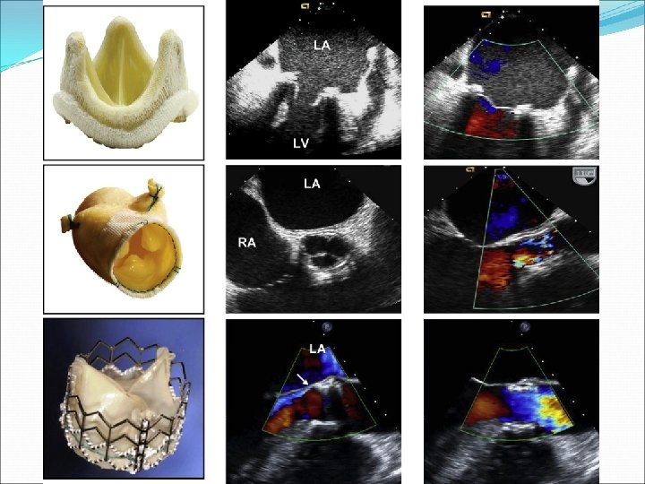
Valves Biologic (Tissue) Mechanical Stented Ball and cage (Starr-Edwards) Single tilting disc (Medtronic -Hall) Bileaflet (St. Jude, Carbo. Medics) Porcine xenograft Pericardial xenograft Stentless Porcine xenograft Pericardial xenograft Homograft Autograft
Mechanical Valves Extremely durable with overall survival rates of 94% at 10 years Primary structural abnormalities are rare Most malfunctions are secondary to perivalvular leak and thrombosis Chronic anticoagulation required in all With adequate anticoagulation, rate of thrombosis is 0. 6% to 1. 8% per patient-year for bileaflet valves
Biological Valves Stented bioprostheses Primary mechanical failure at 10 years is 15 -20% Preferred in patients over age 70 Subject to progressive calcific degeneration & failure after 6 -8 years Stentless bioprostheses Absence of stent & sewing cuff allow implantation of larger valve for given annular size->greater EOA Uses the patient’s own aortic root as the stent, absorbing the stress induced during the cardiac cycle
Biologic Valves Continued Homografts Harvested from cadaveric human hearts Advantages: resistance to infection, lack of need for anticoagulation, excellent hemodynamic profile (in smaller aortic root sizes) More difficult surgical procedure limits use Autograft Ross Procedure
Caged-Ball Valve
Single-Leaflet Valve
Bileaflet Valve
Stentless Aortic Graft Valve
Stented Biologic Mitral Valve
Approach to Valve Evaluation Clinical data including reason for the study and the patient’s symptoms Type & size of replacement valve, date of surgery BP & HR HR particularly important in mitral and tricuspid evaluations because the mean gradient is dependent on the diastolic filling period Patient’s height, weight, and BSA should be recorded to assess whether prosthesis-patient mismatch (PPM) is present
Echo Imaging of Prosthetic Valves should be imaged from multiple views, with attention to: Opening & closing motion of the moving parts (leaflets for bioprosthesis and occluders for mechanical ones) Presence of leaflet calcification or abnormal echo density attached to the sewing ring, occluder, leaflets, stents, or cage Appearance of the sewing ring, including careful inspection for regions of separation from native annulus & for abnormal rocking motion during the cardiac cycle
Echo Imaging Mild thickening is often the 1 st sign of primary failure of a biologic valve Occluder motion of a mechanical valve may not be well visualized by TTE because of artifact and reverberations
Evaluation of the Prosthetic Aortic Valve (AV)
Imaging Considerations Identify the sewing ring, valve or occluder mechanism, and surrounding area Ball or disc is often indistinctly imaged, whereas leaflets of normal tissue valves should be thin with an unrestricted motion Stentless or homograft may be indistinguishable from native valves One can use modified views (lower parasternal) to keep the artifact from the valve away from the LV outflow tract
Doppler of Prosthetic AV Doppler velocity recordings across normal PVs usually resemble those of mild native aortic stenosis Maximal velocity usually > 2 m/s, with triangular shape of the velocity contour Occurrence of maximal velocity in early systole With increasing stenosis, a higher velocity and gradient are observed, with longer duration of ejection and more delayed peaking of the velocity during systole
Doppler Velocity Index (DVI) Dimensionless ratio of the proximal velocity in the LVO tract to that of flow velocity through the prosthesis: DVI= VLVO/ VPr. AV DVI is calculated as the ratio of respective VTIs and can be approximated as the ratio of respective peak velocities Incorporates the effect of flow on velocity through the valve and is much less dependent on valve size
DVI Helpful measure to screen for valve dysfunction, particularly when the CSA of the LVO tract cannot be obtained or valve size is unknown DVI is always < 1 DVI < 0. 25 is highly suggestive of significant obstruction DVI is not affected by high flow conditions through the valve, including AI
Doppler & Prosthetic AV High gradients may be seen with normal functioning valves with: Small size Increased stroke volume PPM Valve obstruction Conversely, a mildly elevated gradient in the setting of severe LV dysfunction may indicate significant stenosis Thus, the ability to distinguish malfunctioning from normal PVs in high flow states on the basis of gradients alone may be difficult
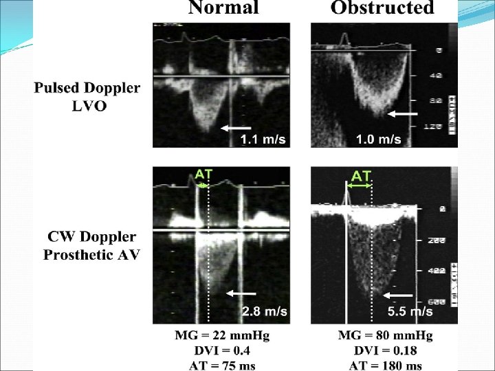
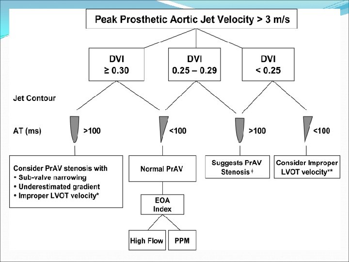
Doppler Continued Other qualitative and quantitative indices that are less dependent on flow should be evaluated Contour of the velocity: In a normal valve, even in high flow, there is a triangular shape, with early peaking of the velocity and short acceleration time (AT) With PV obstruction, a more rounded velocity contour is seen, with velocity peaking almost in mid-ejection, prolonged AT Cutoff of AT of 100 ms differentiates well between normal and stenotic PVs
Effective Orifice Area (EOA) EOA Pr. AV = (CSA LVO x VTI LVO) / VTI Pr. AV EOA is dependent on size of inserted valve Should be referenced to the valve size of a particular valve type For any size valves, significant stenosis is suspected when valve area is < 0. 8 cm 2 However, for the smallest size valve, this may be normal because of pressure recovery Largest source of variability is measurement of the LVO tract
Doppler Parameters of Prosthetic AV function in Mechanical and Stented Biologic Valves in Conditions of Normal Stroke Volume Parameter Normal Possible Stenosis Suggests Significant Stenosis Peak Velocity (m/s) <3 3 -4 >4 Mean Gradient (mm. Hg) <20 20 -35 >35 DVI ≥ 0. 30 0. 29 -0. 25 <0. 25 EOA (cm 2) >1. 2 -0. 8 <0. 8 Contour of Jet velocity in PV Triangular, early peaking Triangular to intermediate Rounded, symmetrical AT (ms) <80 80 -100 >100
Patient-Prosthesis Mismatch (PPM) When the EOA of the inserted prosthesis is too small in relation to the patient’s BSA A given valve area acceptable for a small, inactive person may be inadequate for a larger physically active individual Main consequence is the generation of higher than expected gradients through a normally functioning valve
PPM Continued Commonly seen in: Patients with small aortic annulus sizes, particularly women Patients whom indication for AVR was AS as opposed to AI Young patients, who outgrow their initially inserted prosthesis Failure of post-op regression of LV mass index at 6 months may be clue to presence of PPM For patients with exertional symptoms without evidence of primary valve dysfunction, stress echo should be entertained to further evaluate
Evaluation of Prosthetic AI With color doppler, one can evaluate the components of the color AI jet Flow convergence, vena contracta, extent in the LVO tract and LV Normal “physiologic” jet are usually low in momentum, depicted by homogenous color jets that are small in extent Ratios of jet diameter/LVO diameter from parasternal long-axis imaging and Jet area/LVO area from parasternal short-axis imaging are best applied for central jets
Prosthetic Valve AI With eccentric AI jets, measurement of jet width perpendicular to the LVO tract will cut the jet obliquely and risk overestimation Entrainment of jet in the LVO tract may lead to rapid broadening of the jet just after the vena contracta-> overestimation
Significant AI, AV Dehiscence
AI in PVs Contrary to native valves, the width of the vena contracta may be difficult to accurately measure in the long-axis in the presence of a prosthesis Imaging of the neck of the jet in short-axis, at the level of the sewing ring allows determination of the circumferential extent of the regurgitation Approximate guide: < 10% of sewing ring suggests mild 10 -20% suggests moderate > 20% suggests severe **Rocking of the prosthesis usually associated with >40% dehisscence
Spectral Doppler and PVAI PHT is useful when the value is <200 ms, suggesting severe AI, or > 500 ms, consistent with mild AI Intermediate ranges may reflect other hemodynamic variables such as LV compliance and are less specific Holodiastolic flow reversal in the descending thoracic aorta is indicative of at least moderate AI Severe is suspected when the VTI of the reverse flow approximates that of the forward flow Holodiastolic flow reversal in the abdominal aorta is usually indicative of severe AI
Parameter Mild Moderate Severe Valve Structure/Function Normal Abnormal LV size Normal or Mild Dilation Dilated Jet width (%LVO diameter) Narrow (≤ 25%) Intermediate (2664%) Large (≥ 65%) Jet density (CW doppler) Incomplete or Faint Dense PHT, ms (CW doppler) >500 Variable (200 -500) Steep (< 200) Diastolic Flow Reversal (Descending Aorta) Absent or Brief early diastolic Intermediate Prominent, holodisatolic Regurgitant Volume (ml/beat) < 30 30 -59 >60 Regurgitant Fraction (%) <30 30 -50 >50
Part II-Evaluation of Prosthetic Mitral Valve
Evaluation of Prosthetic MV A major consideration with echo is the effect of acoustic shadowing by the prosthesis on assessment of MR Problem is worse with mechanical valves On TTE, LV function is readily evaluated, but the LA is often obscured for imaging and doppler interrogation TEE provides visualization of the LA and MR but shadowing limits visualization of the LV Thus, comprehensive assessment of PMV requires both TTE & TEE when valve dysfunction is suspected
Prosthetic MV Imaging Considerations In the parasternal long-axis view, the prosthesis may obscure portions of the LA and its posterior wall MR may be difficult to evaluate Parasternal long-axis views allows visualization of the LVO tract, which can be impinged by higher profile prostheses Apical views allow visualization of leaflet excursion for both bioprosthetic and mechanical valves May allow detection of thrombus or pannus Vegetations can be seen but are often masked by acoustic shadowing
Doppler Evaluation of PMV Complete exam should include: Peak early velocity Estimate of mean pressure gradient Heart Rate Pressure half-time (PHT) Determination of whether regurgitation is present DVI and/or EOA as needed LV/RV size and function LA size if possible PA systolic pressure
Peak Early Mitral Velocity Peak E velocity is easy to measure Provides simple screen for prosthetic valve dysfunction Can be elevated in: hyperdynamic states, tachycardia, small valve size, stenosis, or regurgitation Inhomogeneous flow profile across caged-ball and bileaflet prostheses can lead to doppler velocity measurements that are elevated out of proportion to the actual gradient For normal bioprosthetic MVs, peak velocity can range from 1. 0 to 2. 7 m/s
MV Peak Velocity In normal bileaflet mechanical valves, peak velocity is usually < 1. 9 m/s but can be up to 2. 4 m/s As a general rule, peak velocity < 1. 9 m/s is likely to be normal in most patients with mechanical valves unless there is markedly depressed LV function
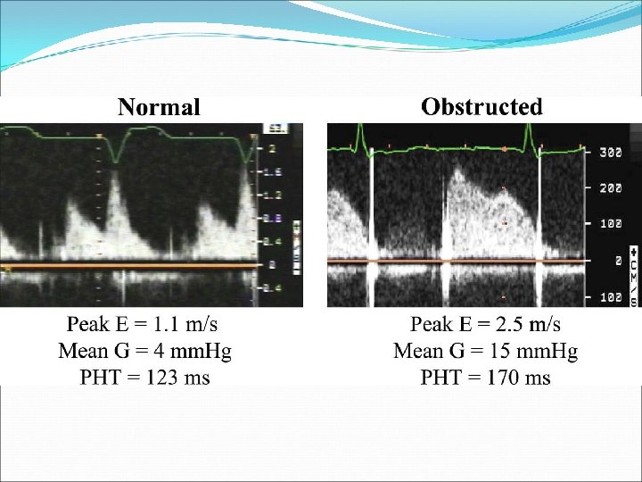
Mean Gradients of MV Normally less than 5 -6 mm Hg Values up to 10 -12 mm Hg have been reported in normally functioning mechanical valves High gradients can be due to: hyperdynamic states, tachycardia or PPM, regurgitation, or stenosis
MV Pressure Half-time (PHT) A large rise in PHT on serial studies or a markedly prolonged single measurement (>200 ms) may be a clue to the presence of: obstruction PHT seldom exceeds 130 ms across normal pv Minor changes in PHT occur as a result of nonprosthetic factors including: Loading conditions Drugs AI PHT should not be obtained in tachycardic rhythms or 1 st degree blocks when the E & A velocities are merged or the diastolic filling period is short
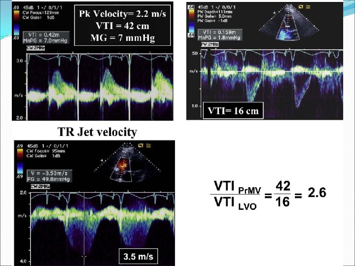
EOA of PMV Calculation from PHT, as traditionally applied in native MS, is not valid in prosthetic valves due to its dependence on LV and LA compliance and initial LA pressure EOAPr. MV= stroke volume/VTIPr. MV Usually reserved for cases of discrepancy between information obtained from gradients and PHT
Prosthetic MV and DVI= VTIPr. MV/ VTILVO DVI can be elevated with stenosis or regurgitation For mechanical valves, a DVI < 2. 2 is most often normal Higher values should prompt consideration of prosthesis dysfunction
Doppler Parameters of Prosthetic MV Function Parameter Normal Possible Stenosis Suggests Significant Stenosis Peak Velocity (m/s) <1. 9 -2. 5 ≥ 2. 5 Mean Gradient (mm Hg) ≤ 5 6 -10 >10 DVI <2. 2 -2. 5 >2. 5 EOA (cm 2) ≥ 2 1 -2 <1 PHT (ms) <130 130 -200 >200
Prosthetic MV Regurgitation Since direct detection of prosthetic MR is often not possible with TTE, particularly with mechanical valves, one must rely on indirect signs suggestive of significant MR Such signs include: Hyperdynamic LV with low systemic output Elevated mitral E velocity Elevated DVI Dense CW regurgitant jet with early systolic maximal velocity Large zone of systolic flow convergence toward the prosthesis seen in the LV Clinical symptoms & presence of the above findings represents a clear indication for TEE
Prosthetic MV Regurgitation Assessment of severity of prosthetic MR can be difficult at times because of the lack of a single quantitative parameter that can be applied consistently in all patients Currently, best method is to integrate several findings from both TTE and TEE that together suggest a given severity of regurgitation
Echo & Doppler Criteria for Severity of Prosthetic MR from TTE/TEE Parameter Mild Moderate Severe LV size Normal NL or Dilated Usually Dilated Valve Usually Normal Abnormal Color Flow Jet Area Small, central jet (usually <4 cm 2 or <20% of LA area) Variable Large, central jet (usually >8 cm 2 or >40% of LA area) Flow Convergence None or Minimal Intermediate Large Jet Density: CW Incomplete/Faint Dense Jet Contour: CW Parabolic Usually Parabolic Early peaking, triangular Pulm Vein Flow Systolic Dominance Systolic Blunting Systolic Flow Reversal VC Width (cm) <0. 3 -0. 59 ≥ 0. 6 R vol (ml/beat) <30 30 -59 ≥ 60 RF (%) <30 30 -49 ≥ 50 EROA (cm 2) <0. 20 -0. 49 ≥ 0. 50
TTE of Prosthetic MV
TEE of Same MV