Pig Dissection http faculty clintoncc suny edufacultyMic hael


 section (=cut) our subject using")


 Superior- toward or closer to")


 • towards the front (head) of the animal")
 • towards the back (tail) of the animal")


 Be creative, use color and")


 in pigs is about 17 weeks (compared")


, the external")
 and")


 enters the posterior portion")


 is the first major artery branching")


 runs ventrally from the aorta to")


")


- Slides: 126
Pig Dissection
• http: //faculty. clintoncc. suny. edu/faculty/Mic hael. Gregory/files/Bio%20102 %20 Laboratory/Fetal%20 Pig. htm
Directional and Anatomical Terminology • Anatomists and morphologists rely on a set of terms to describe structural positions, These may not all be immediately obvious to you, so you should practice using them. The terms are generally presented to you in pairs, as terms are often used to indicate opposing directions.
Planes of section. • We can figuratively (or actually) section (=cut) our subject using planes. There are several particular planes of section that are useful for the purposes of discussing anatomy.
1. 2. 3. 4. 5. 6. 7. 8. 9. 10. 11. 12. 13. 14. 15. 16. Anterior - near or toward the head Posterior - near or toward the tail Dorsal - referring to the back Ventral - referring to the belly Lateral - referring to the side Median - referring to the midline Cranial - referring to the head Caudal - referring to the tail Proximal - toward the attached end of a structure Distal - toward the free end of a structure Longitudinal - in the axis from head to tail Transverse - across the longitudinal axis Pectoral - chest or shoulder area Pelvic - hip region Inferior- toward or closer to the tail (caudal region) Superior- toward or closer to the head region
Page Pig Book Use color on every page! Points 1 Title Page Names of authors (max of 2) 10 Be creative, use color and make it fun!!! 2 Basic Anatomical terms 37 Diagram showing 16 terms from the lab color 5 pts 3 External anatomy: Diagram of head, neck, trunk, tail. 29 Label=Thorax, Abdomen, Sacral What is inside and out of these sections? How can you tell male from female? color=5 pts 4 Internal Anatomy Diagram with parts labeled and umbilical cord drawing 5 Circulatory System: Purpose, flow of blood, structures/functions, diagrams of arteries and veins X section, Heart diagram, path of blood flow, color coded Red = oxygenated Blue= deoxygenated 6 Respiratory System: Purpose, flow of air, structures and functions. Diagram of inspiration and expiration 7 Digestive System: Purpose, path of food, structures and functions 8 Excretory System: Purpose, organs of excretion. Diagram of a cross section of the kidney. Path of flow of nitrogen waste as it is formed and removed from the pig’s body 9 Reproductive System: Purpose, structures and functions Female: structures and how do they work. Male and how do they work 10 Nervous System: Purpose, How is it broken into parts?
Anterior - near or toward the head
Posterior - near or toward the tail or back
Dorsal - referring to the back
Ventral - referring to the belly
Lateral - referring to the side
Median - referring to the midline
Cranial - referring to the head Caudal - referring to the tail
Proximal - toward the attached end of a structure Distal - toward the free end of a structure
Longitudinal - in the axis from head to tail
Transverse - across the longitudinal axis • is an imaginary plane that divides the body into superior and inferior parts. It is perpendicular to the coronal and sagittal planes.
Pectoral - chest or shoulder area
Pelvic - hip region
Inferior- toward or closer to the tail (caudal region) Superior- toward or closer to the head region • As with vertebrate directional terms, superior and inferior can be used in a relative sense in humans, but can not be uniformly applied to other organisms with varying normal anatomical positions. For example, the shoulders are superior to the navel, but inferior to the eyes in humans. In any tetrapod, the shoulders are cranial to the belly, but caudal to the eyes.
sagittal section • The sagittal section, or plane, divides the subject into left and right portions.
Mid-sagittal • The mid-sagittal section splits the subject exactly into left and right sides.
transverse • A transverse plane, also known as an axial plane or cross-section, divides the body into cranial and caudal (head and tail) portions.
Directional Terms: • Anatomists rely on a set of directional terms to explain the orientation of subject material. Although some of the terms are unfamiliar, with practice they will become easier to use, and you will see that “proximal to” is lesser cumbersome than “up the extremity and closer to the middle of the body. ”
left and right • stated relative to the subject, not relative to the people viewing it
anterior (cranial) • towards the front (head) of the animal
posterior (caudal) • towards the back (tail) of the animal
superior • higher on the subject- typically used on bipeds such humans
inferior • lower on subject- also used on subjects such as humans
dorsal • towards the back
Ventral • towards the belly
medial • towards the mid-sagittal section
lateral • away from the mid-sagittal section
proximal • towards the mid-sagittal section along a limb
distal • away from the mid-sagittal section along a limb
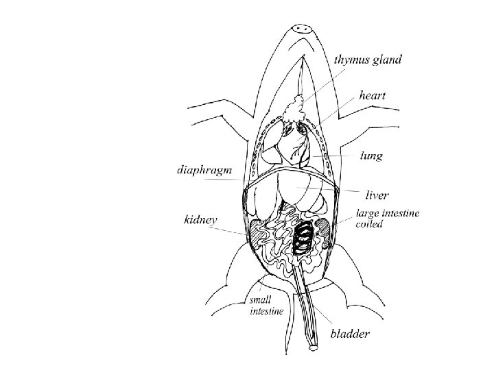
External Anatomy • The body consists of the following regions: head, neck, trunk, and tail. The 2 pairs of appendages present on the trunk are the fore legs and hind legs. The cord projecting from the ventral surface is the umbilical cord. This cord connects the fetal pig to the placenta within the uterus of the mother pig.
Pig Book
1 Title Page Names of authors (max of 2) Be creative, use color and make it fun!!! 2 Basic Anatomical terms Diagram showing 16 terms from the lab 3 External anatomy: Diagram of head, neck, trunk, tail. Thorax (what is inside? ) Abdomen (what is inside? ) Sacral (what is here? ) How can you tell male from female?
4 Internal Anatomy Diagram with parts labeled and umbilical cord drawing 5 Circulatory System: Purpose, flow of blood, structures/functions, diagrams of arteries and veins X section, Heart diagram, path of blood flow, color coded Red = oxygenated Blue= deoxygenated 6 Respiratory System: Purpose, flow of air, structures and functions. Diagram of inspiration and expiration 7 Digestive System: Purpose, path of food, structures and functions 8 Excretory System: Purpose, organs of excretion. Diagram of a cross section of the kidney. Path of follow of nitrogen waste as it is formed and removed from the pig’s body. 9 Reproductive System: Purpose, structures and functions Female: structures and how do they work. Male and how do they work 10 Nervous System: Purpose, How is it broken into parts?
Fetal Pig Dissection • Objectives: Upon completion of this lab students should • Be able to identify, and know the names and functions major structures of the fetal pig's external anatomy. • Be able to identify, and know the names and functions of the major structures of the fetal pig's internal gross anatomy.
• The fetal pigs that we will use in lab were purchased from a Biological supply company. They obtain fetal pigs from processing plants - the unborn pigs are removed from the uteri of slaughtered sows.
• The period of pregnancy (gestation) in pigs is about 17 weeks (compared to 40 weeks in humans). • The fetal pigs we will use in class are 3 -4 weeks from birth. You will work in groups on the fetal pigs. Each group of students will be given a fetal pig to be used for the labs on pig dissection. • Attach a tag with your names (in pencil) to one of the hind legs for future identification.
• Note the slit in the skin in the neck region of the pig. (ours were not injected) • This is the area where the circulatory system of the pig was injected with latex (red latex in the arterial system, blue latex in the venous system) to make it easier to see and trace the blood vessels.
Page 2 1. 2. 3. 4. 5. 6. 7. 8. 9. 10. 11. 12. 13. 14. 15. 16. Anterior - near or toward the head Posterior - near or toward the tail Dorsal - referring to the back Ventral - referring to the belly Lateral - referring to the side Median - referring to the midline Cranial - referring to the head Caudal - referring to the tail Proximal - toward the attached end of a structure Distal - toward the free end of a structure Longitudinal - in the axis from head to tail Transverse - across the longitudinal axis Pectoral - chest or shoulder area Pelvic - hip region Inferior- toward or closer to the tail (caudal region) Superior- toward or closer to the head region
External Anatomy • Page 3 draw a diagram of the main parts • Place the pig on its side in the pan and note that the body consists of the following regions: • head, neck, trunk, and tail. • The 2 pairs of appendages present on the trunk are the fore legs and hind legs. The cord projecting from the ventral surface is the umbilical cord. • This cord connects the fetal pig to the placenta within the uterus of the mother pig.
• The head bears the mouth and jaws, the snout (nose), the external nostrils (nares), the eyes, and the external ears. Feel the relatively thick neck in the fetal pig. This thickness is due to the presence of welldeveloped neck muscles that will eventually be used for rooting.
• The cranial portion of the trunk is called the thorax (chest) and is encased by the ribs. Feel the ribs under the skin and determine the posterior border of the thorax. The thorax contains the lungs, heart, and major blood vessels. • The fore legs are found in the thoracic region. The parts of each of these appendages as well as those of the hind legs (in the sacral region) are the upper leg, lower leg, wrist, foot, and toes (digits). • Examine the digits present on the legs and note that only 2 of the 5 digits found in most terrestrial vertebrates are present. The first toe (corresponding to our thumb) has been lost; the second and fifth toes are reduced, with only the third and fourth toes being fully developed.
• The caudal portion of the trunk is called the abdomen. • In contrast to the thorax, the ventral portion of the abdomen is soft. The umbilical cord is located near the posterior end of the abdomen. There are 2 rows of teats (mammary papillae), one on either side of the umbilical cord. The stomach, intestine, kidneys, and other viscera (soft internal organs) are found within the abdominal cavity.
• The sacral region includes the hind legs, pelvic bones, and their attachment to that area of the vertebral column. • The anus (posterior opening of the digestive tract) is located under the tail. • In female pigs, the vulva (openings of the reproductive and urinary tracts) is found just below the anus. • In male pigs, the external opening of the penis is located posterior to the umbilical cord. The scrotum (scrotal sacs) are found on either side of the midline of the anus.
Page 3
Internal Anatomy Page 4 • For the dissection of the fetal pig you will need string, a scissors, a sharp scalpel, a blunt probe, a forceps, and dissecting pins. • The definition of dissecting is to separate the body into parts for the purpose of study. This means that your scissors and scalpel should be used sparingly and with care. The most useful dissecting instrument is a blunt probe, which can be used to separate organs from membranes.
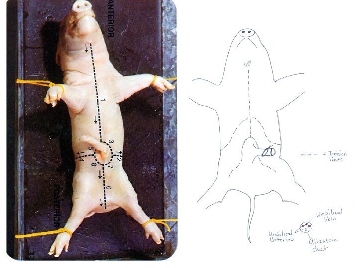
1. 2. 3. Place the pig on its dorsal surface in the dissecting pan. Tie a string around one fore leg and passing the string under the pan tie the other end to the other fore leg. The string should be tied tight enough to spread the forelegs apart. Tie a string in the same fashion to the hind legs. Do not remove the string from the appendages at the end of the lab; simply slip the strings out from under the pan at the end of each lab period. Mark a line on the skin with a permanent marker from the tip of the lower jaw to a point 12 millimeters (mm), (about 2 inch), in front of the umbilical cord. Divide the line around the umbilical cord and mark a pair of parallel lines about 12 mm apart back to the posterior boundary of the abdominal wall. Make sure that you understand where to draw these lines - if you are not sure ask the lab instructor.
4. 5. Examination of the Skin: Use your scalpel to cut through the skin and into the underlying connective tissue along the line extending from in front of the umbilical cord to the tip of the jaw. Recall that the dermis of the skin is a dense connective tissue, while the subcutaneous layer is a loose connective tissue. Use your probe to separate thick layer of skin (epidermis and dermis) from the underlying loose connective tissue for about 25 mm (1 inch) on one side of the incision in the region posterior to the fore legs. Use your scissors to cut out a piece of skin about an inch square from that area. Examine the skin and note its leathery texture. The skin of pigs contains a large number of hair follicles, which will only appear as whitish lines in the skin of your fetal pig. Examine the slide of skin when you are finished with the gross dissection of the fetal pig in this lab.
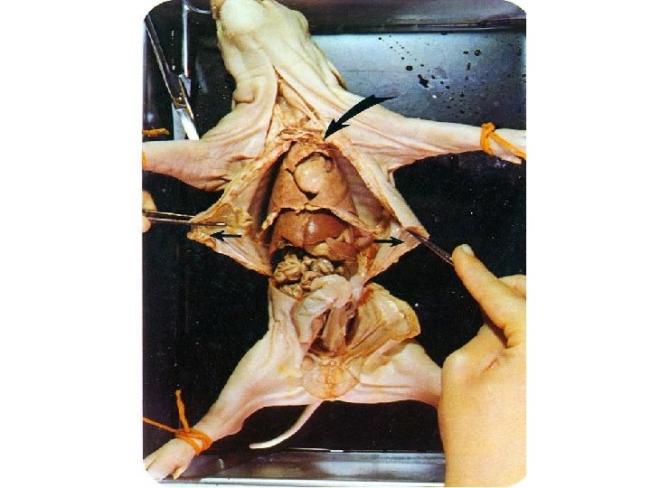
ABDOMINAL REGION 1. In order to open the body cavity, use your scissors to cut completely through the body wall beginning just in front of the umbilical cord (follow the line cut previously). As you cut anteriorly you will cut through the sternum (breastbone) - keep the tip of your scissors up so that you do not damage the underlying structures. 2. Next, use your scissors to make the parallel posterior cuts through the body wall.
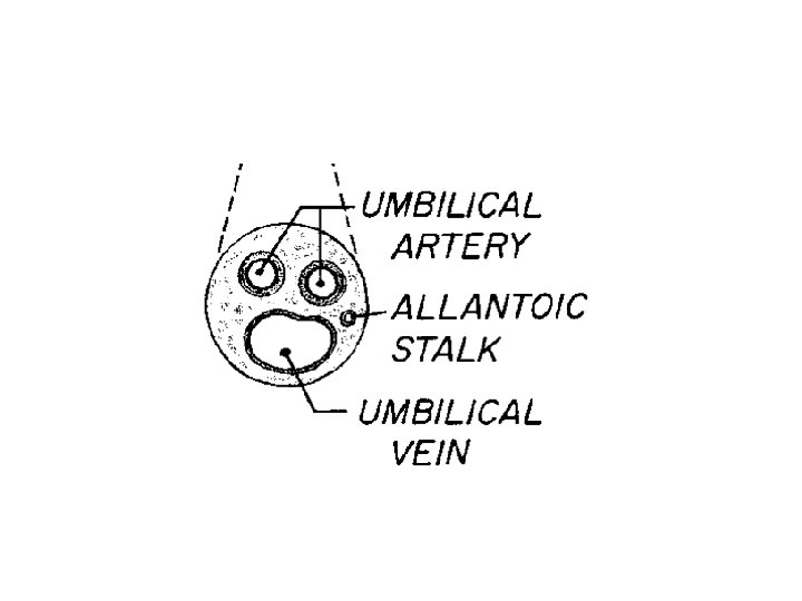
3. Umbilical Structures: A cord will be seen in the abdominal cavity extending anteriorly from the umbilical cord. This cord is the umbilical vein, which carries fetal blood from the placenta to the liver. Cut this vein about half way between the liver and the umbilical cord. Keep the position of this vein in mind as it will be traced later. Pull the flap of the body wall containing the umbilical cord posteriorly to expose the underside of the flap. On the underside note the 3 cords entering the body cavity by way of the umbilical cord. The lateral pair are the umbilical arteries which carry fetal blood to the placenta. The large sac in the center is the urinary bladder. The duct extending from the urinary bladder into the umbilical cord is the allantoic duct. This duct carries nitrogenous wastes from the bladder to the placenta. The fetus receives oxygen and food from the mother by way of the placental circulation and gives up carbon dioxide and nitrogenous wastes.
4. Cut off about 12 mm of the umbilical cord in order to observe in cross-section the 3 blood vessels and the allantoic duct. The blood vessels in the umbilical cord consist of 2 umbilical arteries (may show red latex) with relatively thick walls and an umbilical vein with a thinner wall. A fourth small vessel, the allantoic duct, is from the urinary bladder. 5. Make a drawing of a cross-section of the umbilical cord showing the 4 vessels - label your drawing.
6. Make a pair of lateral incisions through the body wall on each side in front of the hind legs (see figure one) to expose the abdominal cavity. Wash out the abdominal cavity with tap water to remove the coagulated blood present. When examining the viscera (soft internal organs) with your fingers be careful not to tear any of the structures. The body cavity (coelom) in which the thoracic and abdominal organs are located is completely lined with an epithelial layer called the peritoneum. Organs are also covered with a layer of peritoneum. This layer of epithelium is derived from the mesoderm germ layer. A double layer of peritoneum is called a mesentery. Mesenteries serve to suspend and hold structures together in the coelom.
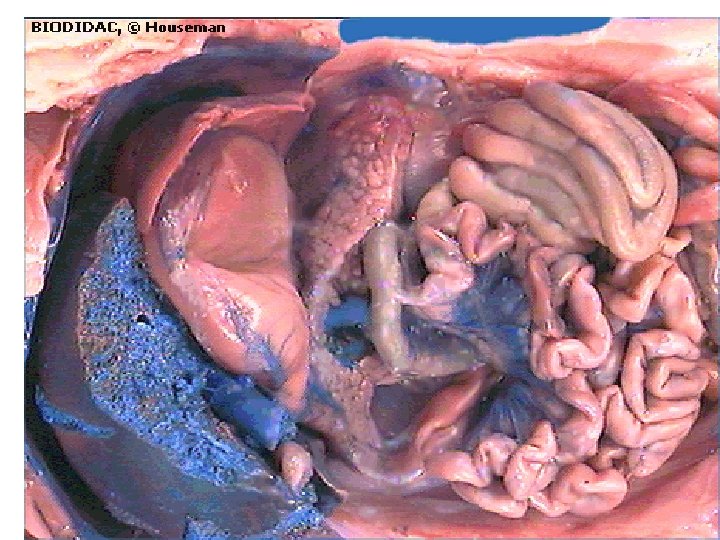
• • Once you have opened the abdomen and washed out the cavity and locate the organs listed in the paragraphs below. The most obvious structure in the abdominal cavity is the liver. The liver is composed of 5 lobes which are attached only at the dorsal and anterior margins. Posterior to the liver are the small intestine and the thicker coiled large intestine (colon). The small and large intestines are suspended from the middorsal body wall by a mesentery. Blood vessels and nerves are found between the two layers of peritoneum making up the mesentery. Carefully lift and push the small intestine forward and find where the posterior part of the small intestine enters the large intestine.
mesentery
• Put the intestines back in their normal positions and lift the liver forward to see the soft, white-walled stomach anterior to the intestines. The dark-colored spleen is located along its left posterior border and attached to the stomach by peritonium. • A light-colored granular structure, the pancreas, is found in the mesentery between the stomach and the first portion of the small intestine. The gall bladder may be seen by lifting up the extreme right lobe of the liver. It appears as a small upside down sac under the lobe. The duct from the gall bladder, the bile duct, opens into the duodenum (first portion of the small intestine). The pancreatic duct also opens into the duodenum at about the same location as the bile duct. Is it possible to find and trace the path of these ducts in your fetal pig?
• Behind the peritoneal lining of the dorsal part of the abdominal cavity are the relatively large kidneys. Cut the peritoneum along the lateral border of the left kidney and pull it off toward the midline. • Locate the muscular diaphragm, which separates the abdominal and thoracic cavities. The diaphragm is thin in the center but thicker at the periphery.
Pancreas
THORACIC REGION • In order to see the organs in the thoracic cavity, it is necessary to cut the attachment of the diaphragm to the body wall on both sides.
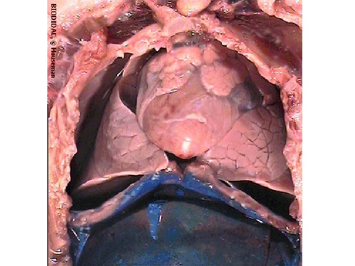
1. 2. 3. 4. 5. Cut the diaphragm away from the body wall on both sides of the pig. Force thoracic cavity open with your fingers to expose the lungs and heart. The lungs appear as solid bodies since they do not contain air in the fetus. The lobes of the left and right lungs, and the heart are surrounded by peritoneum - note the peritoneum as you force thoracic cavity open. This means that each organ is enclosed in a separate sac. The sac enclosing the hearts is called the pericardial sac, and the sac enclosing each lung is called a pleural sac. Force thoracic cavity open further to examine the lungs more closely. Note that the left lung is divided into 3 lobes and the right lung into 3 lobes plus a fourth lobe that passes ventral to a large vein and is directly posterior to the heart. The 2 large lobes of whitish granular tissue concealing part of the heart and the anterior blood vessels is the thymus gland. The thymus extends anteriorly for a short distance into the neck region. This gland is relatively large in young animals but degenerates after sexual maturity. The thyroid gland is a small gland located against the trachea just in front of the thorax. This gland is brownish in color in preserved fetal pigs.
Slide of Human Skin • The skin is composed of 2 layers; an outer, thin epidermis and an inner, thick dermis. Examine the slide under scanning and low power, and identify these 2 layers. Refer to the handout. • The dermis consists of a dense connective tissue. Identify the nuclei of the fibroblasts and the fibers in your section. Blood vessels are present in the dermis but not in the epidermis. Can you identify a blood vessel in your section? It is sometimes possible to see the red blood cells within the small blood vessels of the dermis. • Sweat glands and hair follicles are present in the dermis and extend up through the epidermis. Find a section through a hair follicle and the associated sebaceous (oil) glands. • The sebaceous glands and follicles are derived from the epidermis. The subcutaneous layer is under the dermis. This is a loose connective tissue containing many fat cells. Identify the fat cells in your section.
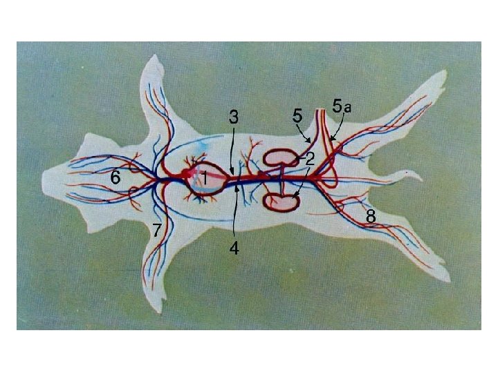
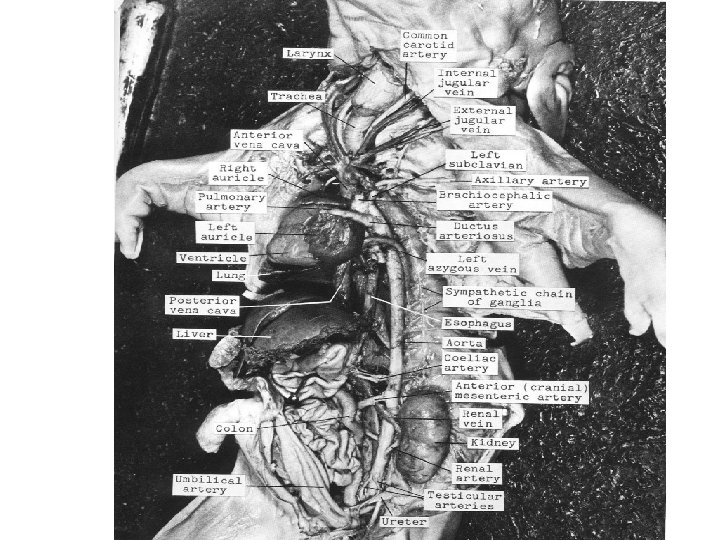
Circulatory System Page 5 • The flow of blood in the circulatory system is as follows: • heart-->arteries-->arterioles-->capillaries->venules-->veins--> heart
• Arteries and arterioles are thick-walled vessels that carry blood away from the heart, whereas veins and venules are relatively thin-walled vessels that carry blood toward the heart. Arterioles subdivide in the various tissues of the body to ultimately form capillaries. Capillaries eventually come together to form venules. The small, thin- walled capillaries are the functional units of the circulatory system. The exchange of materials between blood and the cells takes place at the capillaries.
• Blood transports oxygen from the lungs to the body tissues and returns carbon dioxide to the lungs. The right auricle and the right ventricle are involved in transporting blood to the lungs (pulmonary circulation) and the left auricle and left ventricle are involved in transporting blood to the body tissues (systemic circulation). In other words, the right side of the heart pumps deoxygenated blood to the lungs and the left side of the heart receives oxygenated blood from the lungs and pumps it to the body tissues. • Examine the demonstration slide of an artery and vein sometime during the lab period.
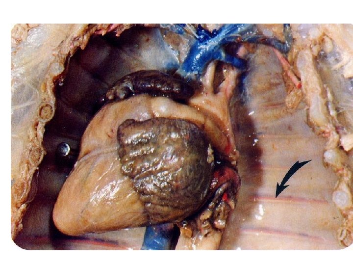
A. Examination of the Heart • Locate a pair of thickened white strands, which are present on either side of the pericardial sac. These are the phrenic nerves. Tease these nerves away from the pericardium and observe their distribution to the diaphragm. • Remove the pericardial sac from around the heart - be careful to not cut blood vessels or nerves in the process. Note that the pericardial membrane is strongly attached where the blood vessels enter and leave the heart. Identify the 4 chambers of the heart: the thin-walled right atrium (auricle), the thin-walled left atrium (auricle), the thick-walled right ventricle and the thick-walled left ventricle. Note the coronary artery and the coronary vein which are present in the diagonal groove between the 2 ventricles.
1. Major Arteries and Veins Heart • Veins • Several veins from the head, neck, shoulders and fore legs join to form the superior vena cava (anterior vena cava, precava), which enters the anterior portion of the right atrium. These veins are located ventral to the arteries supplying the same regions, but the veins are thin-walled and more difficult to trace. Pressing on the auricle with your fingers will force fluid into the veins and may help in tracing them. The muscles extending from the sternum to the larynx and head may have to be removed in order to expose the veins.
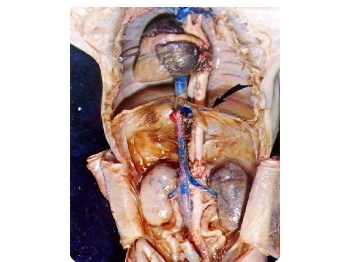
• The inferior vena cava (posterior vena cava, postcava) enters the posterior portion of the right atrium. Find where this vein penetrates the center of the diaphragm, passes through a groove in the small median lobe of the lung, and into the right atrium (it may be necessary to lift up the posterior portion of the heart). This vein returns all the blood from the posterior part of the body to the heart.
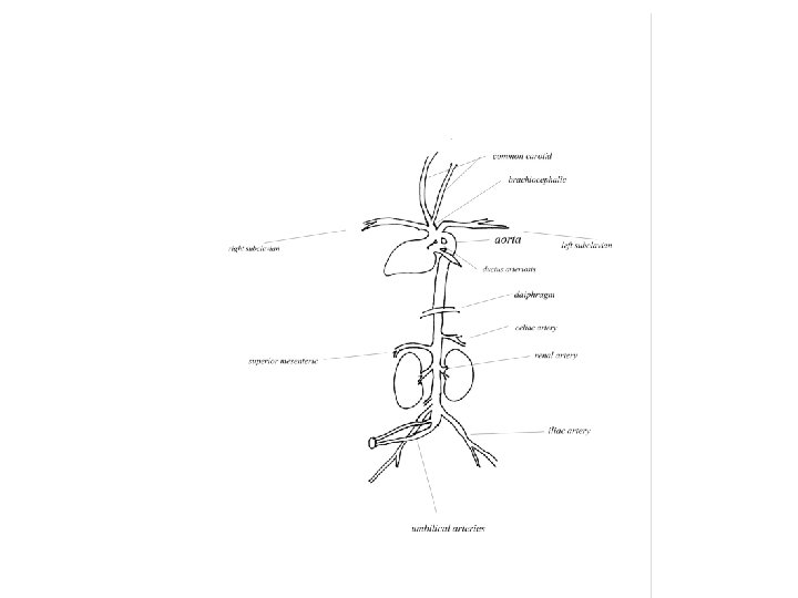
• Hidden partially behind the pulmonary artery, the aorta is a large vessel that branches into the brachiocephalic(3) and the left subclavian artery (7)
1. Pulmonary Artery
Arteries • Two large arterial trunks leave the ventricles anteriorly. The most ventral trunk is the pulmonary trunk, which transport blood directly to the lungs. This artery will be traced later. The other trunk, the aortic arch, gives off 2 main arteries, which send branches to the fore legs, shoulder, neck, and head regions. Lift the left lung to trace the aortic arch dorsally. It turns posteriorly and runs along the dorsal midline as the dorsal aorta and eventually delivers blood to the entire posterior part of the body.
Other Closely Associated Structures • Ventral to the dorsal aorta is a thick, white tube, the esophagus, which leads from the pharynx through the diaphragm and into the stomach. Along the esophagus run 2 main branches of the vagus nerve. The vagus nerves innervate thoracic and abdominal viscera. Another pair of nerve cords, bearing a series of small swelling along their course, may be seen behind the peritoneum dorsal and lateral to the aorta on either side. These are the main trunks of the sympathetic nervous system.
B. Systemic Circulation • You will identify the main branches of the systemic arteries and veins, including the hepatic portal system. This will be done in order to understand where the major organs obtain their blood supply and where the blood goes after leaving them
Systemic Arteries -Diagram • The brachiocephalic artery (innominate) is the first major artery branching off the aortic arch. Find this artery, which branches to give rise first to the right subclavian artery and then to the right and left common carotid arteries. The carotids carry all blood going to the head region. In the head they branch into the external carotids, which supply the face, and the internal carotids, which supply the skull cavity and brain (we will not trace these arteries). The second major artery to arise from the aortic arch is the left subclavian artery - note that the left subclavian artery arises directly from the aortic arch.
• Separate one of the nerve cords which run parallel to the common carotids in a common sheath. The nerves are the main trunks of the vagus nerve and sympathetic nervous system. These were seen before as separate nerves in the thoracic region. Trace the nerves on one side to the point where they separate.
• The dorsal aorta gives off a series of small segmental arteries dorsally between the ribs, and one or more small arteries ventrally to the esophagus. In order to trace the aorta into the abdominal cavity, cut directly through the diaphragm to the aorta. At the point where the aorta penetrates the diaphragm, the celiac artery arises from the aorta sending branches to the spleen, pancreas, stomach, and liver. The anterior mesenteric artery branches from the aorta and is found posterior to the celiac artery. The anterior mesenteric artery sends branches to the small intestine and the coiled portion of the large intestine.
• Dissect the left kidney free from the dorsal body wall and turn it to the right to expose more of the dorsal aorta. Locate the renal artery. Put the kidney back in its original position and continue to explore the aorta posteriorly. If your fetal pig is a female, be careful not to damage the ovaries, oviduct, and uterus during the upcoming exercises. These structures are suspended by mesenteries within the posterior region of the abdominal cavity.
• The posterior mesenteric artery (inferior mesenteric) runs ventrally from the aorta to the colon. A pair of genital arteries arise laterally to supply the reproductive organs. A pair of relatively large iliac arteries arise next and extend laterally to the hind legs. Slightly posterior to these, locate the umbilical arteries which supply blood to the bladder and placenta. After birth the umbilical arteries degenerate into a pair of small vessels supplying only the urinary bladder. The most posterior extension of the aorta, the tiny caudal artery, will be seen when the reproductive system is examined.
Systemic Veins - • Dorsal to the iliac arteries are the iliac veins which unite to form the postcava. As the postcava runs anteriorly it receives the segmental veins from the body, genital veins from the reproductive structures, and renal veins from the kidneys. Note that the postcava turns to the right around the dorsal aorta and comes to lie almost ventral to it at the level of the renal veins.
• From this point on the postcava is not easily followed since it is embedded dorsally in the extreme right lobe of the liver. Turn the intestines and liver to the left, and scrape the liver tissue away from the point where the postcava enters the liver to the point where it penetrates the diaphragm. In the anterior part of the liver, it receives several hepatic veins - at least one from each lobe - and also a small connection from the umbilical vein. This connection, which is difficult to find in the liver tissue, is lost after birth. The umbilical vein distributes itself to the lobes of the liver, where it is continuous with parts of the hepatic portal vein.
• The hepatic portal vein begins in capillaries in the small and large intestines, and ends in capillaries in the liver. The liver capillaries collect into the hepatic vein already mentioned. Find the hepatic portal vein as it leaves the center coil of the large intestine dorsally. It soon receives a large branch from the small intestine, and later a smaller one from the spleen, pancreas, and stomach. Shortly thereafter, the hepatic portal is joined by the umbilical vein and branches into the lobes of the liver. The entrance of the postcava and precava into the right atrium has already been observed.
C. Heart and Pulmonary Circulation • Removal of the Heart • If removal and dissection of the heart is done properly, the heart will retain its normal shape and the observations given below can be repeated. Keep the heart in the plastic bag with your fetal pig for review. • Determine where the precava enters the right atrium and cut this vein. Lift the heart forward to find the postcava and cut this vein about 12 mm from the atrium. Find the pulmonary veins from the left lung and trace their entry into the left atrium. Cut the pulmonary veins, which will expose the left pulmonary artery. Cut the left pulmonary artery at the surface of the lung. The aorta and its branches are now the only remaining vessels attached to the heart. Cut the brachiocephalic (innominate) and left subclavian arteries between the aorta and their first branches. Cut the aortic arch about 25 mm beyond the left subclavian artery, and remove the heart from the body.
Flow of Blood through the Heart • This next part should be done with a sheep heart, if available. If no sheep hearts are available, make sure you know the main structures that reside in the heart as well as the flow of blood through the heart.
• Find the roots of the precava and postcava. Cut through a line connecting them to open the right atrium. You can now look directly into the right ventricle through the tricuspid valve, which is usually open. Make a straight cut through the wall of the atrium, the tricuspid valve, and the outer muscular wall of the right ventricle. Examine the cavity of the ventricle and the remaining flaps of the tricuspid valve. Make another cut through the ventral wall of the right ventricle and into the pulmonary artery in one continuous line. Examine the tricuspid valve from behind, and the 3 semilunar valves at the base of the pulmonary artery.
• Probe into the right and left pulmonary arteries from the pulmonary trunk. The blood passes from the arteries into the lung capillaries which unite eventually into the pulmonary veins. Find the entrances of the pulmonary veins into the left atrium (on either side of the cut ends of the pulmonary arteries). Cut on a line between the pulmonary veins, and on through the left atrium, the same as you did through the right. Look first at the bicuspid valve, usually closed, guarding the entrance to the left ventricle. • Cut through the bicuspid valve and the outer wall of the left ventricle to the tip of the heart. You must look behind the remaining wall of the bicuspid valve to see the entrance to the aortic arch. Cut through this valve and on out through the wall of the aorta. Note in the base of the aorta, the 3 semi-lunar valves. Just above 2 of these valves will be found the openings of right and left coronary arteries, which supply blood to the tissues of the heart.
Respiratory System Page 6 • The mammalian respiratory system takes in air rich in oxygen (inhalation, inspiration) and releases air rich in carbon dioxide (exhalation, expiration). The exchange of O 2 and CO 2 in the lungs is at the level of the blood capillaries and the alveoli (air sacs). Air entering the mouth or nostrils passes into the pharynx. The pharynx is located at the back of the oral cavity and is the region where the food and air passageways cross. Air passes ventrally into the larynx and food passes dorsally into the esophagus.
Procedure 1. To expose the pharynx insert your scalpel into the corner of the mouth and cut back to the jaw bone on each side. This cuts the muscles that hold the jaws closed. 2. Separate the jaws by pushing down on the tongue and inspect the oral cavity and tongue. The tongue is attached at the back of the oral cavity. Note the small, undeveloped teeth in the upper and lower jaws. The ridged roof of the mouth is the hard palate, which separates the oral cavity from the nasal cavities. Posterior to the hard palate, the roof of the mouth becomes smooth and is called the soft palate. 3. Use your scissors to cut completely through the midline of the lower jaw and tongue. This will separate the lower jaw into 2 equal halves. Separate the halves of the jaw and examine the posterior region of the pharynx. Find the flap of tissue attached to the ventral side of the pharynx. This is the epiglottis and serves to cover the glottis (opening of the pharynx into the larynx) during swallowing. 4. Use your scissors to continue the mid-ventral cut into the larynx and trachea. Determine the relationship of the larynx to the esophagus. Expose the trachea back into the thorax as far as the right lung. Cut the trachea open for a short distance in order to see the rings of cartilage in the tracheal wall. These rings serve to hold the trachea open. 5. Cut the bronchial tubes (which connect the trachea to the lungs) close to the lung and remove the right lung from the chest cavity by cutting any remaining mesenteries. Identify at the root of the lung the cut ends of the pulmonary veins and arteries and the bronchial tubes. Note thickness of the wall and the diameter of the vessels and tubes. Also, note that the median lobe of the lung, which partially surrounds the postcava, is actually a part of the right lung.
VENTILATION • Bronchial tubes in the lungs contain smooth muscle, but no skeletal muscle, therefore, the expansion and contraction of lungs during breathing results from movement of the ribs, diaphragm, and other muscles.
• During inspiration the thoracic cavity is expanded by: • Contraction of the intercostal muscles between the ribs. Contraction of these muscles enlarges the thoracic cavity by lifting the ribs upward and outward. • Contraction of the muscles of the diaphragm. Contraction of these muscles lowers the diaphragm to enlarge thoracic cavity. The enlargement of the thoracic cavity results in decreased air pressure in the lungs. Air enters the lungs from the outside to equalize the pressure.
• During expiration the thoracic cavity returns to normal by: • Gravity, which pulls the ribs down when the intercostal and diaphragm muscles relax. • Contraction of the abdominal muscles (and certain chest muscles other than the intercostals), which force the diaphragm up and the thoracic cavity down. Rapidly exhale some air in order to feel the chest constrict and the abdominal muscles tighten.
Digestive System Page 7 • The mammalian digestive system is concerned with processing and absorbing food and water for the body. The system begins with the mouth and ends with the anus. Ingested food is digested and the products are absorbed in the digestive tract; undigested material is expelled from the anus.
Procedure 1. Examine the region of the pharynx - locate the origin of the esophagus and follow it posteriorly. Cut through the diaphragm to find the point where the esophagus joins the stomach. The sac-like stomach is a somewhat J-shaped organ. It is divided into 2 regions; a larger cardiac region into which the esophagus opens and a smaller pyloric region which opens into the small intestine. 2. Note the greater curvature of the stomach and open the stomach by cutting along this curvature. Examine the stomach internally and note that a gross difference between the cardiac and pyloric regions is not evident. 3. Cut through the pyloric valve and into the anterior portion of the small intestine, the duodenum. Note the heavy muscle in the wall of the pyloric valve; this is called the pyloric sphincter. This sphincter controls the amount of partially digested food passing into the small intestine from the stomach.
4. The first 50 -75 mm of the small intestine is called the duodenum. Recall that the pancreatic duct and the bile duct empty into the duodenum. Begin the removal of the small intestine by carefully cutting the mesentery holding the coils together. This will allow a gradual unwinding of the intestine. Estimate the length of the small intestine in your fetal pig. The small intestine in man is approximately 20 feet long. The portion of the small intestine following the duodenum is the jejunum, which is about 2 of the length of the intestine. The remainder of the intestine is called the ileum.
5. Cut open a short length of the ileum and examine the lining. Note that the lining is folded to form macroscopic folds. The folds are covered with numerous small finger-like projections, which are barely visible to the naked eye. These projection are called villi. The folds and villi serve to greatly increase the surface area of the intestine. 6. Locate where the small intestine unites with the large intestine. Extending to the left of this junction is a blindly ending pouch (1225 mm long), the cecum. Unravel the large intestine and estimate the length of this portion of the digestive tract.
Excretory System Page 8 • Nitrogenous waste products produced by cell metabolism are removed from the blood stream by the kidneys. The kidneys, urinary bladder, and associated ducts make up the excretory system. The process of removing nitrogenous wastes from the blood is called excretion. Recall that blood is supplied to each kidney by a renal artery. The blood leaves each kidney by a renal vein.
Procedure 1. Remove the left kidney and cut it lengthwise into 2 equal halves. The kidney will appear in gross section to be composed of 3 regions: an outer granulated region, the cortex; a radially striated layer, the medulla; and an inner cavity, the renal pelvis. Each kidney contains over 1 million functional units called nephrons (renal tubules). The blind end of each nephron is located in the cortex where it forms a cup-like structure called a Bowman's capsule. The capsule encloses a ball of capillaries, the glomerulus. A renal corpuscle is a glomerulus surrounded by a Bowman's capsule. The nephrons extend through the medulla to the renal pelvis where the urine is collected. 2. The renal pelvis empties into the urinary bladder through the ureter. The ureter runs posteriorly to the urinary bladder. Expose this duct without damaging the structures crossing over it. These include the umbilical arteries and the reproductive ducts in both sexes, and the ovaries just posterior to the kidneys in females. The bladder empties to the outside through another duct, the urethra. This duct will be observed when the reproductive system is dissected. In the case of the fetus, wastes are transported via the allantoic duct to the placenta, where they are removed by the mother's blood.
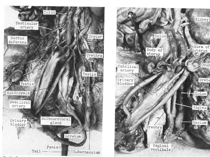
Reproductive Systems Page 9 • The reproductive and excretory systems of vertebrates have a close connection with one another. In the males, the same duct transports the sperm and urine. The male and female reproductive organs of vertebrates have the same embryonic origin; it is during development that they become different in structure and function. The male and female reproductive systems are composed of the sex glands (gonads) and their associated ducts and glands.
• Each student will be responsible for knowing both the male and female reproductive systems.
Female Reproductive System • The female gonads are the 2 ovaries, which are located posterior to the kidneys. Each ovary is suspended in the abdominal cavity by a mesentery. A small convoluted tube, the oviduct, is located lateral to each ovary. This tube is also called the Fallopian tube. The oviduct continues on each side as a slightly larger tube forming one of the 2 horns of the uterus. Pig embryos develop in the uterine horns, which become greatly enlarged at maturity and during pregnancy. The 2 horns of the uterus unite to form the body of the uterus, which lies dorsal to the urethra. In humans the fetus develops in the body of the uterus and there are no uterine horns.
Procedure 1. In order to separate the hind legs, use your scalpel to cut through from the ventral midline between the legs, . The cut will pass through muscles and through the pelvic bones. Be careful when you are cutting the pelvic bone it is easy to cut structures you need to identify. One way to do this is to cut half way through the pelvic bone and spread the legs apart to break the bone the rest of the way. Then use your blunt probe to push away the muscles and other tissue. If you have any trouble doing this ask for help from your lab instructor. 2. Three tubes will be exposed which are from ventral to dorsal - the urethra, part of the reproductive tract, and the rectum. The urethra and the reproductive tract unite about 12 mm before the vulva and form a common passage, the vestibule (urogenital canal). The tube connecting the vestibule and the body of the uterus is called the vagina. 3. Open the vestibule and vagina along one side and locate the opening of the urethra and the constriction of the vagina at the base of the uterus. 4. Expose the rectum and open it by cutting anteriorly starting with the anal opening. Locate the anal sphincter muscle, which is located at the distal end of the large intestine. Find the caudal artery which is against the spinal column and dorsal to the rectum.
Operation of the Female Reproductive System: • Ova develop in follicles within the ovaries. When fully developed, the ova are released from the follicles and enter the oviduct, where fertilization may take place. After fertilization, cleavage of the zygote begins and eventually the embryo becomes implanted in the uterine horn (or uterus in humans). The placenta is formed from uterine tissue and from 2 extraembryonic membranes (chorion and allantois) formed by the embryo. Fetal development continues in the uterine horn (uterus in humans), until birth. During birth, the offspring pass through the uterine horn to the body of the uterus, to the vagina, and to the vestibule on their way out of the body.
Male Reproductive System • The location of the male gonads, the testes, depends on the age of the fetus. The testes begin their development in the same location as the ovaries, but prior to birth start to descend into the scrotal sacs (scrotum). They will be found somewhere along this path in your fetal pig. The sperm ducts (vas deferens) will be seen looping over the umbilical arteries and the ureters, and then joining together dorsal to the urinary bladder. Posteriorly, the sperm duct pass through the abdominal wall on either side of the midline via the inguinal canals. Identify these structures before you complete the following procedure.
Procedure 1. Cut through the skin of one of the scrotal sacs and extend the cut to the point find where the sperm duct leaves the abdominal cavity. This will open the inguinal canal and the testis will be exposed. 2. Open the sac that surrounds the testis. The convoluted tubule looping around the testis is the epididymis, which empties into the single sperm duct. 3. Cut through the skin slightly to one side of the ventral midline to expose the slender penis extending from the anal region to the urinary orifice. The central tube of the penis is the urethra. This tube arises from the urinary bladder. 4. To expose the remaining structures of the reproductive system, it will be necessary to cut through the tissues in the midline between the legs and break the pelvic bone (refer to step 1 for the female reproductive system). The legs can be spread apart as the cut is deepened to the level of the urethra. The urethra appears somewhat thicker in males than in females due to the presence of accessory glands that are closely associated with the urethra. The Bulbourethral glands, which are about 12 mm long, are lateral to the urethra. The seminal vesicles are located where the ductus deferans and the urethra unite. 5. Dissect the urethra and the accessory glands away from the rectum to determine where the sperm ducts enter the urethra. The urethra transports both sperm and urine in the male reproductive system. 6. Expose the rectum (which is located just dorsal to the urethra) and open it by cutting anteriorly starting at the anal opening. Locate the anal sphincter muscle which is located at the distal end of the large intestine. Is it possible to find the small caudal artery which is located against the spinal column and dorsal to the rectum?
Operation of the Male Reproductive System • In the male reproductive system, sperm (spermatozoa) are formed in the seminiferous tubules of the testes. Sperm produced in these tubules are stored in a connecting tubule, the epididymis. The sperm, together with secretions from the accessory glands (Bulbourethral glands, seminal vesicles), form the semen. Semen is released via the urethra in a process called ejaculation. After the semen is deposited in the vagina of the female, some of the sperm eventually reach the distal end of the Fallopian tube where fertilization of the ova may take place.
Female reproductive organs
Nervous System Page 10 • The nervous system is made up of the central nervous system (brain and spinal cord) and the peripheral nervous system (cranial, spinal, and autonomic nerves). The nervous system can be subdivided into 2 distinct parts based on function. One part of this system is the somatic (voluntary) nervous system. It is under conscious control and is composed of nerve cells of the brain, nerve cells receiving stimuli from major sense organs, and nerve cells that stimulate striated muscles. The other part of the nervous system is the autonomic nervous system, which is not under voluntary control. The autonomic system controls activities such as digestion, excretion, secretion, and circulation. The organs that are controlled by this system are the heart, smooth muscles, and glands. Activities of these organs are also influenced by hormones.
• The autonomic nervous system is subdivided into the sympathetic and parasympathetic nerve systems. Both of these systems innervate the internal organs, and the actions of these 2 systems oppose one another. In general, the sympathetic nervous system stimulates the heart and decreases the activity of the digestive system and associated organs. On the other hand, the parasympathetic system tends to decrease heart activity and stimulate activity of the digestive system and associated organs.
• Procedure • First remove the remaining skin from the head and neck of your pig and cut off the ears. Remove what remains of the upper and lower lips. • Turn the pig on its side with the left side up. • Dissect away the membrane of the dorsal surface of the lower jaw in order to see the teeth still present in the jaw. Remove one of the teeth and note that the cavity of the tooth is filled with a jellylike substance.
• The top and side of the skull must be removed to see the brain. Start at the top of the skull and carefully remove the bone, a small piece at a time. In order to keep the brain from being damaged, free the tough covering of the brain from the skull bone before it is completely broken away. The coverings of the brain are called the meninges. The tough outer layer is the dura mater. The finer inner layer, which closely follows the contours of the brain, is the pia mater.
• Several bones of the skull will be identified at this time. A pair of frontal bones are located immediately between the orbits (a single bone in man). Posterior to the frontal bones are the parietals. The cerebrum of the brain is underneath the frontal and parietal bones. The cerebrum is composed of 2 cerebral hemispheres. The nasal bones are anterior to the frontal bones and cover the olfactory organs. Parts of the sphenoid bone will be removed in the region of the orbits as well as the temporal bone, which is posterior to the orbits. The occipital bones make up the posterior region of the skull. It will be necessary to remove the neck muscles to expose and remove the occipital bones. The cerebellum and medulla regions of the brain are located underneath the occipital bones.
• Identify the skull bones listed above on the human skeleton located in the room. Refer to the figure of a human skull provided. • Make a vertical cut in the olfactory organ after the nasal bones have been removed. The numerous folds of olfactory organ are covered with sensory epithelium. Cut into one of the nasal cavities from above and follow the cavity posteriorly to the olfactory organ and then to the pharynx. • Following exposure of the cerebrum, cerebellum and medulla, cut the brain into 2 equal halves and remove the left half. It is now possible to observe the relationship of the brain stem (including the medulla) to the cerebrum and cerebellum. The pituitary gland, which is located in a small pit in the floor of the skull, may be observed immediately in back of the optic nerves.