The Integumentary System Integument means COVERING The integumentary


")

 Composed of epithelial")


 Keratinocytes (covering) 2)")
 Keratinocytes most # cells n Produce a fibrous protein called KERATIN n Formed")
 the pigment Melanocytes n Melan-black n Can transfer melanin to")
 Melanin")
 Everyone has the same number of melanocytes but make")


 Langerhans’ cells Participating in the immune response by stimulating defense against: 1) microorganisms")
 Merkel cells n Has a spiked appearance n Connected to nerve cells from")

")


 – Softens and lubricates hair and skin – Slows")


 – 5%")


- Slides: 82
The Integumentary System Integument means COVERING.
The integumentary system n Many functions, most but not all are PROTECTIVE § Insulates & cushions the deeper body organs & protects entire body from mechanical damage (bumps & bruises), chemical damage (such as acids & bases), thermal damage (heat & cold) ultraviolet damage (sunlight) and bacteria. The uppermost layer is keratin & cornified, or hardened, to help prevent water loss from body surface.
n The skin’s rich capillary network & sweat glands (both controlled by nervous system) play an important role in regulating heat loss from the body surface. It acts as a mini excretory system: urea, salts & water are lost when we sweat. It manufactures protein important to immunity and synthesizes Vitamin D (modified cholesterol molecules located in skin are converted to Vitamin D). Finally, cutaneous sensory receptors (touch, pressure, temperature & pain) provide us with information from external environment.
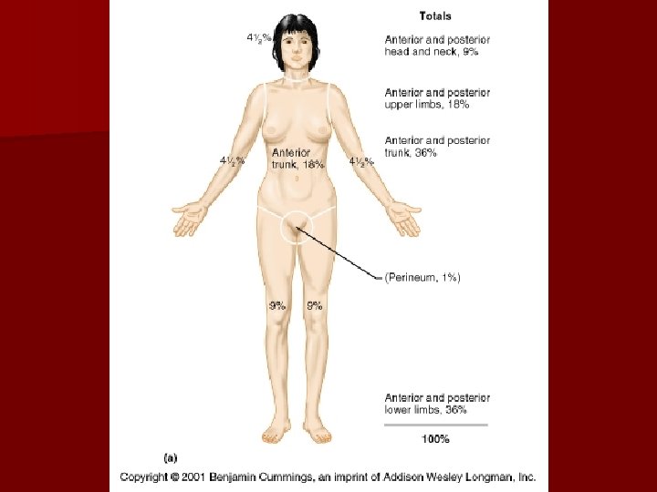
What are the major characteristics of the skin? n Waterproof, stretchable, washable, and permanent-press, that automatically repairs small cuts, rips and burns and is guaranteed to last a lifetime. n Surface area of up to 2. 2 square meters n 11 pounds n 7% of total body weight n Pliable yet tough
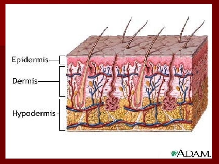
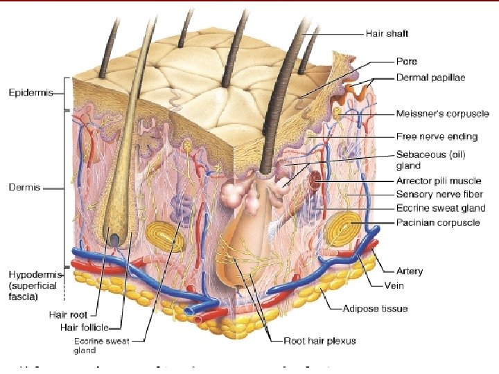
What are the major layers of the skin? n Epidermis (epi-upon) Composed of epithelial tissue (stratified squamous) Non-vascularized n Dermis – underlies the epidermis Tough leathery layer composed of fibrous connective tissue, blood & lymph vessels, sensory Tissue and glands (sweat & sebaceous) n Hypodermis (not considered skin) – Made of adipose and areolar tissue – Stores fat, anchors skin, protects against blows
Epidermis Dermis Basement membrane
n The epidermis and dermis are firmly connected. However a burn or friction may cause them to separate allowing interstitial fluid to accummulate in the cavity between the layers which results in a blister.
Epidermis Dermis Hypodermis
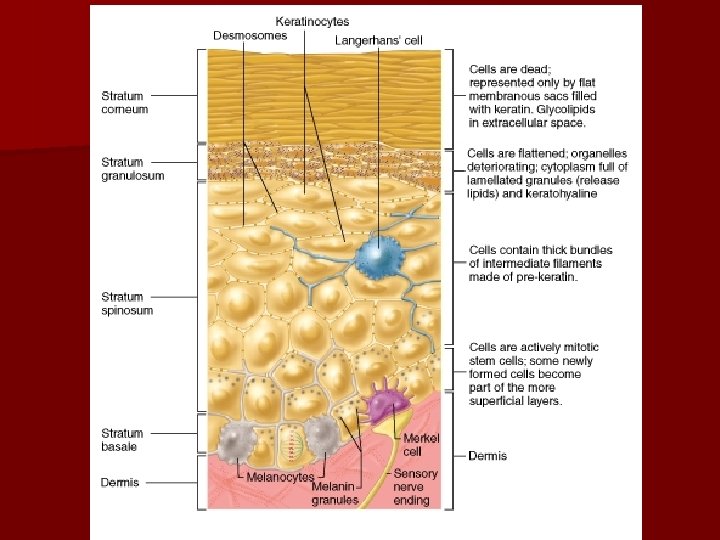
The epidermis n Composed of 5 layers or strata n Avascular * This explains why a man can shave daily and not bleed even though he cuts off many cell layers each time he shaves.
Thin or thick skinned? We all have thin & thick skin !!! Thick skin Areas of presssure or friction. Palms of hands Soles of feet Tips of fingers Has all 5 strata Thin skin Covers rest of body More flexible Each stratum contains fewer layers of cell
Thick & thin only refers to epidermis n In areas of excessive friction or pressure the outer most strata of the epidermis increases in layers causing a callus. § The skin over bony prominences may develop a cone shaped structure called a corn.
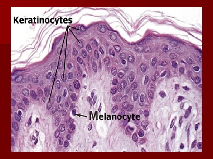
What are the different types of cells in the epidermis? 1) Keratinocytes (covering) 2) Melanocytes (color) 3) Langerhans’ cell (immunity) 4) Merkel cells (receptors)
1) Keratinocytes most # cells n Produce a fibrous protein called KERATIN n Formed in the lowest levels of the epidermis. n Pushed upward by the production of new cells beneath them. n Become dead and scale-like making a tough protective layer n Millions rub off everyday
n Synthesizes melanin 2) the pigment Melanocytes n Melan-black n Can transfer melanin to keratinocytes n Protects skin from ultraviolet light. Sun stimulates production of melanin- tanning n Freckles & moles are accummulated melanin melanocyte Melanin in keratinocytes
What causes the color of skin? 3 pigments contribute to skin color 1) Melanin 2) Carotene 3) Hemoglobin
Melanin- protein pigment (natural sunscreen) Everyone has the same number of melanocytes but make varying amounts and colors (differences in skin color) Carotene-yellow to orange pigment found in carrots. § Most commonly found in the palms or soles. Most intense when large amounts of carotene-rich foods are eaten. Hemoglobin- Red blood gives a pinkish hue to fair skin
n When hemoglobin is poorly oxygenated both the blood and skin of Caucasians appears blue. This is called CYANOSIS. In darker skinned people it is not as apparent due to the masking effects of melanin. It is apparent in the mucous membranes and nail beds.
Other color variations n Redness or erythema- inflammation, allergy, fever, embarrassment n Pallor- emotional stress some people become pale. Also could be due to anemia, impaired blood flow to area. n Jaundice- abnormal yellow skin tone due to liver disorder.
Bruises or black & blue marks- blood has escaped from the circulatory system and clotted in tissue. aka HEMATOMA
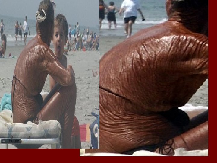
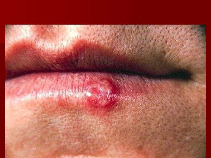
Homeostatic imbalance & the SUN n Despite melanin’s protective effects excessive sun exposure eventually damages the skin. It causes the elastic fibers to clump, leading to leathery skin. n It also depresses immune system. Those infected with the herpes simplex, or cold sore, virus are more likely to have an eruption after sunbathing. n DNA can also be altered leading to skin cancer.
3) Langerhans’ cells Participating in the immune response by stimulating defense against: 1) microorganisms 2) superficial skin cancers Langerhans’ cell
4) Merkel cells n Has a spiked appearance n Connected to nerve cells from dermis n Function as sensory receptors for touch.
The layers of the epidermis n deepest layer of the epidermis, undergoes rapid cell division. n intermediate layer, contain spiny shaped keratinocytes. n outermost layer 20 -30 cells thick of dead keratinized cells. – Dandruff- excessive shedding of scalp cells – Average person shed 40 pounds of these cells in their lifetime. – Everything you see on a human is dead!
What are the characteristics of the dermis? n Divided into 2 layers Papillary (upper) & reticular (lower) layer n Made up of connective tissue n Richly supplied with blood vessels and lymph vessels n Has hair follicles, sensory receptors, sebaceous (oil) and sweat glands. n Ridges formed from the papillary layer can form finger prints.
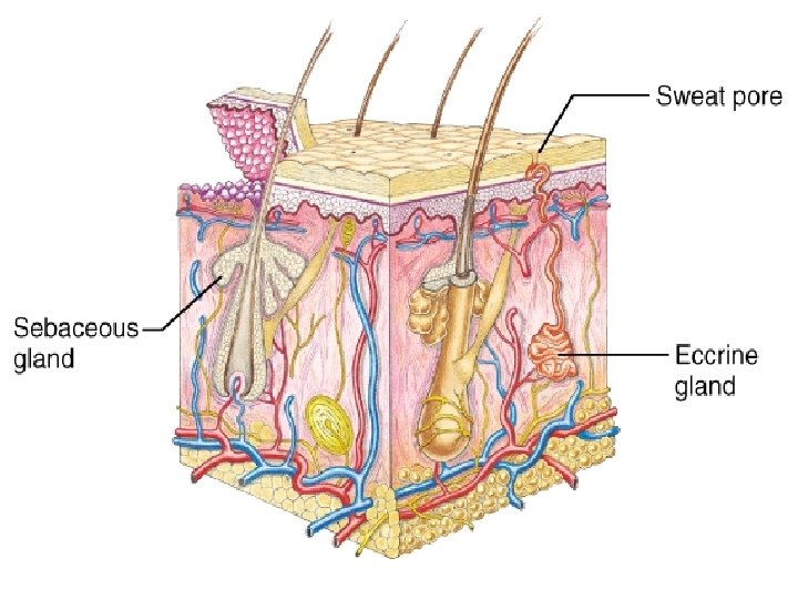
What are the major appendages of the skin? n Sweat glands What is sweat: water, salts, vitamin C, urea & even lactic acid It is acidic with a p. H from 4 -6 n Sebaceous glands n Hairs n Nails
What are the types of glands found in the skin? n Sweat glands- sudoriferous glands – Eccrine- common sweat glands § thermoregulation – Apocrine- produce sweat plus a milky or yellowish substance composed of fat and protein. § Found in the arm pits and genitalia § Thought to be scent glands.
Sebaceous glands- oil glands (sebum) – Softens and lubricates hair and skin – Slows water loss and kills bacteria Ceruminous glands- produce cerumen (ear wax) Mammary glandsproduce milk
Whiteheads & blackheads
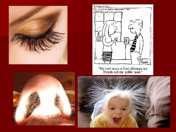
Why is hair useful? n Senses insects that land on the skin. n Hair on the head protects the head from a blow, sunlight and heat loss. n Eyelashes shield the eye n Nose hairs filter the air
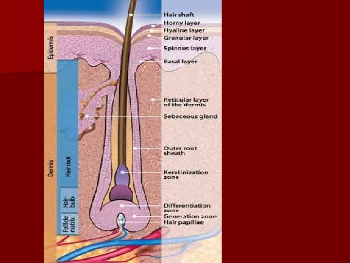
What are hairs? n Made from hair follicles n Made of dead keratinized skin cells n Two parts shaft and root n Shaft has 3 layers of cells – Medulla(central core) – Cortex (bulky layer) – Cuticle (heavily keratinized; protects hair)
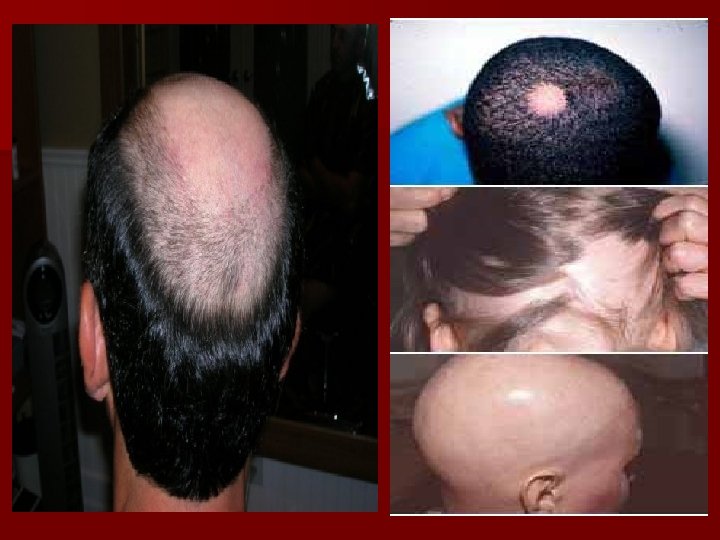
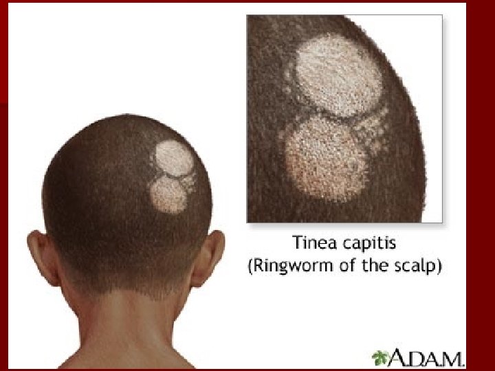
ALOPECIA- LOSS OF HAIR n DUE TO ANXIETY, PROTEIN DEFICIENT DIETS, THERAPIES LIKE CHEMOTHERAPY, RADIATION, EXCESSIVE VITAMIN A, CERTAIN FUNGAL DISEASES (RINGWORM), IMMUNITY, n MAY ALSO BE DUE TO GENETICS AS IN MALE PATTERN BALDNESS (ANDROGENETIC ALOPECIA) n RECENT STUDIES HAVE INDICATED THE CHROMOSOME # 20 AND THE X AS CONTRIBUTERS TO GENETIC ALOPECIA
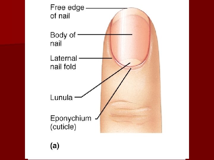
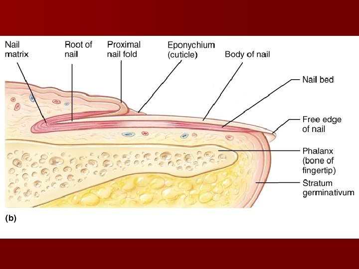
What are the parts of nails? n. A nail is a scale like modification of the epidermis n Made of tightly compressed keratinized cells n Useful tools to pick up small objects or scratch an itch. n Nail matrix is the region responsible for nail growth.
What are the primary functions of the Integumentary System? n Protection: provides 3 types of barriers – Chemical barriers: low p. H of skin secretions slows bacterial growth. Human defensin is an antibiotic that destroys bacteria (produced by human skin)
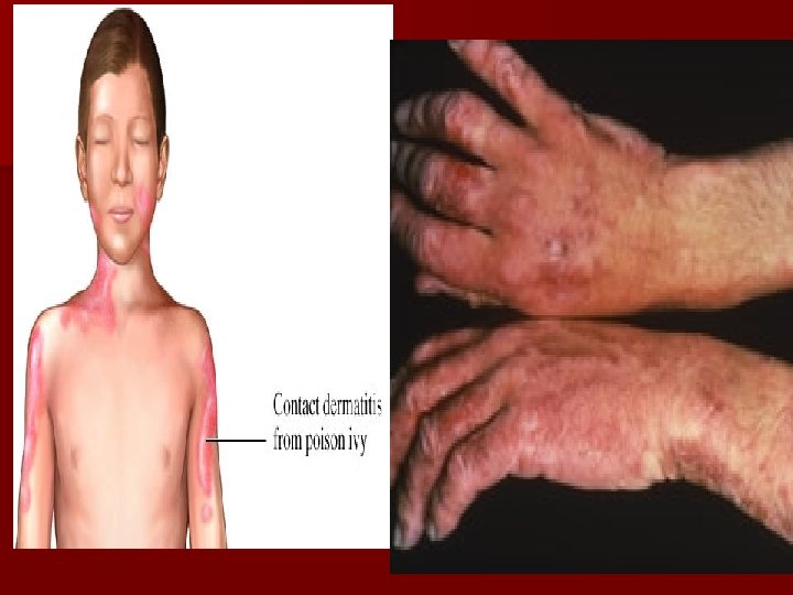
Physical barriers – Physical barriers: very few substance are able to enter the skin. Substances able to pass. § Lipid-soluble substances: oxygen, carbon dioxide, some vitamins § Oleoresins- poisons (poison ivy) § Organic solvents- dry-cleaning fluid, paint thinner § Salts of heavy metals- lead, mercury, nickel § Penetration enhancers- drug agents that help substances into the body.
Biological barriers n Langerhans’ cells- act as macrophages police the epidermis for viruses and bacteria.
Functions cont. Thermoregulation- skin contains sweat glands that secrete watery fluid, that when evaporated, cools the body. n Sensation- Skin contains sensory receptors that detect cold, touch, and pain. n Vitamin D synthesis- cholesterol in the skin is bombarded by sunlight and converted to vitamin D (calcium cannot be absorbed from digestive tract) n
Functions cont. n Blood reservoir- blood will be moved from skin to muscles during strenuous activity. n Excretion- Sweating is an important outlet for wastes such as salt and nitrogen containing compounds. (urine)
Skin Cancer n Benign tumors such as warts and moles are not serious. n Malignant tumors can start on the skin and invade other body areas. n Crucial risk factor- overexposure to UV radiation
Types of Skin Cancer n Basal cell carcinoma- most common, 30% of all white skin people get it. – – – n Arises from the stratum basale layer of the skin 99% curable if caught early Dome shaped nodules that form an ulcer in the center. Squamous Cell carcinoma– – – Arise from stratum spinosum Grows rapidly and metastasizes if not removed Small red rounded elevation on the skin
Skin Cancer Types cont. n Melanoma – Cancer of melanocytes (very dangerous) – 5% of skin cancers but rising fast – Can arise from preexisting moles – Appears as a spreading brown or black patch – Chance of survival is poor if the lesion is greater than 4 mm thick
Basal Cell Carcinoma
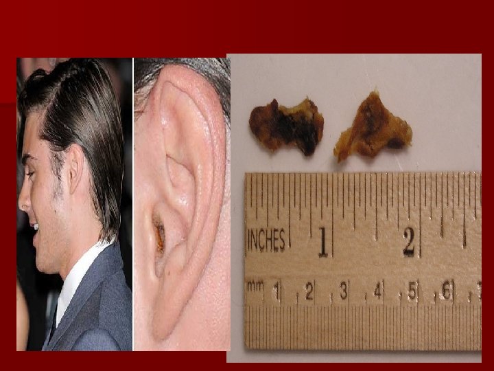
Lesion removed from patient Basal Cell Carcinoma
Squamous cell carcinoma
Melanoma
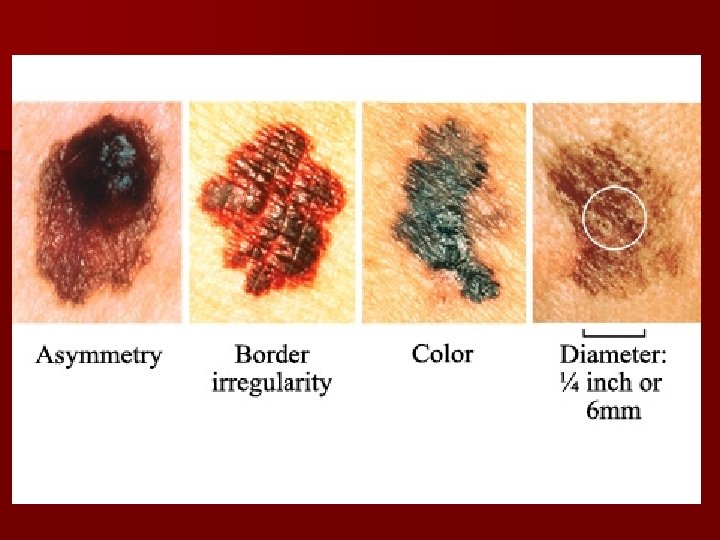
What is the ABCD rule? n Used for recognizing melanoma n A-Asymmetry: two sides of the pigmented mole do not match n B-Border irregularity: borders are not smooth n C- Color: lesion has a multiple of colors n D- Diameter the spot is larger than 6 mm in diameter (size of a pencil eraser)
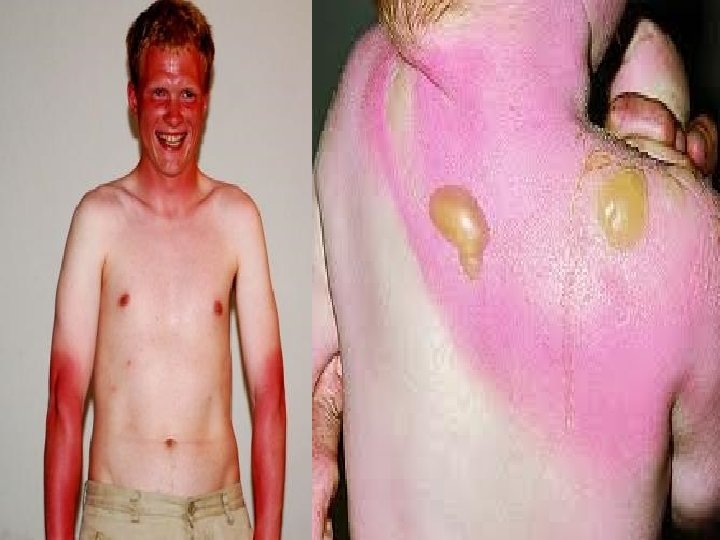
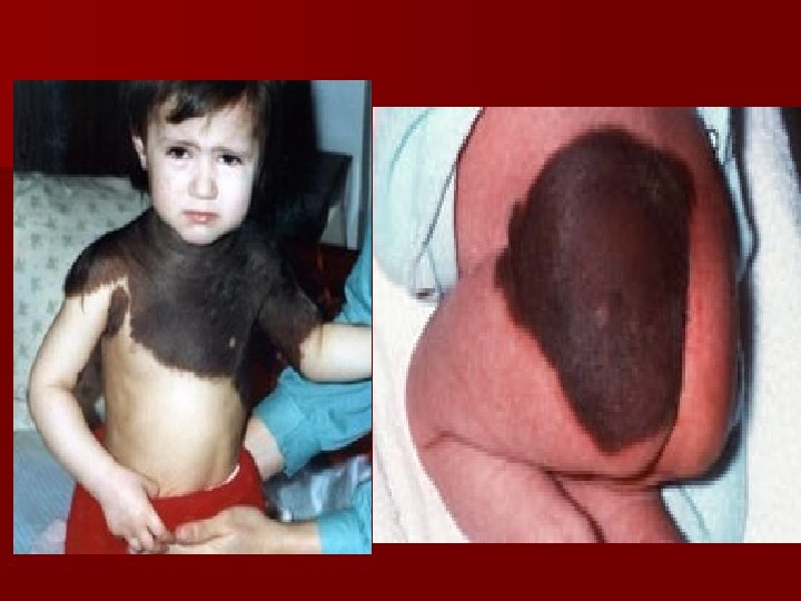
What are the 3 types of burns? n First-degree burns: only the epidermis is damaged. Redness, swelling and pain are common. (sunburn) 2 -3 days to heal n Second-degree burns: epidermis and upper layers of dermis. Blistering can occur. 3 -4 weeks to heal. n Third-degree burns: involves the entire thickness of the skin. (pg. 164 -165)
Second-degree burns Third-degree burn
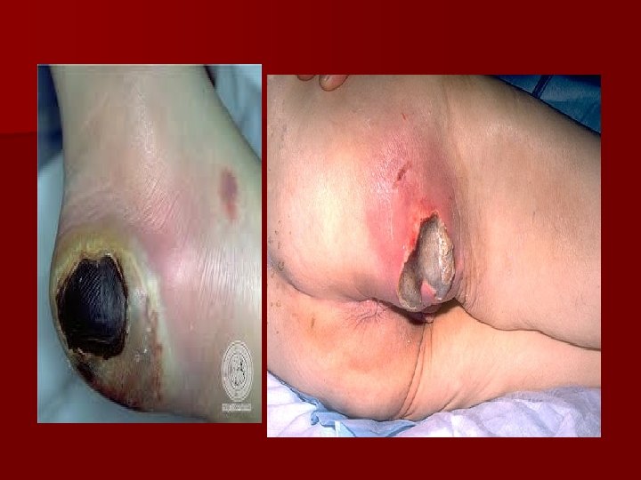
Decubitus ulcer
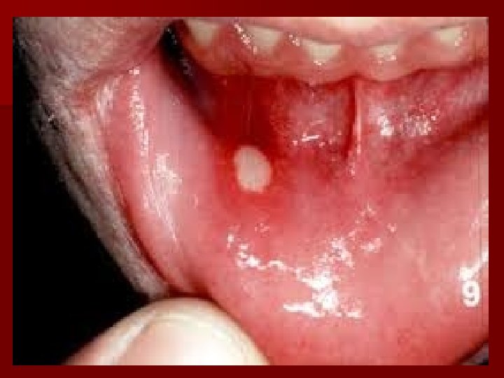
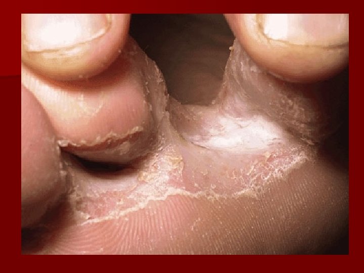
IMPETEGO
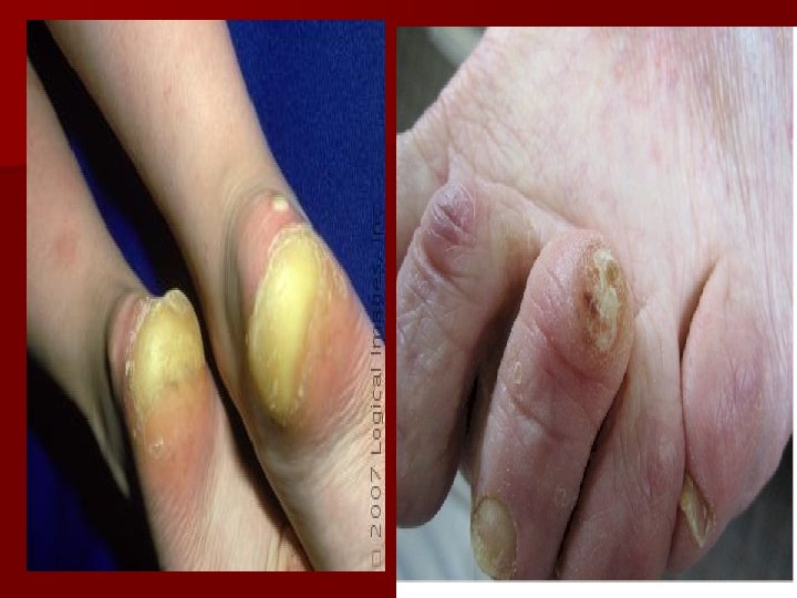
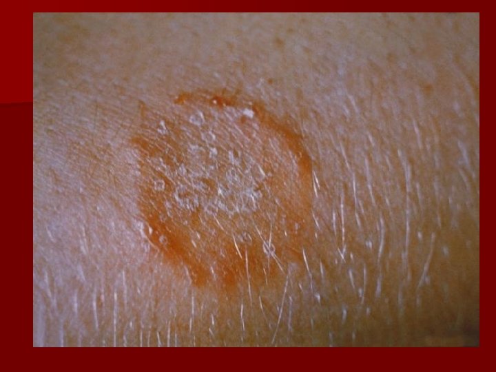
psoriasis
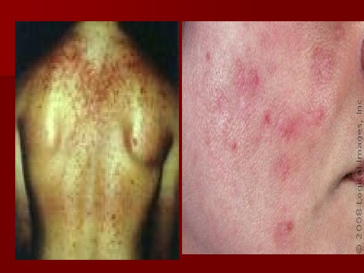
ECZEMA
Lipoma