SKIN Definition Skin is a soft outer covering


- Slides: 47
SKIN
Definition • Skin is a soft outer covering of an animal, in particular a vertebrate. It is continuous with the mucous membrane at the orifices of the body. • It is having many functions , so it is regarded as an organ of the body.
Synonyms • Cutis – Cutaneous. • Derma – Dermatology, Dermatomes. • Integumemt.
Surface area & thickness • Surface area in adult – 1. 5 to 2 sq meters. • Thickness varies from 0. 5 to 3 mm. • In assessing burns rule of nine is used – head & neck 9%, each upper limb 9%, front of trunk 18%, back of trunk 18%, each lower limb 18%, perineum 1%.
Pigmentation of skin • • • Melanin – Brown colour. Melanoid - Brown colour. Carotene – Yellow to orange colour. Haemoglobin – Purple colour. Oxyhaemoglobin – Red colour. Colour is red where keratin is thin & white where keratin is thick.
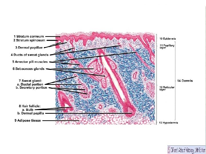
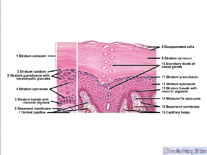
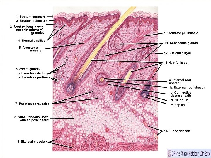
Layers of skin • Mammalian skin is composed of two primary layers: • • The epidermis, which provides waterproofing and serves as a barrier to infection; • • The dermis, which serves as a location for the appendages of skin.
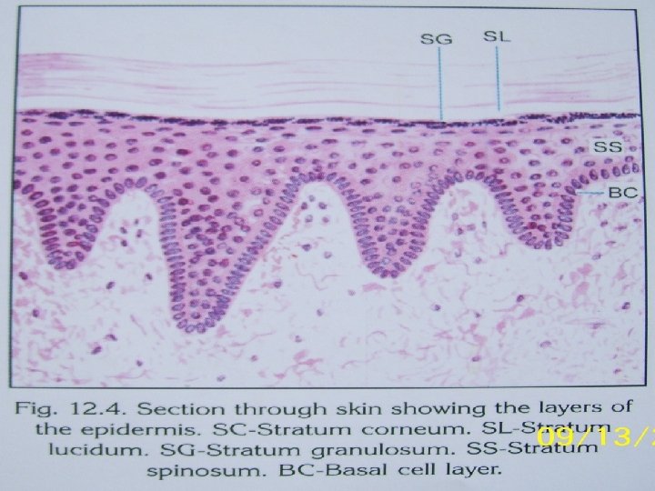
Epidermis • Epidermis, "epi" coming from the Greek meaning "over" or "upon", is the outermost layer of the skin. It forms the waterproof, protective wrap over the body's surface and is made up of stratified squamous epithelium with an underlying basal lamina. • It is avascular layer with stratified squamous epithelium.
Sublayers of Epidermis • Epidermis is divided into the following 5 sublayers or strata: • • Stratum corneum • • Stratum lucidum • • Stratum granulosum • • Stratum spinosum • • Stratum germinativum (also called "stratum basale")
Dermis • The dermis is the layer of skin beneath the epidermis that consists of connective tissue and cushions the body from stress and strain. The dermis is tightly connected to the epidermis by a basement membrane. • It is vascular layer made up of connective tissue.
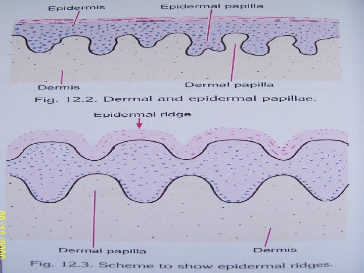
Parts of dermis • The dermis is structurally divided into two areas: a superficial area adjacent to the epidermis, called the papillary region, and a deep thicker area known as the reticular region.
Papillary region • The papillary region is composed of loose areolar connective tissue. This is named for its fingerlike projections called papillae, that extend toward the epidermis. The papillae provide the dermis with a "bumpy" surface that interdigitates with the epidermis, strengthening the connection between the two layers of skin.
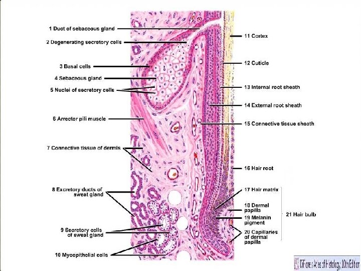
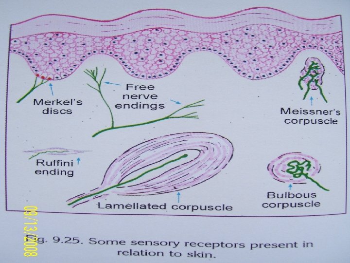
Reticular region • The reticular region lies deep in the papillary region and is usually much thicker. It is composed of dense irregular connective tissue, and receives its name from the dense concentration of collagenous, elastic, and reticular fibers that weave throughout it. These protein fibers give the dermis its properties of strength, extensibility, and elasticity. Also located within the reticular region are the roots of the hair, sebaceous glands, sweat glands, receptors, nails, and blood vessels.
Surface irregularities of skin • Tension lines • Flexure lines OR skin creases • Papillary ridges OR friction ridges (Fingerprints – Loops, whorls, arches)
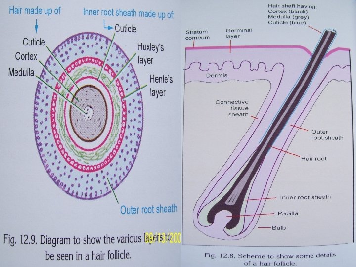
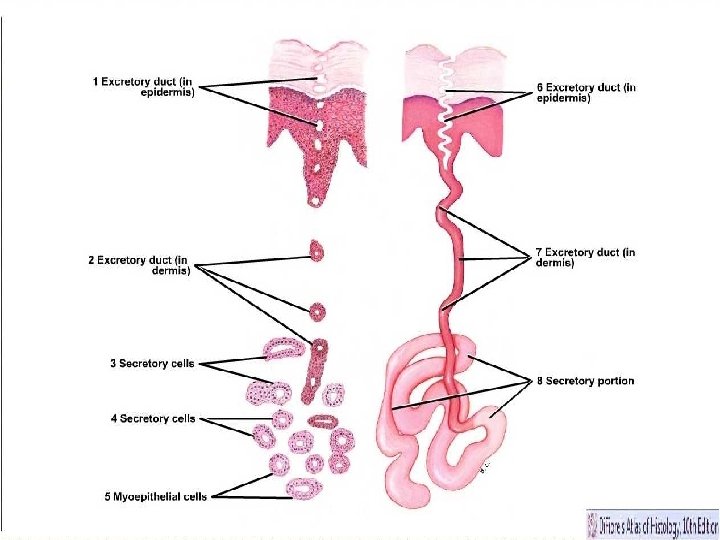
Appendages of skin • Nails – Having 3 parts root, free border, body. • Hair – Having root & shaft. • Sweat glands – Having body & duct. • Sebaceous gland – Produce oily secretion.
Nail
Functions of skin • • • Protection Sensory Temperature regulation Absorption Secretion Excretion p. H regulation Synthesis Storage Reparative.
Protection, Sensation • Protection: an anatomical barrier from pathogens and damage between the internal and external environment in bodily defense; Langerhans cells in the skin are part of the adaptive immune system. • Sensation: contains a variety of nerve endings that react to heat and cold, touch, pressure, vibration, and tissue injury; see somatosensory system and haptic perception.
Heat regulation • Heat regulation: increase perfusion and heatloss, while constricted vessels greatly reduce cutaneous blood flow and conserve heat. Erector pili muscles are significant in animals. • Control of evaporation: the skin provides a relatively dry and semi-impermeable barrier to fluid loss.
Absorption • Water resistance: The skin acts as a water resistant barrier so essential nutrients aren't washed out of the body. • Absorption: Oxygen, nitrogen and carbon dioxide can diffuse into the epidermis in small amounts, some animals uses their skin for their sole respiration organ (contrary to popular belief, however, humans do not absorb oxygen through the skin).
Storage and synthesis • Regulation of p. H: Excreting acid with sweat. • Reparative: Wounds of skin heals quickly. • Storage and synthesis: acts as a storage center for lipids, chlorides and water.
Fascia Superficial fascia Deep fascia
Superficial fascia • It is a general coating of the body beneath the skin, made up of loose areolar tissue with varying amount of fat. • Fat is abundant in gluteal region, abdominal region, mammary gland etc. • Fat is absent in eyelid, external ear, penis, scrotum.
Adipose tissue in mammary gland.
Types of fats • Yellow fat • Brown fat • Fat fills axilla, orbits etc • Fat also present around kidneys to support it.
Features • • Most distinct - Anterior abdominal wall Very thin – Dorsal part of hand & feet, face. Very dense – Scalp, palm & soles. It contains : Deep sweat glands, subcutaneous muscles , lymph nodes, cutaneous nerves & vessels.
Functions • Facilitates movements of skin. • Conserves body heat. • Acts as a soft medium for vessels & nerves.
Deep fascia • It is a fibrous sheet covers the body under the superficial fascia. • It is not having fat. • It is tough & inelastic.
Distribution • It is well defined in the limbs and neck. • It is ill defined on the face & trunk.
Features • Extensions forms intermuscular septa & fibroareolar sheaths for muscles. • Thickenings forms retinacula & aponeuroses. • Deep fascia blendes with a subcutaneous bone.
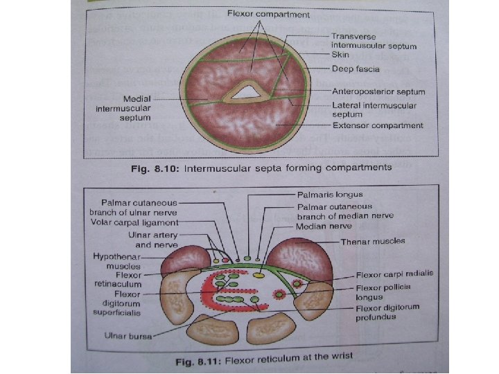
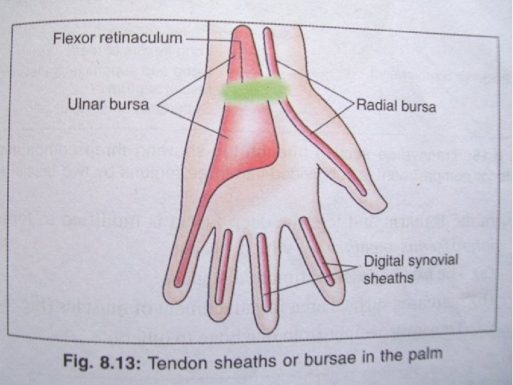
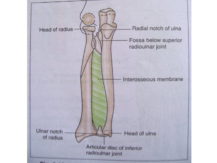
Modifications • Intermuscular septa • Muscle coverings: Epimysium, perimysium, endomysium. • Nerve coverings: Epineurium, perineurium, endoneurium. • Sheaths covering vessels. • Sheaths covering joints: Capsule, synovial membrane, bursae. • Sheaths covering tendons. • In palm & soles aponeuroses. • In between bones interosseous membrane.
Functions • Keeps underlying structures in position. • Provides extra surface for muscular attachments. • Helps in venous & lymphatic return. • Assists muscles in their action. • Retinacula acts as pulleys for tendons.