Good morning Testis Epididymis and Spermatic cord Dr


 Ø Pampiniform plexus leaves the posterior border of testis. Ø At superficial")


")


 Ø Cord-like thick-walled muscular duct Ø Transports spermatozoa from the")


 lymph nodes Autonomic nerves Sympathetic fibers")


- Slides: 56
Good morning
Testis, Epididymis and Spermatic cord Dr. Qudsia Sultana
Objectives Ø Introduction of Testis Ø Parts of Testis Ø Coverings of the Testis Ø Structure of the testis Ø Blood Supply, nerve supply and lymphatic drainage Ø Descent of testis Ø Applied anatomy Ø Spermatic cord.
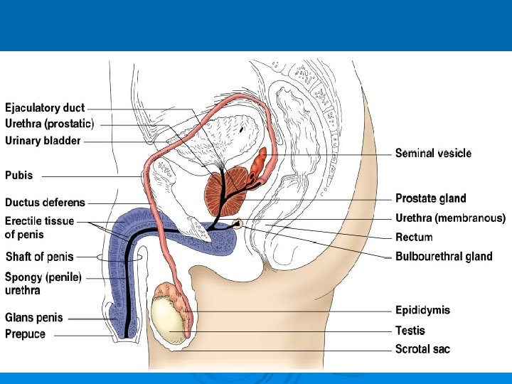
Male Reproductive System Ø Testis Ø Epididymis Ø Ductus deferens Ø Seminal vesicles Ø Ejaculatory ducts Ø Penis Accessory structures : Prostate and Bulbo-urethral glands
Testis Ø Ø Ø The testis are the male gonads homologous to ovary in females. The primary functions of the testes are to produce sperm (spermatogenesis) and to produce androgens, primarily testosterone Ellipsoid in shape. Suspended into the Scrotum by spermatic cord. Oblique in direction-laterally
MEASUREMENTS Ø LENGTH : 5 cms Ø BREADTH : 3 cms Ø THICKNESS : 2. 5 cms Ø WEIGHT : 10 -14 gms
PARTS OF THE TESTIS Presenting Parts : • Upper End • Lower End • Anterior Border • Posterior Border • Medial Surface • Lateral Surface
Upper endØ Spermatic cord Ø head of epididymis, connected to it by efferent ductules. Ø Fibrofatty bodyappendix of testis. Lower end – Ø tail of epididymis. Anterior bordersmooth and convex
Posterior borderØ broad and flat, Ø laterally occupied by body of epididymis. Ø Medially-Vas deferens Medial surfaceØ Smooth and convex
Lateral surfaceØ Smooth and convex Ø Overlapped posteriorly by epididymissemilunar recess“Sinus of epididymis”. Ø (helpful in side determination)
Coverings
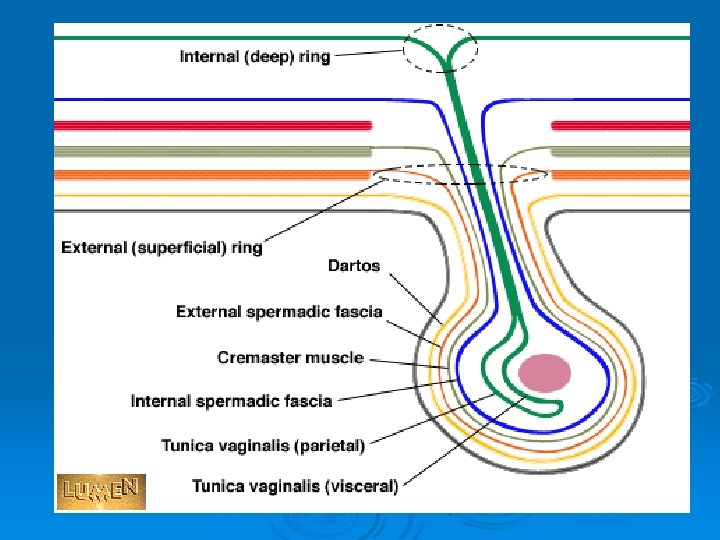
Extrinsic COVERINGS of testis • SKIN • DARTOS MUSCLE • EXTERNAL SPERMATIC FASCIA • CREMASTERIC MUSCLE & FASCIA • INTERNAL SPERMATIC FASCIA • INTRINSIC COVERING-
Intrinsic Coverings of the testis From outside inwards 1. Tunica vaginalis parietal and visceral 2. Tunica albuginea 3. Tunica vasculosa
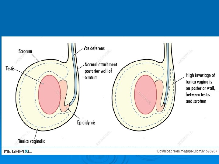
Tunica vaginalis Ø It is a peritoneal extension covering the testis except at the posterior border. Ø It is having two layer with a cavity between them.
Tunica albuginea It is the thick fibrous membrane covering of the testis. Ø It is thickened along the posterior border of the testis to form mediastinum testis. Ø Number of fibrous septa arising from this mediastinum divide the substance of the testis into 200 – 300 lobules. Ø Each lobule is occupied by seminiferous tubules and interstitial cells of leydig. Ø
Tunica Vasculosa Ø It is the vascular layer of the testis, consisting of a plexus of blood vessels, held together by delicate areolar tissue.
STRUCTURE OF TESTES
MACROSCOPIC STRUCTURE Ø SEMINIFEROUS TUBULES Ø COILED TUBULES Ø STRAIGHT TUBULES Ø RETE TESTIS Ø EFFERENT DUCTULES Ø CANAL OF EPIDIDYMIS
ARTERIAL SUPPLY Ø Testicular arteryabdominal aorta Ø Artery to vas deferens. Superior or inferior vesical artery Ø Cremasteric artery Inferior epigastric artery
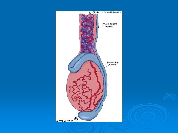
Testicular veins(Vine) Ø Pampiniform plexus leaves the posterior border of testis. Ø At superficial inguinal ring unite to form four veins. Ø At deep inguinal ring unite to form two veins and finally a single testicular vein is formed. Ø Left side vein drain into left renal vein Right side vein drain into inferior vena cava.
Ø Counter current heat exchange Ø The heat conveyed by the arteries of testis-absorbedveins of pampiniform plexus. Ø Scrotal temp of testis-3 to 40 c less than abdomen.
LYMPHATIC DRAINAGE Ø PRE-AORTIC LYMPH NODES Ø PARA-AORTIC LYMPH NODES.
NERVE SUPPLY -Pain sensation. T 10 -T 11 Ø Referred pain from testis to umbilicus can be due to segmental innervation.
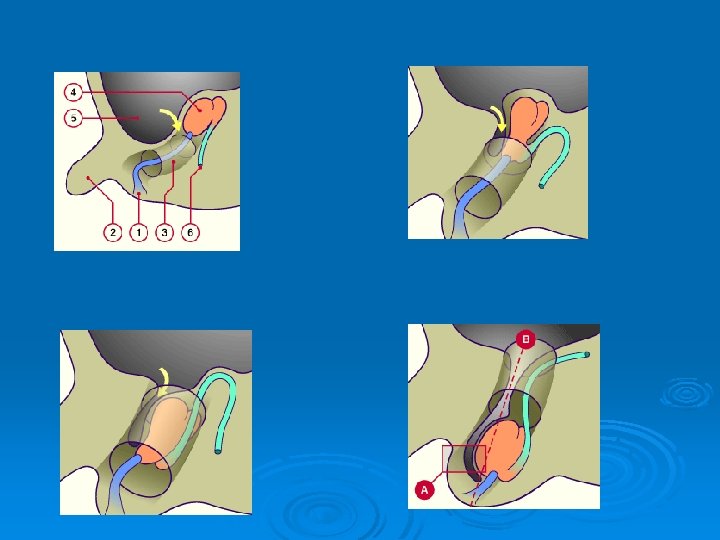
DESCENT OF TESTIS v. TESTIS develops retro peritoneal in dorsal abdominal wall at the level of T 10 - T 12. STARTS descending from second month of intra uterine life. v
Ø 3 rd month- iliac fossa Ø 6 th month- deep inguinal ring Ø 8 th monthsuperficial inguinal ring Ø 9 th month at birthscrotum
Factors for the decent Ø Gubernaculum testis Ø Intra abdominal pressure. Ø Intra abdominal temperature. Ø Hormones-foetal testis
APPLIED ANATOMY Ø MONORCHISM- unilateral absence of testis Ø ANORCHISM –bilateral absence of testis
ECTOPIC TESTIS Lower part of abdomen Base of penis Saphenous opening Anterior superior iliac spine Behind the scrotum
HYDROCOELE: - accumulation of fluid in tunica vaginalis Ø VAGINAL Ø INFANTILE Ø CONGENITAL Ø ENCYSTED
HAEMATOCELE: - COLLECTION OF BLOOD IN TUNICA VAGINALIS.
VARICOCELE Veins of pampiniform plexus become dilated and tortous -Usually on left side
LYMPH VARIX -Lymphatic vessels of the cord becomes dilated and tortous -Caused by obstruction due to filariasis
TORSION TESTIS CAUSES: -INVERSION OF TESTIS -SUDDEN CONTRACTION OF CREMASTERIC MUSCLE COMPLICATIONS: -EDEMA -HAEMORRHAGE -ARTERIAL OBSTRUCTION
TESTICULAR TUMOURS MALIGNANT TUMOURS: -SEMINOMA -NON SEMINOMA
EPIDIDYMIS · Comma-shaped, tightly coiled tube · Found on the superior part of the testis and along the posterior lateral side · Functions to mature and store sperm cells (at least 20 days) · Expels sperm with the contraction of muscles (in its walls) to the vas deferens Copyright © 2003 Pearson Education, Inc. publishing as Benjamin Cummings Slide 16. 5
PARTS OF EPIDIDYMIS Ø HEADHead fits over part of the upper pole of the testis. Ø The head is formed by highly convoluted continuations of efferent ductules. Ø Appendix of epididymis
PARTS OF EPIDIDYMIS BODY and TAILBody extends around the posterolateral border of testis. Ø Tail reaches near the lower pole of testis. Ø The coils are held together by bands of connective tissue. Ø CANAL OF EPIDIDYMIS-Vas Deferens Ø
DUCTUS DEFERENS(Vas Deferens)
Runs from the epididymis through the inguinal canal into the pelvic cavity Ø Its joins the duct of the seminal vesicle to form the ejaculatory duct Ø Propels sperm from the epididymis to the urethra Ø Vasectomy – cutting and ligating the ductus deferens, which is a nearly 100% effective form of birth control Ø
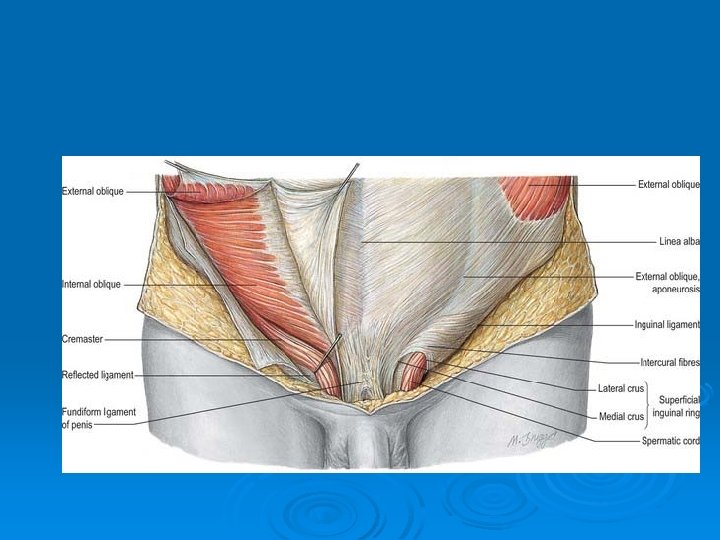
Spermatic cord Cord in males -collection of structures that pass through inguinal canal. Round ligament in females. It suspends the testis in scrotum Spermatic cord
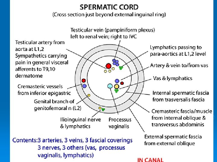
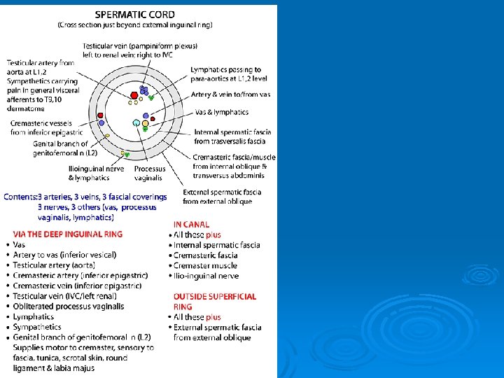
COVERINGS OF SPERMATIC CORD Three concentric layers of fascia. Ø Internal spermatic fascia Ø Cremasteric muscle and fascia Ø External spermatic fascia
components of spermatic cord§ § § § § Vas deferens Remains of processus vaginalis Testicular artery Cremasteric artery Artery to vas deferens Pampiniform plexus of veins Testicular lymph vessels Autonomic nerves Genital branch of genitofemoral nerve
Vas deferens (ductus deferens) Ø Cord-like thick-walled muscular duct Ø Transports spermatozoa from the epididymis to urethra through ejaculatory duct. .
Testicular artery Ø Branch of abdominal aorta. Traverses inguinal canal, supplies testis and epididymis. Cremasteric artery Ø Inferior epiastric artery Ø Cremasteric muscle Ø Artery to vas deferens Ø Superior or inferior vesical artery
Testicular veins Ø Pampiniform plexus leaves the posterior border of testis. Ø At superficial inguinal ring unite to form four veins. Ø At deep inguinal ring a single testicular vein is formed. Ø Left side vein drain into left renal vein Right side vein drain into inferior vena cava.
Lymph vessels Testicular lymph vessels – lumbar (para-aortic) lymph nodes Autonomic nerves Sympathetic fibers from renal or aortic sympathetic plexuses Genital branch (L 2) of genitofemoral nerve PROCESSUS VAGINALIS
Applied anatomy Ø Torsion Ø Varicocele of veins of spermatic cord Ø Encysted hydrocele