The complement system Complement has three functions Opsonin


 • C 1 s")
 • In the presence of")


 • Cleavage of C 3 and activation of the remainder")
 • Molecules of C 3 undergo cleavage at continuous low")
 • Action by the 4 serum proteins on C 3")


 binds")


 mannose sugars")
- Slides: 40
The complement system
• Complement has three functions: – Opsonin – Chemoattractant – Membrane Attack Complex (MAC) • Complement functions in two (three? ) systems: – Alternative – Classical – Lectin-based
The complement system • A defensive system consisting of over 30 proteins produced by the liver and found in circulating blood serum. • Complement kills microbes in three different ways – 1. opsonization – 2. inflammation – 3. Cytolysis
• Complement refers to a complex set of 14 distinct serum proteins (nine components) that are involved in three separate pathways of activation. – Components numbered in order of discovery. – Sequence of activation is not in numerical order. – Components circulate in inactive precursor form, develop activity upon activation. • Complement proteins designated by “C” followed by numbers and letters.
• Promote the inflammatory response by opsonization which enhances susceptibility of coated cells to phagocytosis. • Alter biological membranes to cause direct cell lysis
General Properties of Complement • Primary role is cell lysis. • Activity of complement destroyed by heating sera to 56 C for 30 minutes. • Ig. M and Ig. G are the only immunoglobulin capable of activating complement (classical pathway). • Complement activation can be initiated by complex polysaccharides or enzymes (alternative or properdin pathway). • Portions of the complement system contribute to chemotaxis, opsonization, immune adherence, anaphylatoxin formation, virus neutralization, and other physiologic functions
A Cascade system • The complement works as a cascade system. – Cascade is when one reaction triggers another reaction which trigger others and so on. These types of systems can grow exponentially very fast.
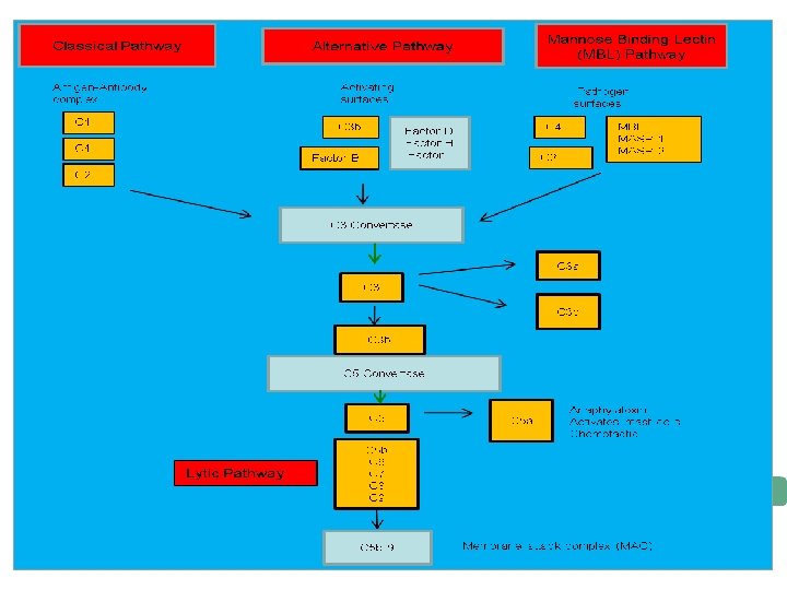
THREE PATHWAYS
Cascade activation • Complement proteins are often designated by an uppercase letter C and are inactive until they are split into products. – Example: C 1 • When the products are split they become active. The active products are usually designated with a lower case a or b. – Example: C 1 a and C 1 b
Two Pathways • The complement pathway can be activated by either of two different pathways. – Classical pathway (specific immune system) – alternative (non-specific immune system)
The Classical Pathway • The classical pathway is considered to be part of the specific immune response because it relies on antibodies to initiate it. • C 1 becomes activated when it binds to the ends of antibodies
• C 1: The Recognition Unit – C 1 consists of 3 subunits: C 1 q, C 1 r, and C 1 s. – C 1 q molecule consists of a collagenous region with six globular head groups globe end serves as recognition unit – When antibody binds to antigen, binding sites for the globular head groups of C 1 q are exposed on the Fc region of the antibody. – For C 1 q to initiate the cascade it must attach to at least 2 Fc fragments, requires at least 2 molecules of Ig. G or one molecule of Ig. M. – C 1 q binding causes C 1 r to enzymatically activate C 1 s.
The Activation Unit (C 4 b 2 a 3 b) • C 1 s cleaves C 4 into C 4 a and C 4 b • C 1 s cleaves C 2 into C 2 a and C 2 b • C 4 b 2 a (C 4 b 2 b in some texts) is enzymatically active and can cleave many molecules of C 3 into C 3 a and C 3 b.
Membrane Attack Unit (C 5, 6, 7, 8, 9) • In the presence of C 5 b, molecules of C 6, C 7, C 8 and a variable number of C 9 molecules assemble themselves into aggregates. • This molecular complex causes a change in membrane permeability. • Exact cause of lysis unknown, one theory is change in lipid membrane causes exchange of ions and water molecules across membrane. • Cells can lyse without C 9 but it’s slower.
The building of a C 3 activation complex • Once C 1 is activated, it activates 2 other complement proteins, C 2 and C 4 by cutting them in half • C 2 is cleaved into C 2 a and C 2 b • C 4 is cleaved into C 4 a and C 4 b • Both C 2 b and C 4 b bind together on the surface of the bacteria • C 2 a and C 4 a diffuse away
C 3 Activation complex • C 2 b and C 4 b bind together on the surface to form a C 3 activation complex • The function of the C 3 activation complex is to activate C 3 proteins. – This is done by cleaving C 3 into C 3 a and C 3 b
C 3 b • Many C 3 b molecules are produced by the C 3 activation complex. • The C 3 b bind to and coat the surface of the bacteria. • C 3 b is an opsonin – Opsonins are molecules that bind both to bacteria and phagocytes – Opsonization increases phagocytosis by 1, 000 fold. Opsonins eria Bact
C 3 a increases the inflammatory response by binding to mast cells and causing them to release histamine
Building the C 5 activation complex • Eventially enough C 3 b is cleaved that the surface of the bacteria begins to become saturated with it. • C 2 b and C 4 b which make up the C 3 activation complex has a slight affinity for C 3 b and C 3 b binds to them • When C 3 b binds to C 2 b and C 4 b it forms a new complex referred to as the C 5 activation complex
The C 5 activation complex • The C 5 activation complex (C 2 b, C 4 b, C 3 b) activates C 5 proteins by cleaving them into C 5 a and C 5 b • Many C 5 b proteins are produced by the C 5 activation complex. These C 5 b begin to coat the surface of the bacteria.
The function of C 5 a • C 5 a disperses away from the bacteria. – Binds to mast cells and increases inflammation. – Most powerful chemotactic factor known for leukocytes
Building the Membrane Attack complex • C 5 b on the surface of bacteria binds to C 6 • The binding of C 6 to C 5 b activates C 6 so that it can bind to C 7 • C 7 binds to C 8 which in turn binds to many C 9’s • Together these proteins form a circular complex called the Membrane attack complex (MAC)
Membrane Attack complex • The MAC causes Cytolysis. – The circular membrane attack complex acts as a channel in which cytoplasm can rush out of and water rushes in. • The cells inner integrity is compromised and it dies • Animation of the classical pathaway
Overview
The alternative pathway • The alternative pathway is part of the nonspecific defense because it does not need antibodies to initiate the pathway. • The alternative pathway is slower than the Classical pathway
Alternative Pathway (Properdin Pathway) • Cleavage of C 3 and activation of the remainder of the complement cascade occurs independently of antibody. • Triggers for the alternative pathway include – Bacterial cell walls – Bacterial lipopolysaccharide – Fungal cell walls – Virally infected cells – Rabbit erythrocytes
Alternative Pathway (Properdin Pathway) • Molecules of C 3 undergo cleavage at continuous low level in normal plasma. • At least 4 serum proteins (factor B, factor D, properdin (P), and initiating factor (IF) function in this pathway. • C 3 b attaches to appropriate site (activating surface) which is actually a protective surface
Alternative Pathway (Properdin Pathway) • Action by the 4 serum proteins on C 3 b proceeds to the C 3 activator stage without participation of C 1, C 4 or C 2. • Activation sequence: C 3, C 5, C 6, C 7, C 8, C 9.
The Alternative complement pathway
Initiation of The Alternative pathway • C 3 contains in unstable thioester bond. • This unstable bond makes. C 3 subject to slow spontaneous hydrolysis to C 3 b and C 3 a • The C 3 b is able to bind to foreign surface antigens. • Mammalian cells which inactivates C 3 b
Factor B • C 3 b on the surface of a foreign cells binds to another plasma protein called factor B
Factor D • The binding of C 3 b to factor B allows a protein enzyme called Factor D to cleave Factor B to Ba and Bb. • Factor Bb remains bound to C 3 b while Ba and Factor D disperse away.
The C 3 activation complex • Properdin, also called factor P, binds to the C 3 b. Bb complex to stabilize it. • C 3 b. Bb. P make up the C 3 activation complex for the alternative pathway
The C 3 activation Complex • The C 3 activation complex causes the production of more C 3 b. • This allows the initial steps of this pathway to be repeated and amplified • 2 X 10⁶ molecules can be generated in 5 minutes
C 5 activation complex • When an additional C 3 b binds to the C 3 activation complex it converts it into a C 5 activation complex. • The C 5 activation complex cleaves C 5 into C 5 a and C 5 b. • C 5 b begins the production of the MAC.
Lectin Pathway • Activation of the lectin pathway begins when mannan-binding protein (MBP) binds to the mannose groups of microbial carbohydrates. • Two more lectin pathway proteins called MASP 1 and MASP 2 (equivalent to C 1 r and C 1 s of the classical pathway) bind to the MBP. • This forms an enzyme similar to C 1 of the classical complement pathway that is able to cleave C 4 and C 2 to form C 44 b. C 2 a, the C 3 convertase capable of enzymatically splitting hundreds of molecules of C 3 into C 3 a and C 3 b
Lectin Pathway The beneficial results are the same as in the classical complement pathway: Trigger inflammation (C 5 a>C 3 a>C 4 a); Chemotactically attract phagocytes to the infection site (C 5 a); Promote the attachment of antigens to phagocytes via enhanced attachment or opsonization (C 3 b>C 4 b; Serves as a second signal for the activation of naive B-lymphocytes (C 3 d); Cause lysis of gram-negative bacteria and human cells displaying foreign epitopes (MAC). And remove harmful immune complexes from the
Overview
Activation of the Lectin Pathway C 3 b mannan binding lectin (MBL) mannose sugars C 3 convertase C 3 MBL C 3 b covalent bond with surface “activating surface” (bacterial or yeast cell surface) -mannan binding lectin (MBL) recognizes mannose sugars on microbial cells -host mannose is hidden and is not accessible to MBL -lectin pathway results in the formation of a C 3 convertase that generates C 3 b -C 3 b forms a covalent bond with the surface of the pathogen and is part of C 5 convertase