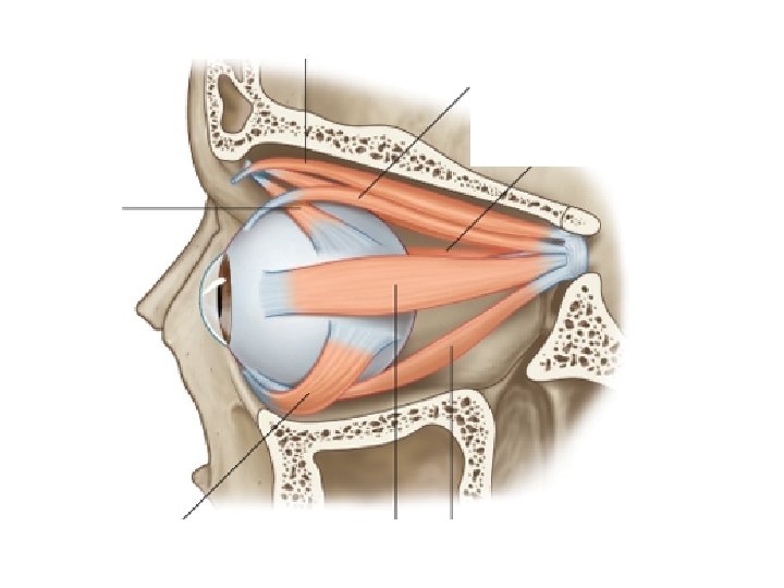
SPECIAL SENSES Agonist and Antagonist Muscles Muscles that


 Glands • secrete lipids - help stabilize the tear film • minimize")
.")


- Slides: 112
SPECIAL SENSES
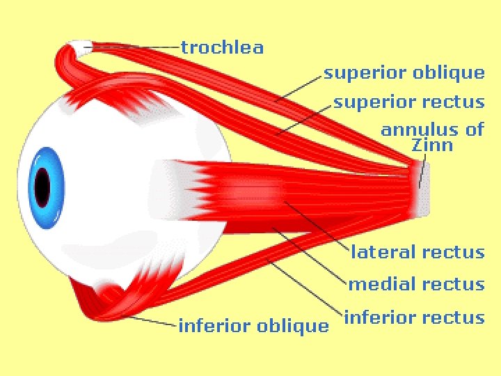
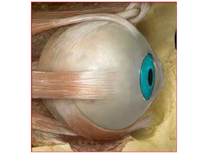
Agonist and Antagonist Muscles • Muscles that work in opposite directions to complete an action.
Agonist Muscle • Reacts in response to voluntary or involuntary stimulus • creates the mvmnt necessary to complete a task.
Antagonist Muscle • Acts against the agonist muscle and • helps to move the body part back in place after the action is completed.
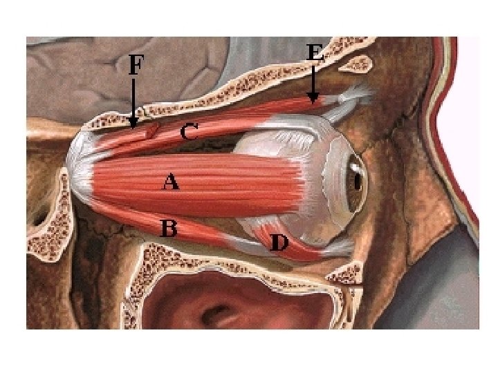
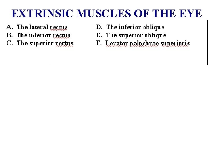
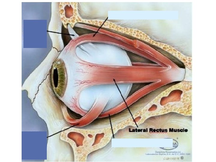
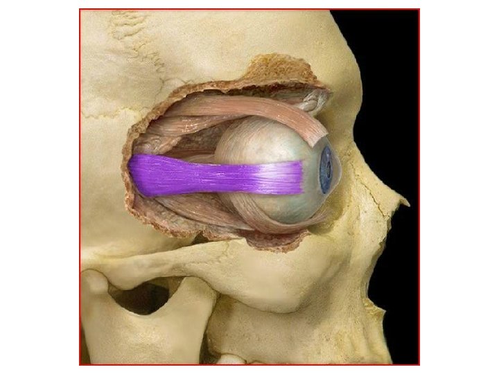
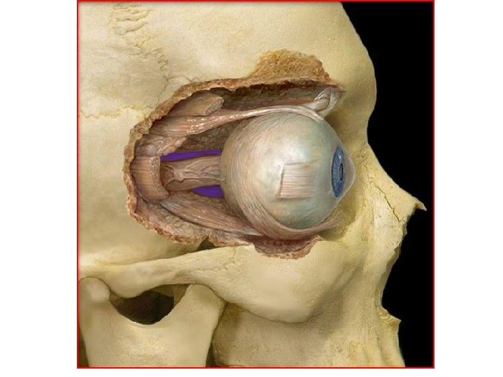
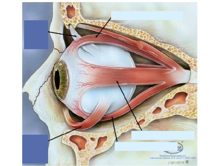
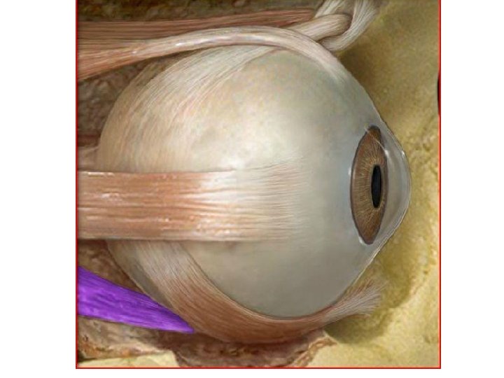
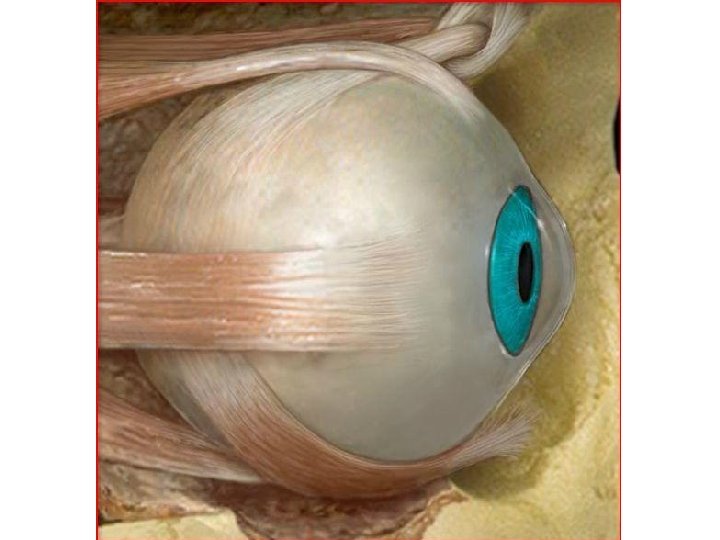
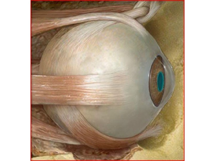
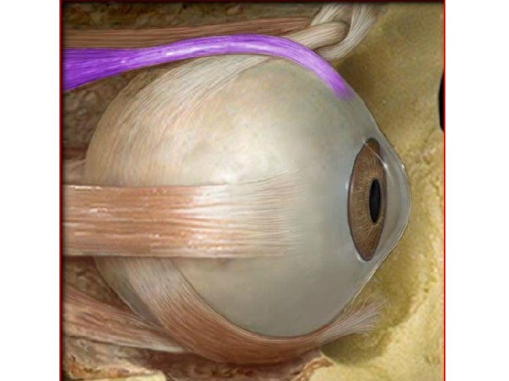
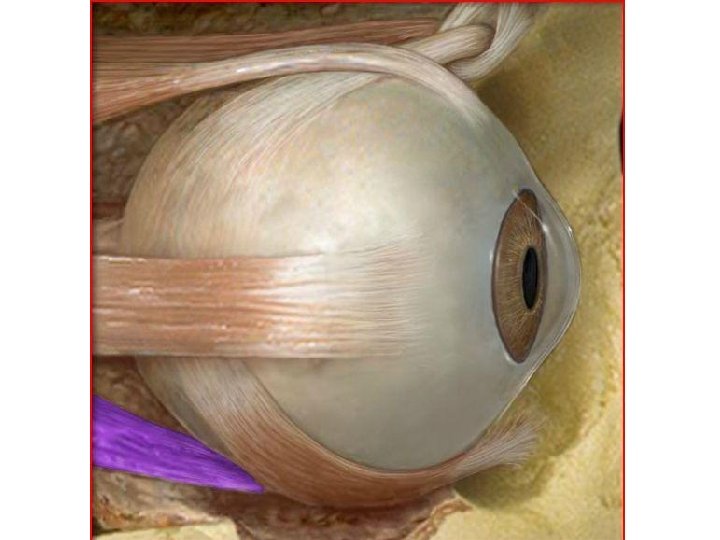
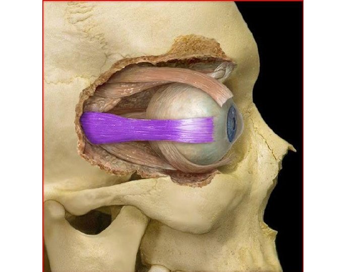
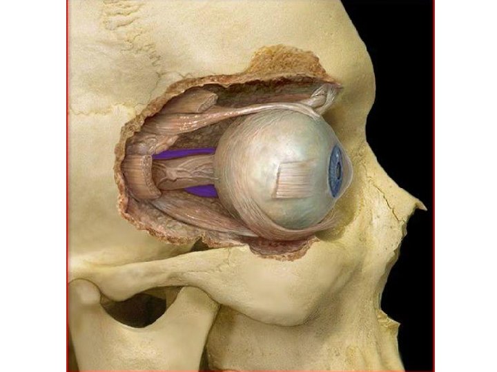
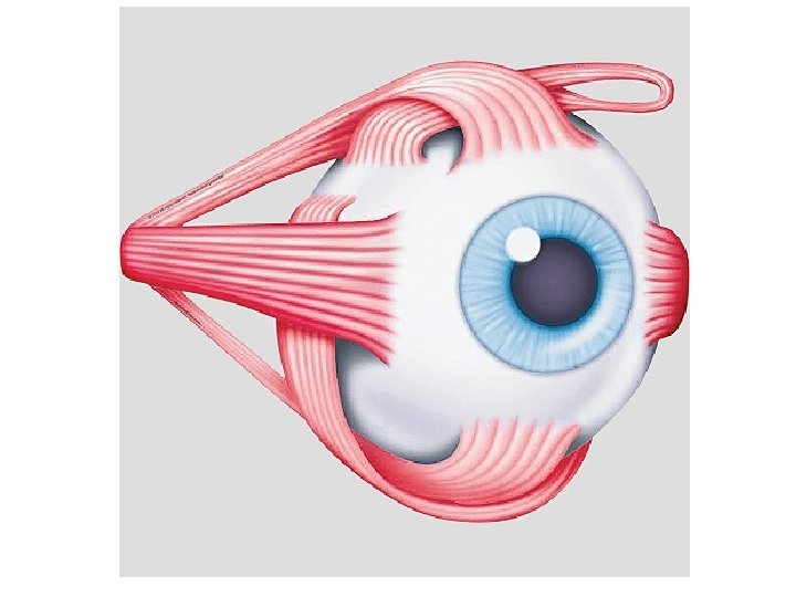
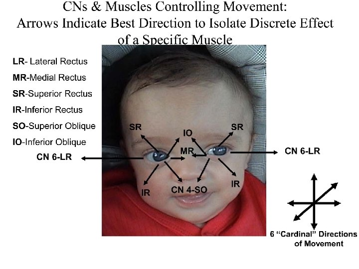
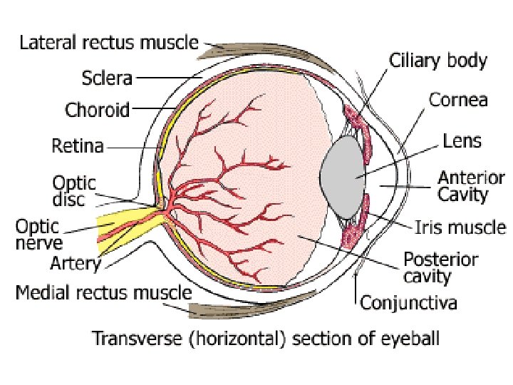
Lateral Rectus Muscle • The agonist of the Medial Rectus Muscle • Moves eye in an outward direction – laterally - away from the nose (Abduction) • Its origin point is the Annulus of Zinn and its insertion point is the Sclera
Medial Rectus
Medial Rectus Muscle • The antagonist of the Lateral Rectus Muscle • Moves the eye in an inward direction – medially - toward the nose (Adduction) • Its origin point is the Annulus of Zinn and its insertion point is the Sclera
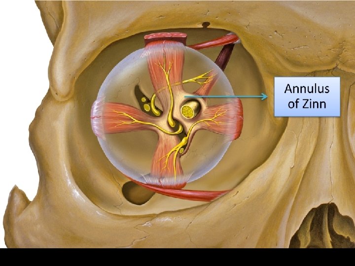
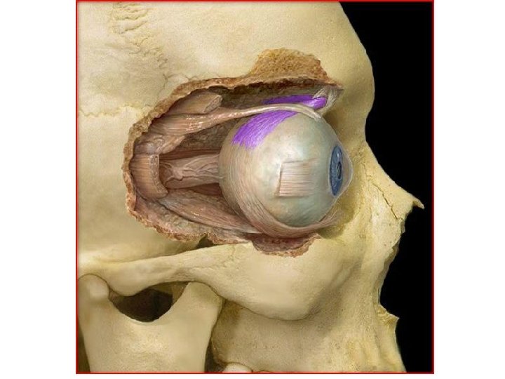
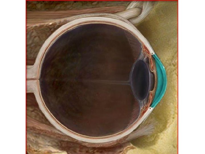
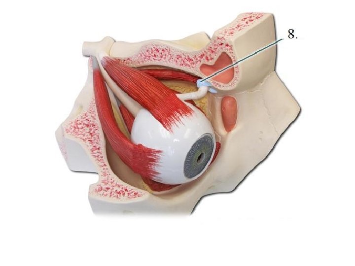
Annulus of Zinn • Fibrous ring located in posterior region of the orbit – around the optic canal • Located around the optic canal and encloses a part of the superior orbital fissure • The common fibrous origin of the 4 recti muscles • Named after Johann Gottfried Zinn
Johann Gittfried Zinnia
2 Parts of Annulus of Zinn • Upper part – Superior Tendon of Lockwood – Gives origin to superior rectus, part of medial rectus, and upper head of lateral rectus • Lower Part – Ligament/Tendon of Zinn – Gives origin to inferior rectus, part of medial rectus, and lower head of lateral rectus
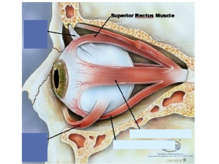
Superior Rectus Muscle • The agonist of the Inferior Rectus Muscle • Responsible for 2 mvmnts of the eye – Upward (Elevation) – Moves the eye inward (Adduction) • Its origin point is from the Annulus of Zinn • Its insertion point is into the Sclera of the eye
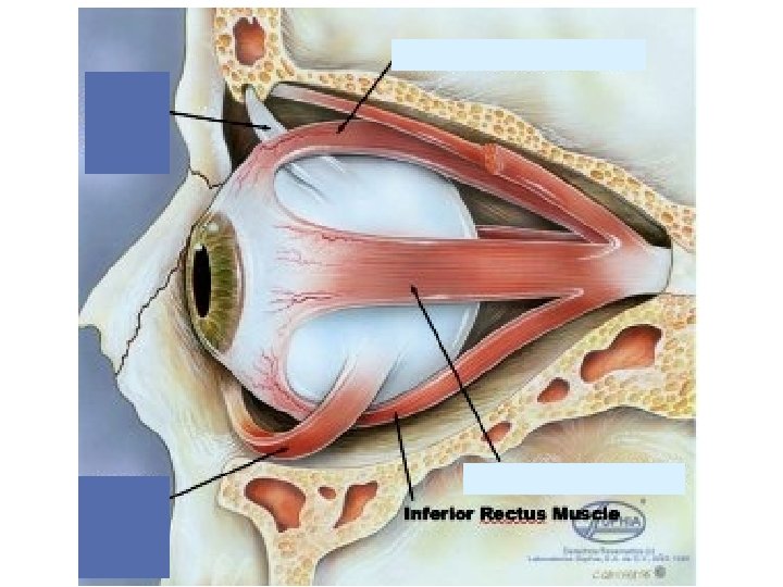
Inferior Rectus Muscle • The antagonist of the Superior Rectus Muscle. • Responsible for 2 mvmnts: – Downward (Depression) – Moves the eye inward (Adduction) • Its origin point is the Annulus of Zinn • Its insertion point is into the sclera of the eye
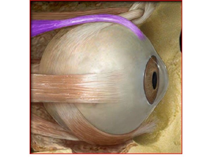
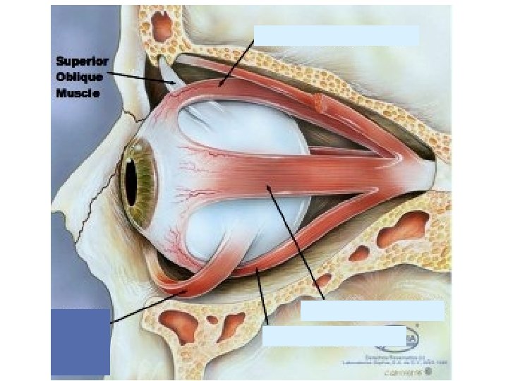
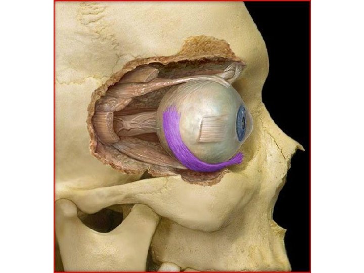
Superior Oblique Muscle • The agonist of the Inferior Oblique Muscle • Responsible for 2 mvmnts: – Moves eye in downward direction (Depression) – Moves the eye in outward direction (Abduction)
• Its origin point is from the posterior of the Annulus of Zinn • it then passes anteriorly and ends in a round tendon. • The tendon extends through a pulley-like loop of fibrocartilaginous tissue called the trochlea (Pulley) in the anterior medial part of the roof of the orbit • and then turns to its insertion point on the posterolateral aspect of the eyeball.
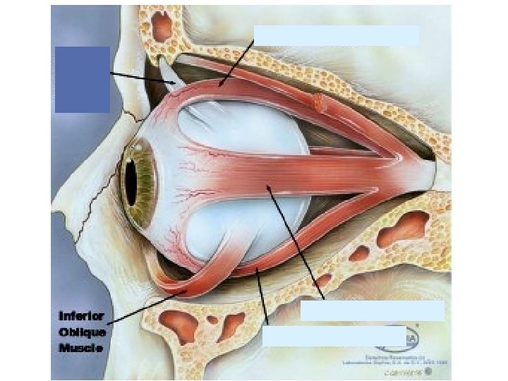
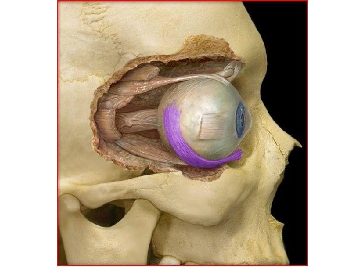
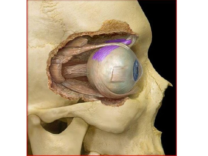
Inferior Oblique Muscle • The antagonist of Superior Oblique muscle • Responsible for 2 mvmnts – Moves the eye in an upward direction (Elevation) – Moves the eye in an outward direction (Abduction)
• Its origin point is from the maxilla at the anteromedial aspect of the floor of the orbit • its insertion point is on the posterolateral aspect of the eyeball
Tapetum
Giraffe Tapetum
3 Accessory Eye Structures
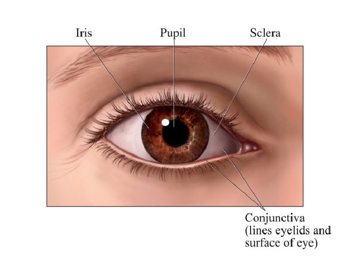
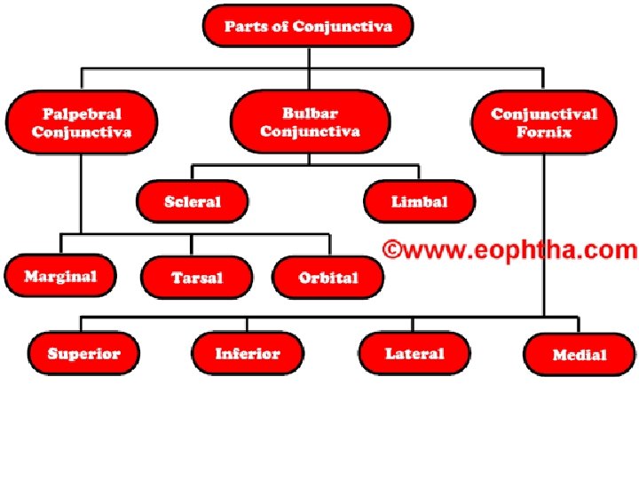
Conjunctiva • A thin, clear, moist membrane - coats inner surfaces of the eyelids (palpebral conjunctiva) and outer surface of eye (ocular, or bulbar, conjunctiva). • Inflammation of conjunctiva is called conjunctivitis (pinkeye) • Secretes mucus
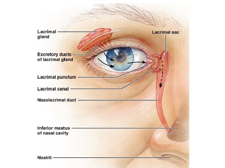
Lacrimal Glands • Secrete a mixture of salt H 2 O, ions and proteins (lysozymes). • Tears that cover surface of eye are essential for maintenance of transparent cornea • film keeps cornea wet; allowing gas XΔ – cleans debris from transparent surface, provides clear path to retina – Protects ocular surface from invasion by bacteria and viruses…Antimicrobial Properties
Tarsal (Meibomian) Glands • secrete lipids - help stabilize the tear film • minimize evaporative loss of tear fluid • Maintain aq tear volume - keeps ocular surface protected thruout blink cycle • Lipids allow tear fluid to maintain its viscosity spreads quickly over eye’s surface after each blink
Meibomian glands are not visible, only their tiny opening in the lid (yellow arrows). Green arrows indicating upper and lower punctum (opening in the lid for the tears flow).
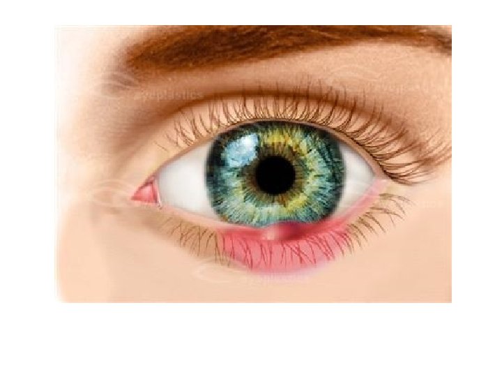
Blocked Meibomian Gland
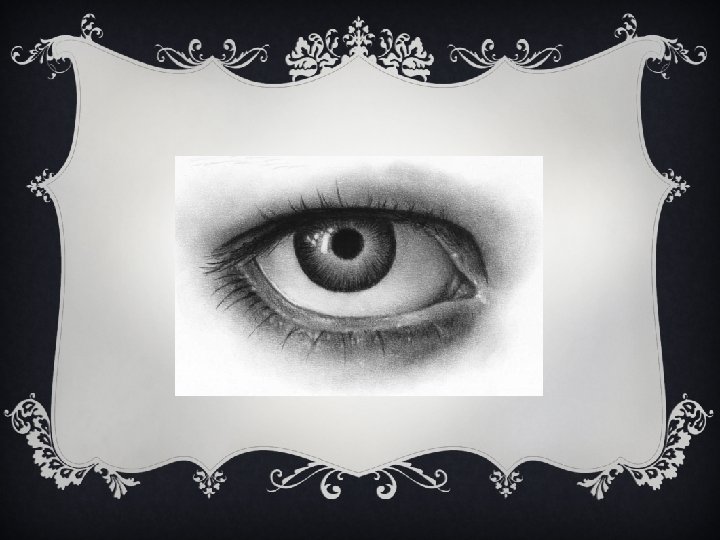
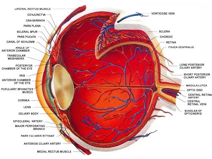
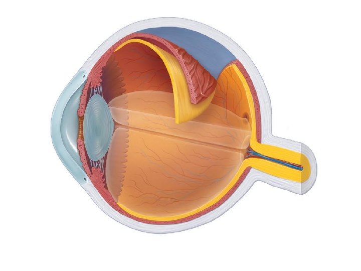
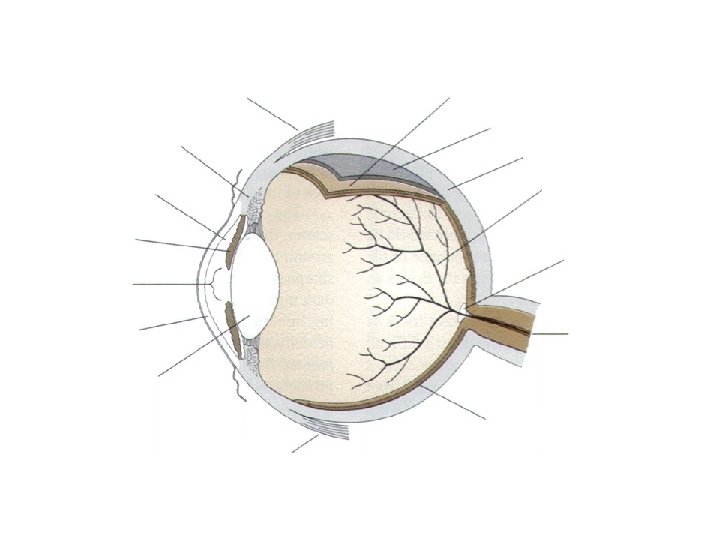
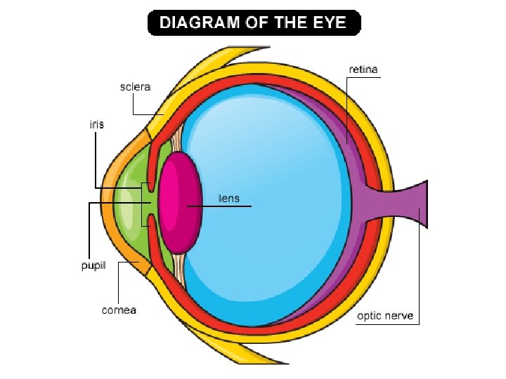
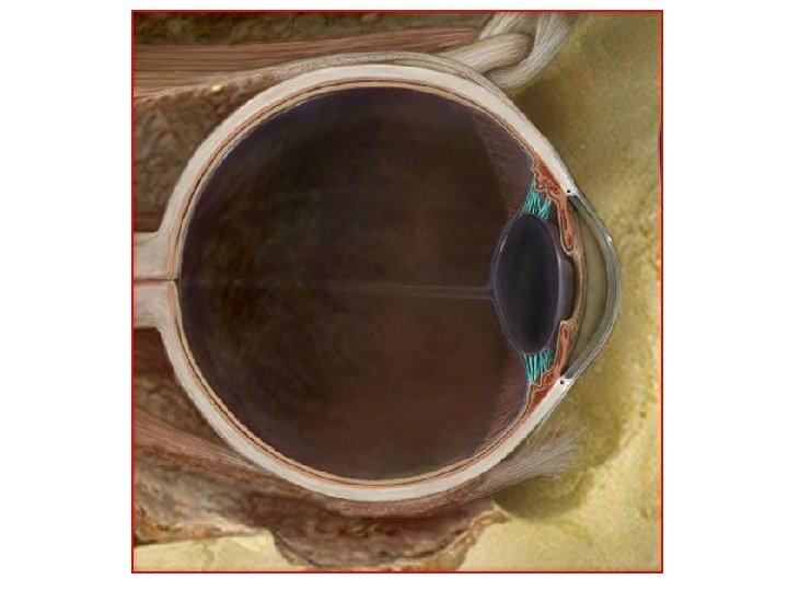
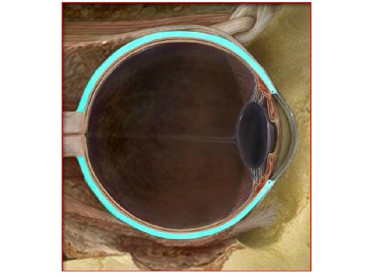
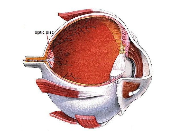
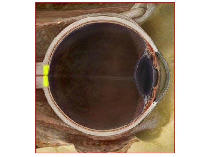
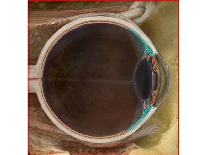
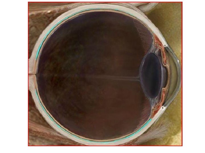
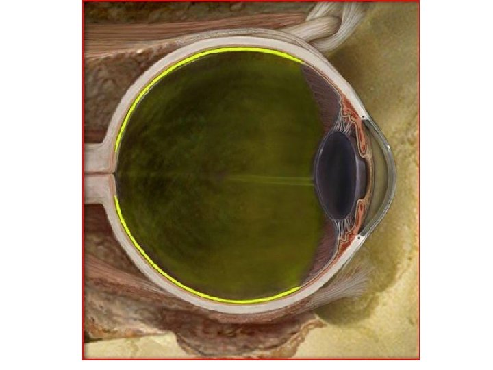
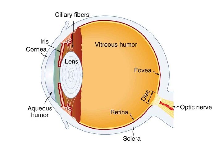
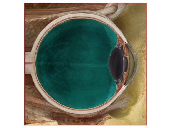
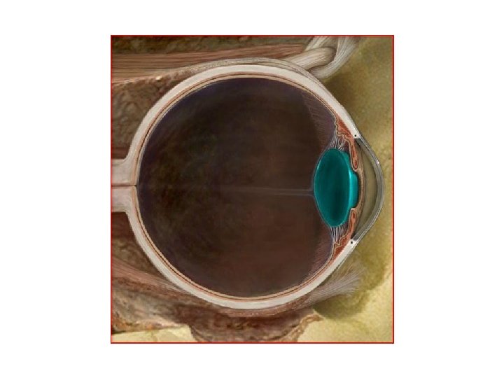
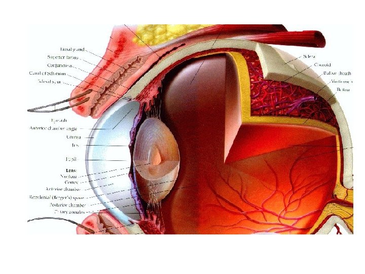
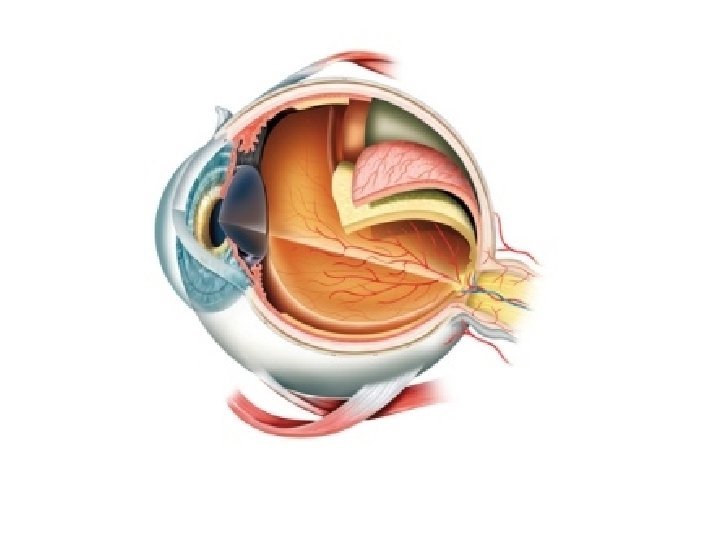
Parts of the Eye
Suspensory Ligament
Aqueous Humor
Sclera
Optic Disk
Ciliary Body
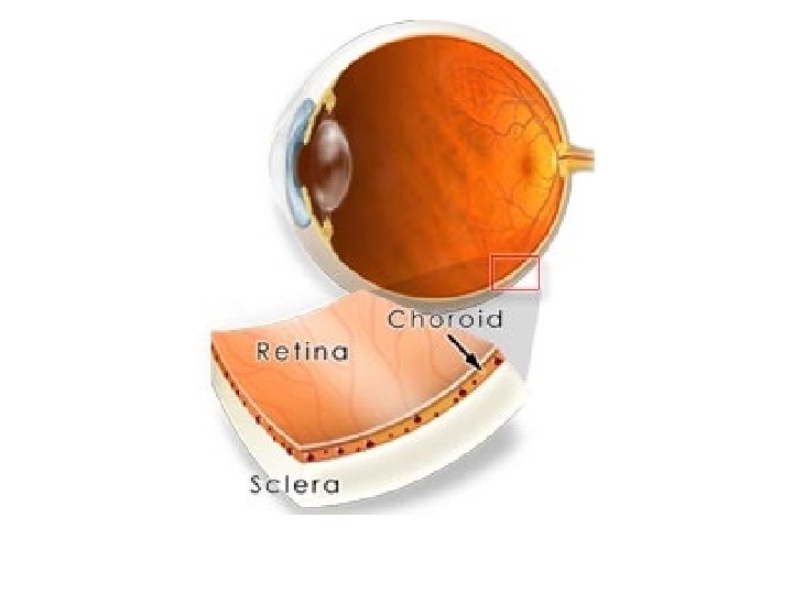
Choroid Coat
Retina
Vitreous Humor
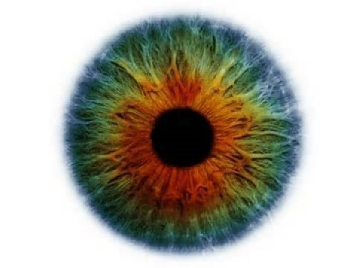
Iris
Pupil
Fovea Centralis
• Macula - Section of the retina that the light hits. • Fovea Centralis – The very center of the macula – The point on the retina that is the absolute center of a person’s direct vision
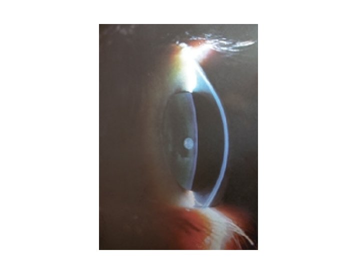
Cornea
Lens
Additional Terms
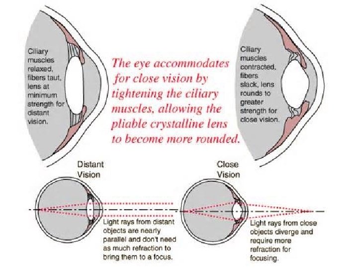
Accomodation Pupillary Reflex • Reflex constriction of the pupils when viewing close objects
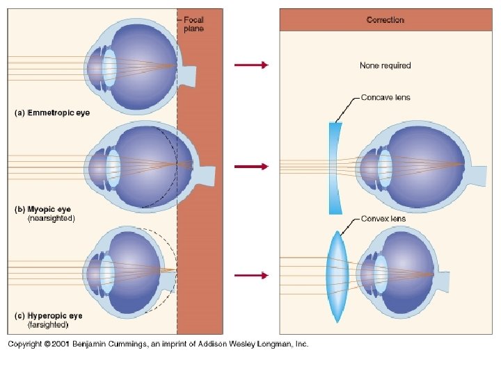
Astigmatism
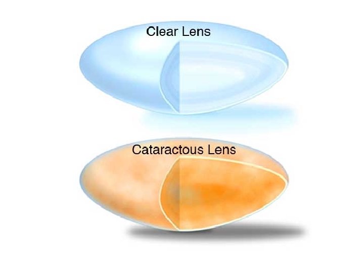
Cataract
Convergence
Glaucoma
Night Blindness • Inability to see well in the dark • Often caused by Vitamin A deficiency
Photopupillary Reflex
Refraction
• What controls the amount of light that goes into the eye? • How does the eye see images? • What do people with brown eyes have that people with blue eyes do not? • How do the lenses of our eyes change when an object is far away? • What parts of the retina help us see at night? • What parts of the retina help us see colors? • What is the first step in making an artificial eye? • How large is the eyeball? CCC! Eyeball…. . Bill Nye