Skeletal System It is very foolish of a


 plates are layers of cartilage that are replaced by bone")


 Articular (Hyaline) Cartilage Fibrous Capsule")


 is a defect that")


 • Talipes equinovarus (TEV) is a compound foot disorder in which")
")
 • Forceful displacement of an articulating bone to an abnormal position, usually")


- Slides: 91
Skeletal System “It is very foolish of a man to be frightened of a skeleton, for Nature has put an insurmountable obstacle against running away from it. ” G. K. Chesterton
Functions of the Skeletal System • Supports the body by providing a framework for the attachment of other tissues and organs. • Storage of calcium and phosphate ions. • Production of red blood cells (in the marrow). • Protection of soft tissues and organs. • Allow movement by providing a place for muscles to attach.
Classification of Bones • Long bones are longer than they are wide, with heads at each end. ▫ Examples include the femur and humerus.
• Short bones are often cubeshaped, and contain higher amounts of spongy bone. ▫ Examples include the bones of the wrist (carpals) and ankle (tarsals).
• Flat bones are thinner, flattened, and often curved. ▫ Made of thin layers of compact and spongy bone. ▫ Examples include the skull, ribs, and sternum.
• Sesamoid bones are embedded within a tendon.
• Irregular bones do not fit into any of the other categories due to their unusual shapes. ▫ Examples include the vertebrae, sacrum, and pelvic bones.
Bone Anatomy • Bone is made of two types of tissue. ▫ Compact bone is relatively solid, while spongy bone has many spaces in between the bony rods or struts.
Anatomy of a Long Bone • Long bones are divided into three sections: ▫ Proximal epiphysis is the end of the bone closest (“approximate”) to the trunk of the body. ▫ Diaphysis is the middle shaft of the bone. ▫ Distal epiphysis is the end of the bone farthest (“distant”) to the trunk of the body.
• The diaphysis is covered by a layer of dense fibrous tissue called the periosteum. • The medullary cavity is the hollowed out area inside the shaft. ▫ Contains yellow marrow (fat storage) in adults. ▫ Red marrow (blood cell formation) in infants.
Anatomy of a Long Bone Spongy Bone Proximal Epiphysis Compact Bone Medullary Cavity Diaphysis Periosteum Distal Epiphysis
Microscopic Anatomy of Bone • Each functional unit of bone is called an osteon. ▫ Thin, calcified plates form the ring-like lamellae. ▫ Each layer of a lamella contain pits called a lacunae, which contains osteocytes (bone cells).
Microscopic Anatomy of Bone
Microscopic Anatomy of Bone • Central canals at the center of each osteon contains the blood vessels to nourish the bone. • Perforating canals join the central canals together.
Microscopic Anatomy of Bone • There are two types of bone cells. ▫ Osteoblasts lay down the minerals needed to build the bone. ▫ Osteoclasts shape the bone into the appropriate form. • The cells are all connected back to the nutrient supply through tiny canals called canaliculi.
Microscopic Bone Anatomy Lamella Central Canal Lamella Lacuna Osteocyte Canaliculus Lacuna
Bone Growth • Bones continuously grow and lengthen throughout childhood, up to about age 25. • In the embryonic stages of development, bones contain much more hyaline cartilage. ▫ This cartilage is gradually replaced by bone in a process called ossification.
• Epiphyseal (growth) plates are layers of cartilage that are replaced by bone and regrow until adulthood. They are also called epiphyseal plates. Hyaline cartilage New center of bone growth Medullary cavity Bone starting to replace cartilage Growth in bone length Bone collar Hyaline cartilage model In an embryo (a) In a fetus Blood vessels
• During adolescence, the bones grow much more quickly, overtaking the cartilage until only articular cartilage remains. Articular cartilage Hyaline cartilage Spongy bone New center of bone growth New bone forming Epiphyseal plate cartilage Growth in bone width Medullary cavity Bone starting to replace cartilage Growth in bone length Bone collar Hyaline cartilage model In an embryo (a) Blood vessels New bone forming Epiphyseal plate cartilage In a fetus In a child
The Skeleton • The skeleton is divided into two regions: • The axial skeleton includes everything around the longitudinal (vertical) center plane of the body. ▫ Skull, spine ▫ 80 bones • The appendicular skeleton includes the appendages: the arms and legs. • Bones of arms and legs • 126 bones
The Axial Skeleton
The Skull • Most of the bones of the skull are flat, designed to be protective. • Each bone is joined by a suture, a joint made of dense fibrous tissue.
Fontanels • The fetal skull has a few sutures that are much wider, called fontanels. ▫ These allow the brain to grow and expand. ▫ Convert to bone within about 2 years. • Fontanels are the soft spots on the heads of infants.
Skull, Lateral View Parietal Bone Sphenoid Bone Frontal Bone Nasal Bone Zygomatic Bone Occipital Bone Temporal Bone Maxilla Mandible
Skull, Inferior View Maxilla Zygomatic Bone Sphenoid Bone Temporal Bone Mastoid Process Parietal Bone Occipital Bone Foramen Magnum Occipital Condyle
Sinuses • Sinuses are hollow bones with thin plates between them designed to drain fluids. ▫ Sinus headaches happen when they get blocked and the fluids overflow into the nasal cavity.
The Hyoid Bone • The only bone in the entire body that does not form a joint with any other bone. • The base of the tongue attaches to this bone, and it aids in swallowing and speech.
Ear • The middle ear is made up of three small bones. ▫ Malleus (hammer) ▫ Incus (anvil) ▫ Stapes (stirrup)
Vertebral Column • There are 24 vertebral bones, each separated by a disk of fibrocartilage. • The vertebrae are named based on their location. ▫ C 1 -C 7 – Cervical vertebrae in the neck �C 1 is called the atlas �C 2 is called the axis ▫ T 1 -T 12 – Thoracic vertebrae in upper back. ▫ L 1 -L 5 – Lumbar vertebrae in the lower back. • Two bones found below the lumbar region, made from nine vertebrae fused together. ▫ Sacrum ▫ Coccyx
Vertebral Column Atlas Axis Cervical C 1 -C 7 Thoracic T 1 -T 12 Lumbar L 1 -L 5 Sacrum Coccyx
Cervical Vertebrae Vertebral Body Vertebral Foramen Spinous Process
Thoracic Vertebrae Vertebral Body Vertebral Foramen Transverse Process Spinous Process
Lumbar Vertebrae Vertebral Body Vertebral Foramen Transverse Process Spinous Process
Ribs and Sternum • Protect major organs of the thoracic cavity. ▫ Heart ▫ Lungs • There are three sets of ribs: ▫ True ribs (pairs 1 -7) are connected directly to the sternum. ▫ False ribs (pairs 8 -12) are connected to the sternum through cartilage or not at all. �Floating ribs (pairs 11 and 12) are false ribs only connected to the thoracic vertebrae.
Sternum and Ribs Manubrium True Ribs: 1 -7 Body of Sternum Xiphoid Process False Ribs: 8 -10 Floating Ribs: 11 -12
The Appendicular Skeleton
The Appendicular Skeleton Parietal Bone Occipital Bone Clavicle Scapula Humerus Frontal Bone Maxilla Mandible Sternum Rib Cage Vertebral Column Coccyx Radius Sacrum Ulna Carpals Metacarpals Phalanges
The Appendicular Skeleton Pelvis Femur Patella Tibia Fibula Talus Metatarsals Tarsals Phalanges
Scapula, Posterior View Coracoid Process Acromion Glenoid Cavity Body
Clavicle, Anterior/Superior View Sternal End Acromial End
Humerus, Right, Posterior view Head Deltoid Tuberosity Medial Epicondyle Capitulum Trochlea
Radius and Ulna Trochlear Notch Head of Radius Ulna Interosseous Membrane Radius
Right Wrist and Hand Distal Phalanges Middle Phalanges Proximal Phalanges II I III IV Metacarpals V Carpals Radius Ulna
Pelvis, Anterior View Sacrum Ilium Coccyx Pubis Acetabulum Ischium
Right Femur, Anterior Surface Lateral Condyle Neck Head Medial Condyle Patellar Surface
Lateral Tibial Condyle Right Tibia and Fibula, Anterior View Head of Fibula Medial Tibial Condyle Interosseous Membrane
Right Foot Calcaneus Tarsals V I Proximal Phalanges Middle Phalanges Distal Phalanges Metatarsals
Articulations • Articulations, also called joints, exist wherever two bones meet. • Joints are classified according to the range of motion they allow. ▫ Synarthrosis are immovable joints. ▫ Amphiarthrosis are slightly moveable. ▫ Diarthrosis are freely moveable.
Synarthroses • Immoveable joints include sutures; bones that are interlocked and bound together with dense connective tissue. • The cartilaginous connections between ribs and sternum are also immoveable.
Amphiarthroses • Bones in an amphiarthrosis are usually farther apart than a synarthrosis and can move slightly. ▫ The interosseous membrane connecting the tibia/fibula and radius/ulna. ▫ The symphysis pubis and the fibrocartilage between the vertebrae.
Diarthroses • Diarthroses are also called synovial joints because of the membrane and fluid that separates the bone, allowing free movement. ▫ Knee, elbow, shoulder, etc.
Compact Bone Synovial Membrane Joint Cavity (synovial fluid) Articular (Hyaline) Cartilage Fibrous Capsule
• In diathroses, bones are not in contact with each other. Instead, they are covered with articular cartilage. ▫ Made of hyaline cartilage tissue. • The joint is surrounded by a fibrous joint capsule, the inner surface of which is lined by a synovial membrane. ▫ This produces the synovial fluid that cushions the joint.
Types of Synovial Joints • Ball and socket joints are found in the shoulder and hip. ▫ Head of one bone rests in a depression in another. ▫ Have the greatest range of motion (360˚)
Types of Synovial Joints • Hinge joints allow an angular movement along a single plane. ▫ Elbow, knee, and between the phalanges.
Types of Synovial Joints • Condylar joints allow angular movement in two planes. ▫ Radius and carpal bones. ▫ Phalanges and metacarpals or metatarsals.
Types of Synovial Joints • Saddle joints allow circular movement and angular movement in two planes. ▫ Between carpal and metacarpal at the base of the thumb.
Types of Synovial Joints • Gliding joints allow multidirectional movement within a single plane. ▫ Between the carpal or tarsal bones.
Types of Synovial Joints • Pivot joints allow rotation in a single plane. ▫ Between the atlas and axis.
Injuries
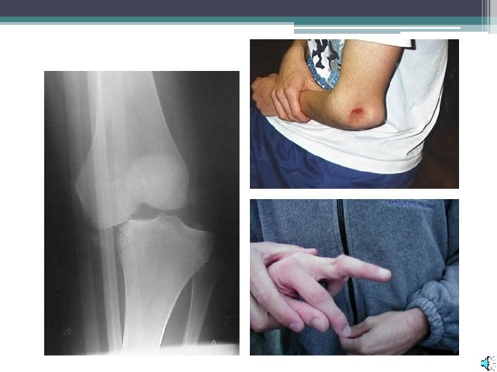
• Sprains are a stretching/tearing of the ligament tissue. ▫ Occur when a joint is forced into an abnormal position
• Dislocations are caused when a bone is completely displaced from its joint.
• A simple or closed fracture is caused when the bone breaks internally but does not come through the skin.
• An open or compound fracture projects through the skin. ▫ Increased bleeding and risk of infection.
• Greenstick fractures do not break through the entire bone. ▫ More likely in children, due to a greater portion of cartilage. • Similar to pulling a branch off a living tree.
Types of Fractures
Skeletal System Disorders • • Leukemia Bursistis Osteoporosis Spina bifida Scurvy Arthritis Rickets • • • Talipes equinovarus Scoliosis Kyphosis Fracture Dislocation (luxation) Subluxation
Leukemia • In people with leukemia, the bone marrow produces abnormal white blood cells. • The abnormal cells are leukemia cells. ▫ At first, leukemia cells function almost normally. In time, they may crowd out normal white blood cells, red blood cells, and platelets. ▫ This makes it hard for blood to do its work.
An abnormal amount of large WBC’s is one indicator of leukemia.
Bursitis • Wherever your bones, tendons, and ligaments move against each other, particularly near joints, the points of contact are cushioned by small fluid-filled sacs called bursae. • By reducing friction, each of the more than 150 bursae in your body helps the joints operate smoothly through the full range of natural movement. • But when a bursa becomes irritated and swollen, it's called bursitis -- or inflammation of the bursa.
Prepatellar bursitis. Inflammation of the patellar bursae directly over the patella.
Osteoporosis • Osteoporosis is a disease in which bones become fragile and more likely to break. • If not prevented or if left untreated, osteoporosis can progress painlessly until a bone breaks. • These broken bones, also known as fractures, occur typically in the hip, spine, and wrist.
Osteoporosis
Spina Bifida • Spina bifida (also called meningocele or myelomeningocele) is a defect that comes from a problem in the very early development of the unborn child. • It happens when some of the back bones (vertebrae) do not close over the center tube of nerves (spinal cord). • As a result, a soft unprotected area is left, which may bulge through the skin as a dark bag. • This 'bag of nerves' is covered by a very thin layer (membrane) which may leak liquid from the spinal cord and brain.
Spina Bifida
Scurvy • A condition in which there is a lack of vitamin C in the diet. • Vitamin C in necessary in the development of connective tissues, lipid and vitamin metabolism, synthesis of neurotransmitters, immunity and wound healing. • Symptoms include fatigue, joint aches, bleeding gums, leg rashes and painful leg edema.
Scurvy was once a problem for sailors who were at sea for months at a time without fresh fruits in their diets.
Arthritis Nearly 40 million Americans have arthritis, which causes joints to become inflamed or swollen. There are more than 100 types of arthritis that affect the joints and connective tissues (tendons and ligaments) of the body.
Arthritis
Rickets • Rickets is caused by a deficiency in vitamin D. During growth, human bone is made and maintained by the interaction of calcium, phosphorus, and vitamin D. • Calcium is deposited in immature bone (osteoid) in a process called calcification, which transforms immature bone into its mature and familiar form. • However, in order to absorb and use the calcium available in food, the body needs vitamin D. • In rickets, the lack of this important vitamin leads to low calcium, poor calcification, and deformed bones.
Rickets
Talipes equinovarus (Clubfoot) • Talipes equinovarus (TEV) is a compound foot disorder in which the primary deforming force is in the talus. • In the TEV foot, the talar head and neck excessively adducted, plantarflexed and medially rotated with respect to the body of the talus. • TEV may be congenital or acquired due to neuromuscular diseases or trauma.
Talipes equinovarus (Clubfoot)
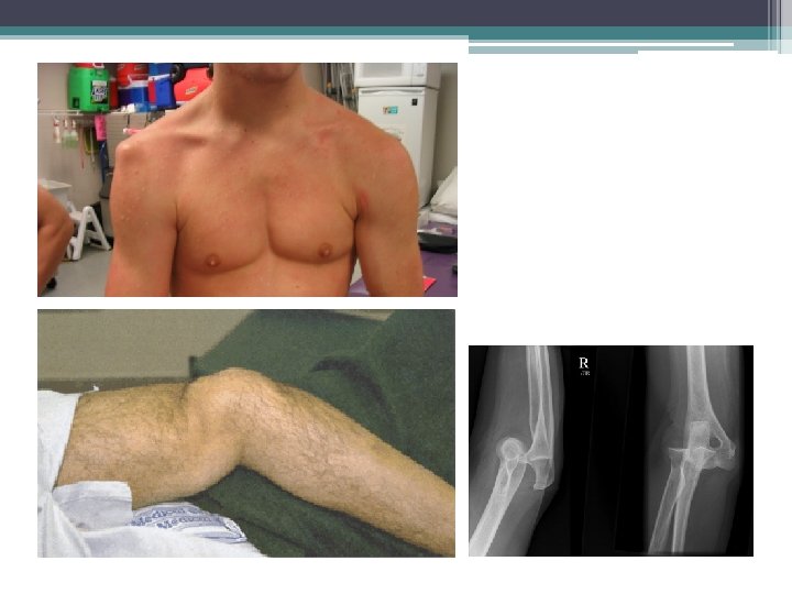
Dislocation (luxation) • Forceful displacement of an articulating bone to an abnormal position, usually accompanied by damage to tendons, ligaments, the articular capsule, or other structures. • A subluxation is a partial dislocation.
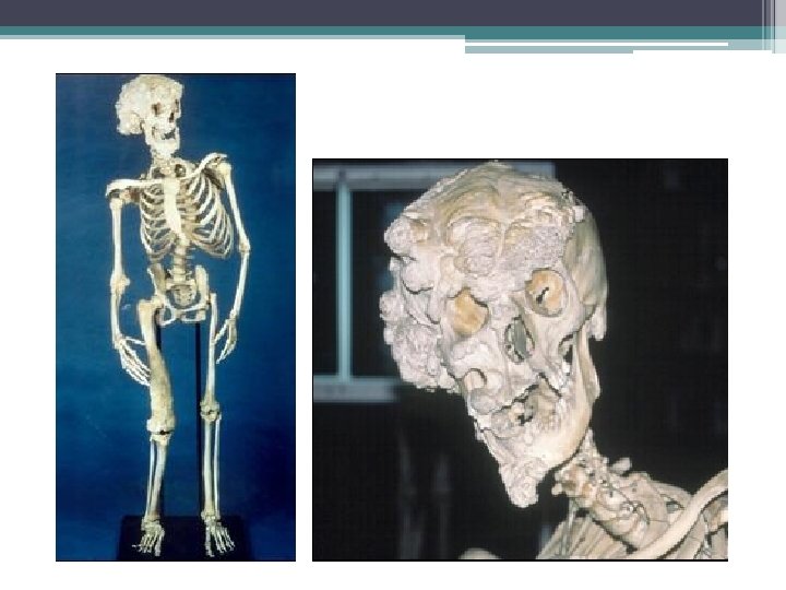
The Elephant Man Joseph Merrick, the Elephant Man, was once considered to have been afflicted with either elephantiasis or neurofibromatosis type I. However, it is now generally believed that Merrick suffered from the very rare Proteus syndrome or perhaps a combination of the two conditions.
Proteus Syndrome • Named for the Greek god who could change his shape, this rare hereditary disorder is characterized by multiple lesions of the lymph nodes (lipolymphohemangiomas), overgrowth of one side of the body (hemihypertrophy), an abnormally large head (macrocephaly), partial gigantism of the feet, and darkened spots or moles (nevi) on the skin. • Merrick's appearance, and especially his skeleton, carry all the hallmarks of the disorder, although apparently an extremely severe case. • His head was so large that the hat he wore measured three feet in circumference.
Based on DNA testing and CT scans, a computerized morph of what Joseph Merrick would have looked like had he not suffered from the disease.