Semaphorins semas and their plexin Plex and neuropilin
















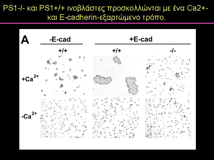
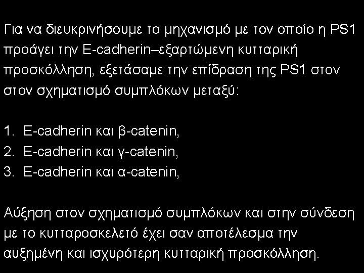

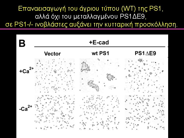

- Slides: 138
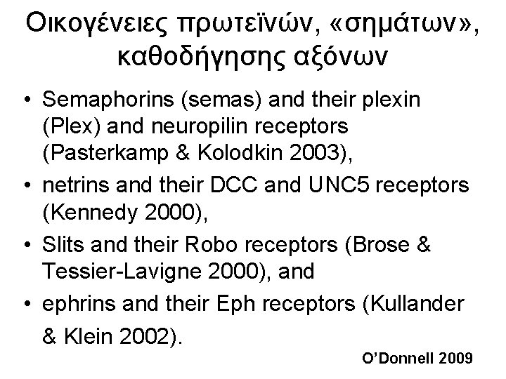
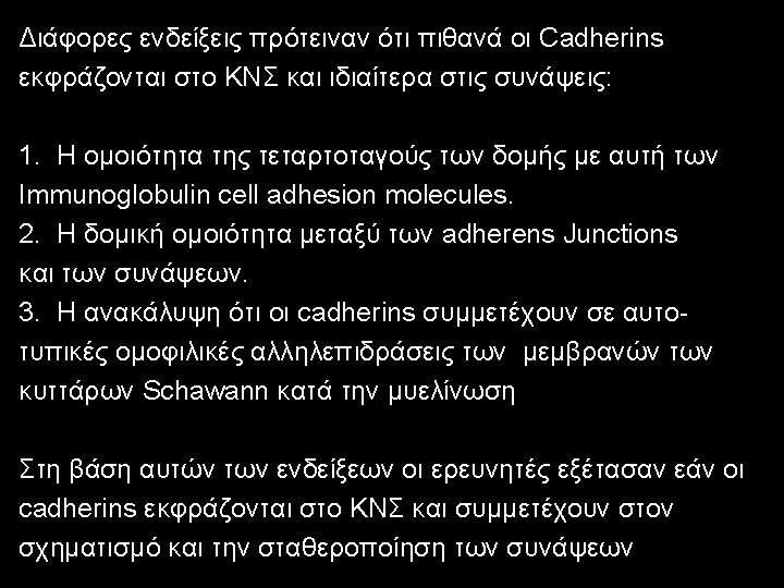
Οικογένειες πρωτεϊνών, «σημάτων» , καθοδήγησης αξόνων • Semaphorins (semas) and their plexin (Plex) and neuropilin receptors (Pasterkamp & Kolodkin 2003), • netrins and their DCC and UNC 5 receptors (Kennedy 2000), • Slits and their Robo receptors (Brose & Tessier-Lavigne 2000), and • ephrins and their Eph receptors (Kullander & Klein 2002). O’Donnell 2009
Άλλες οικογένειες πρωτεϊνών που λειτουργούν στην καθοδήγηση αξόνων • Sonic hedgehog (Shh) (Charron et al. 2003), • Βone morphogenetic proteins (BMPs) (Butler & Dodd 2003), and • Wingless-type (Wnt) proteins (Lyuksyutova et al. 2003, Yoshikawa et al. 2003). O’Donnell 2009
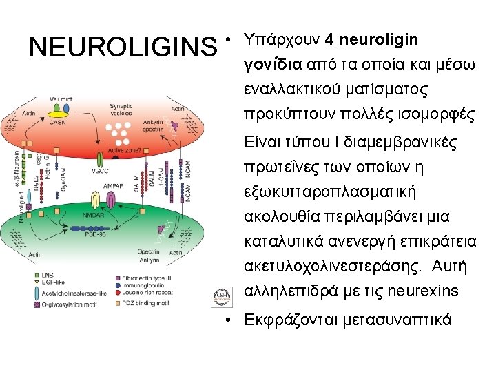
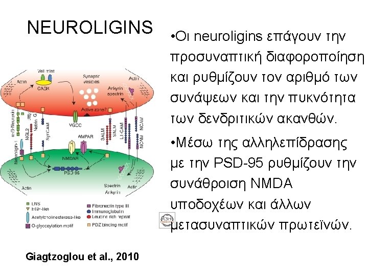
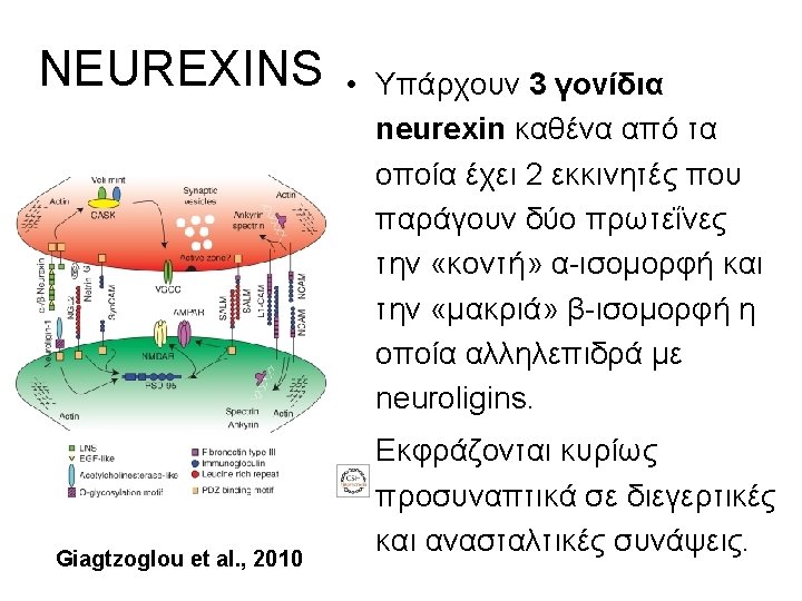
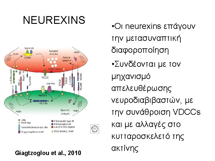
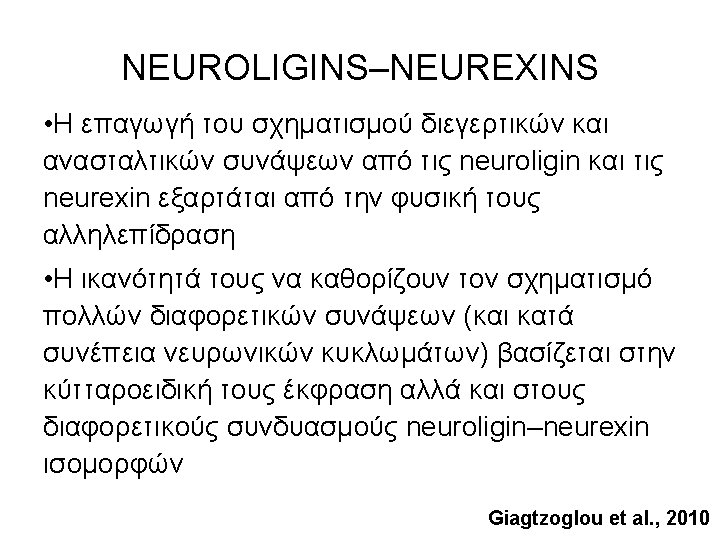
NEUROLIGINS–NEUREXINS
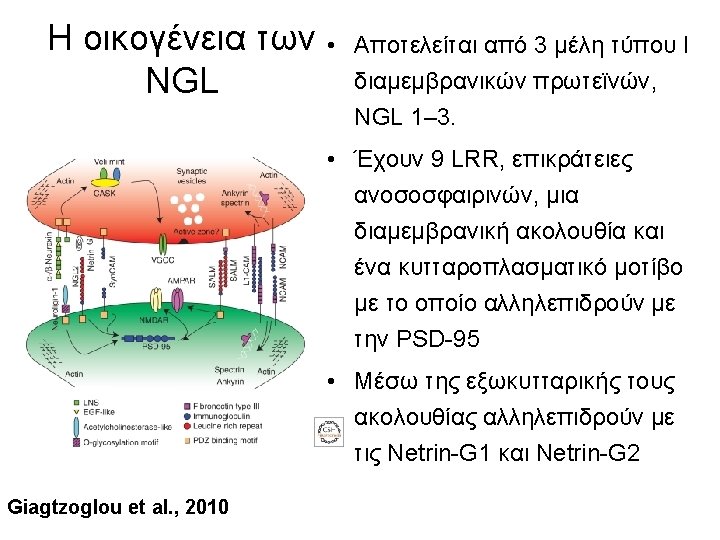
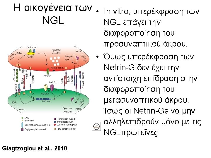
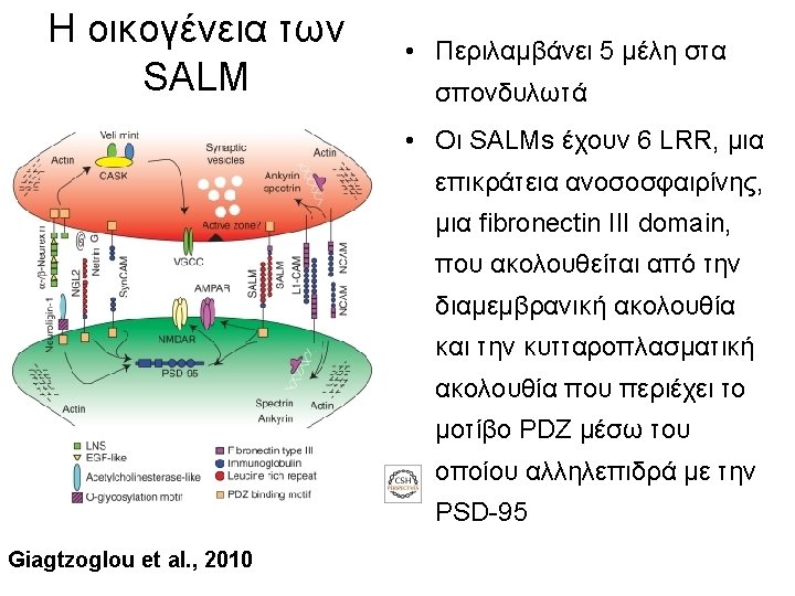
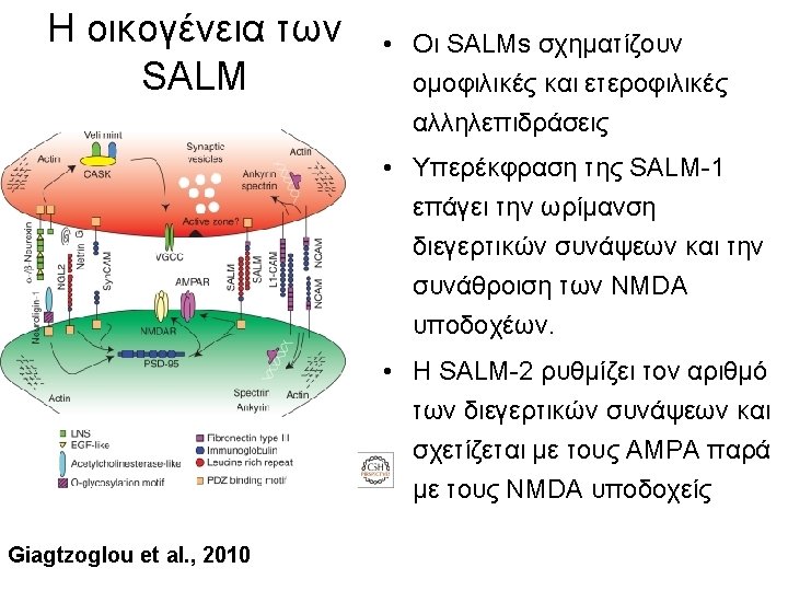
LEUCINE RICH REPEAT (LRR) ΠΡΩΤΕΪΝΕΣ The netrin-G ligand (NGL) and the synaptic-like adhesion molecule (SALM)
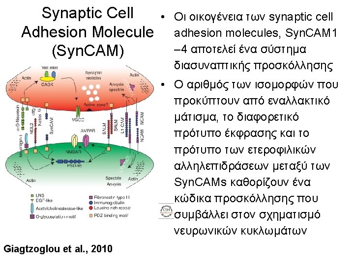
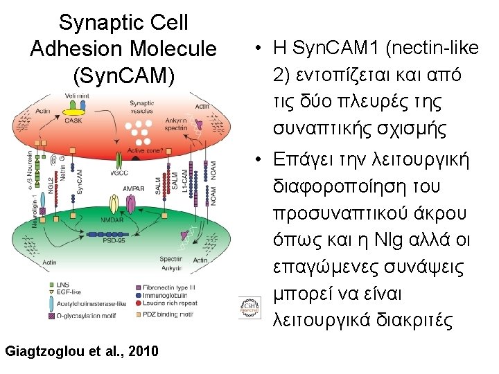
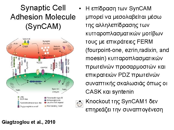
The Synaptic Cell Adhesion Molecule Syn. CAM)
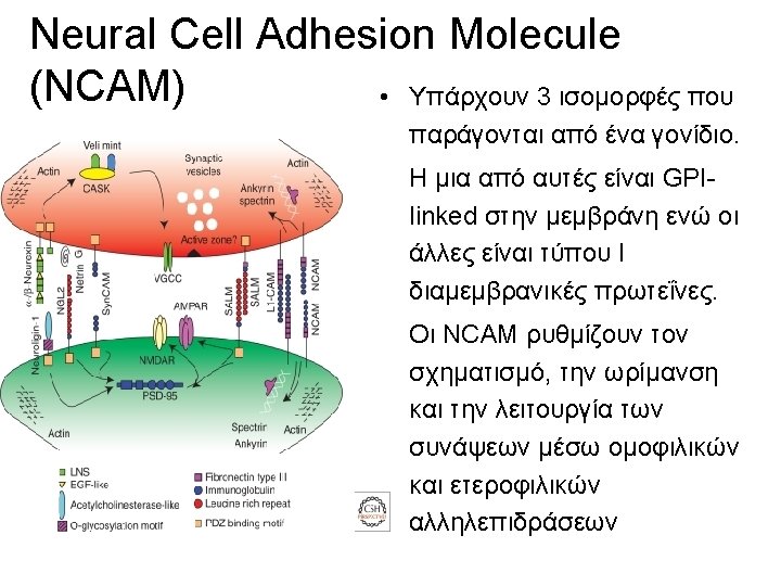
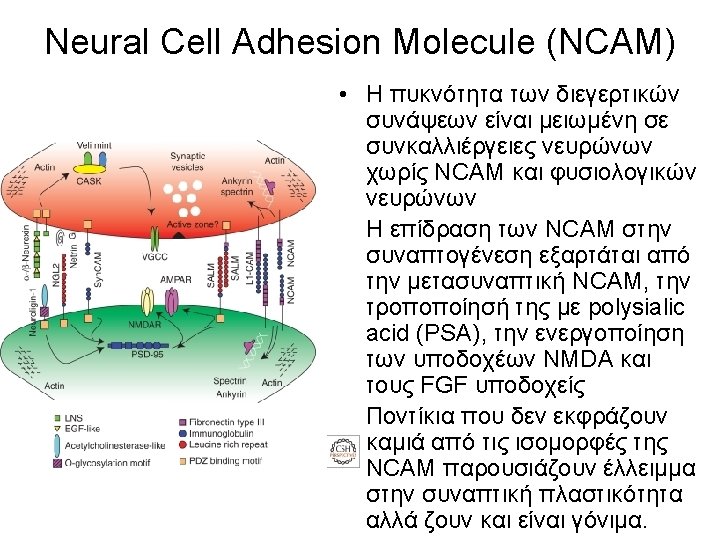
Neural Cell Adhesion Molecule (NCAM)
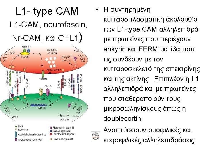
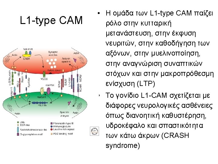
L 1 -type CAM
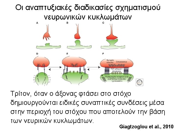
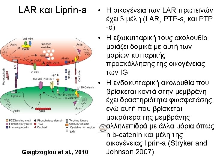
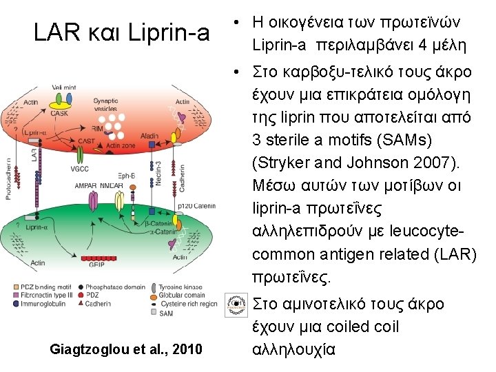
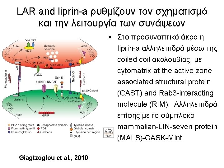
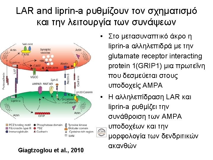
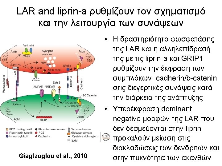
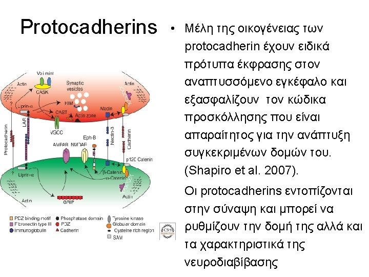
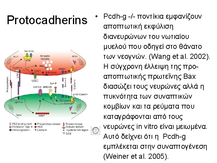
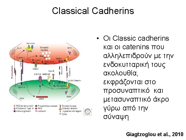
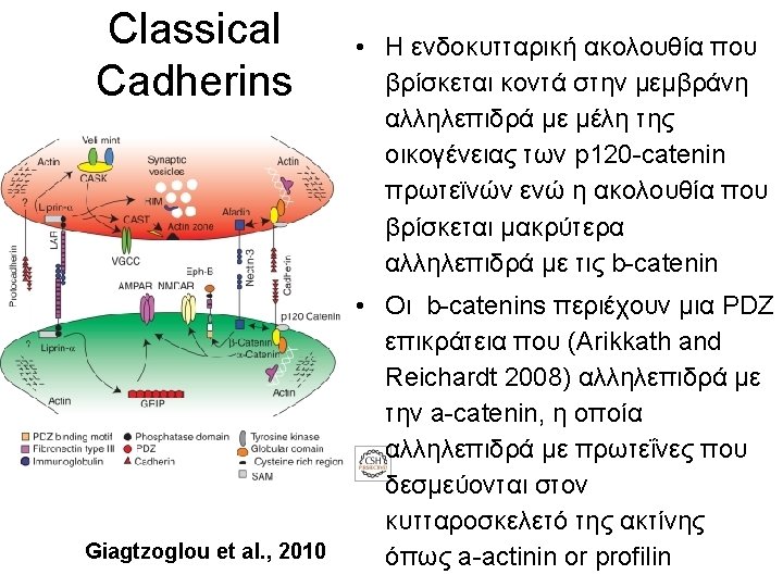
LAR and liprin-a ρυθμίζουν τον σχηματισμό και την λειτουργία των συνάψεων • Στο προσυναπτικό άκρο η liprin-a αλληλεπιδρά μέσω της coiled coil ακολουθίας με cytomatrix at the active zone associated structural protein (CAST) and Rab 3 -interacting molecule (RIM). Αλληλεπιδρά επίσης με το σύμπλοκο mammalian-LIN-seven protein (MALS)-CASK-Mint Giagtzoglou et al. , 2010
Nonclassical Cadherins: Protocadherins
Brain tissue expresses several cadherins. N- and E-Cadherin are localized to synaptic complexes in a mutually exclusive distribution Fannon and Colman, Neuron 17: 423 -434 1996
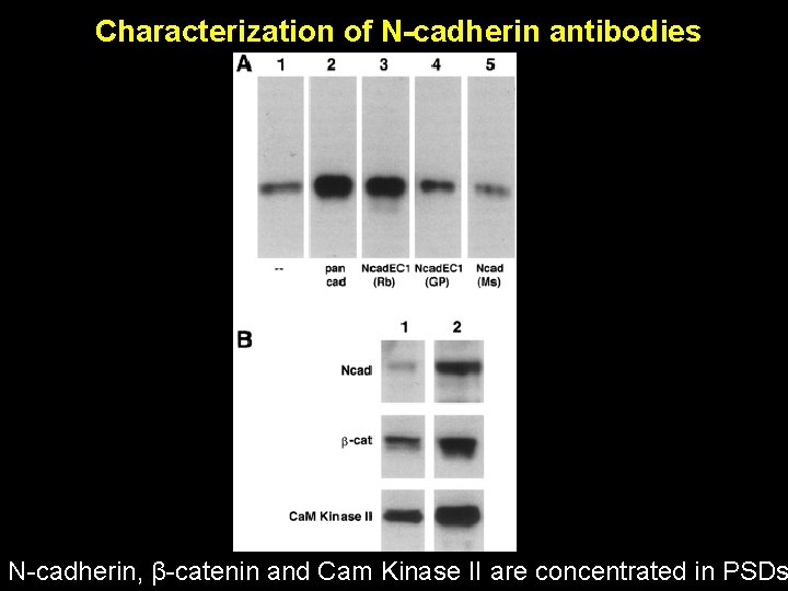
Characterization of N-cadherin antibodies N-cadherin, β-catenin and Cam Kinase II are concentrated in PSDs
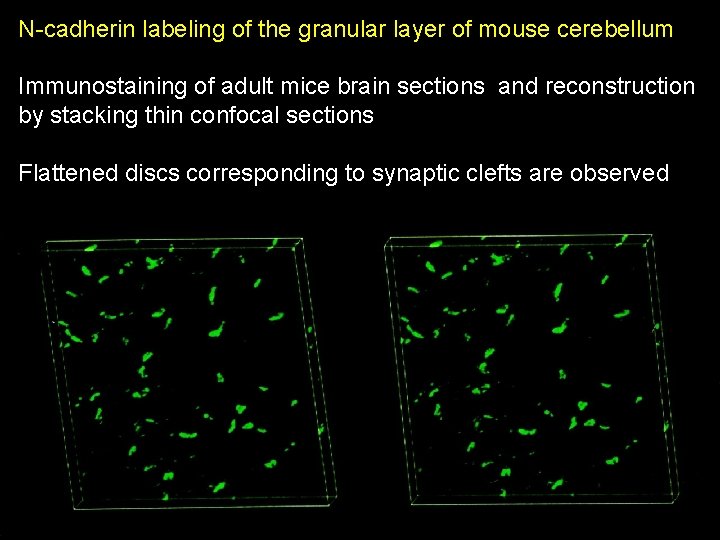
N-cadherin labeling of the granular layer of mouse cerebellum Immunostaining of adult mice brain sections and reconstruction by stacking thin confocal sections Flattened discs corresponding to synaptic clefts are observed
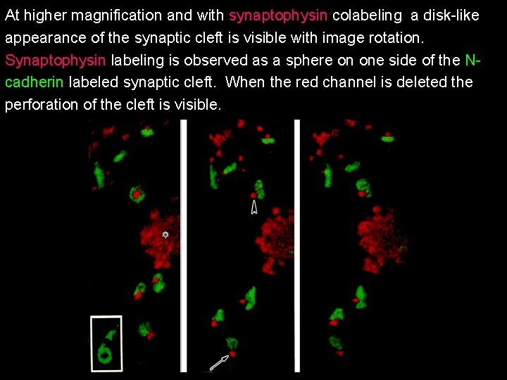
At higher magnification and with synaptophysin colabeling a disk-like appearance of the synaptic cleft is visible with image rotation. Synaptophysin labeling is observed as a sphere on one side of the Ncadherin labeled synaptic cleft. When the red channel is deleted the perforation of the cleft is visible.
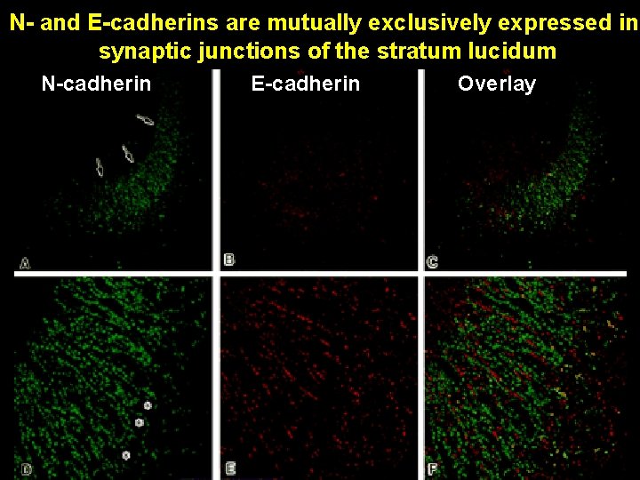
N- and E-cadherins are mutually exclusively expressed in synaptic junctions of the stratum lucidum N-cadherin E-cadherin Overlay
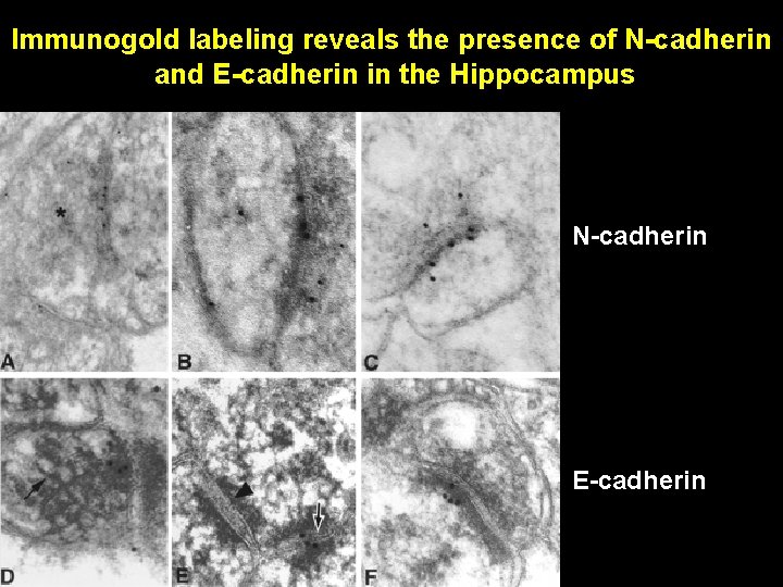
Immunogold labeling reveals the presence of N-cadherin and E-cadherin in the Hippocampus N-cadherin E-cadherin
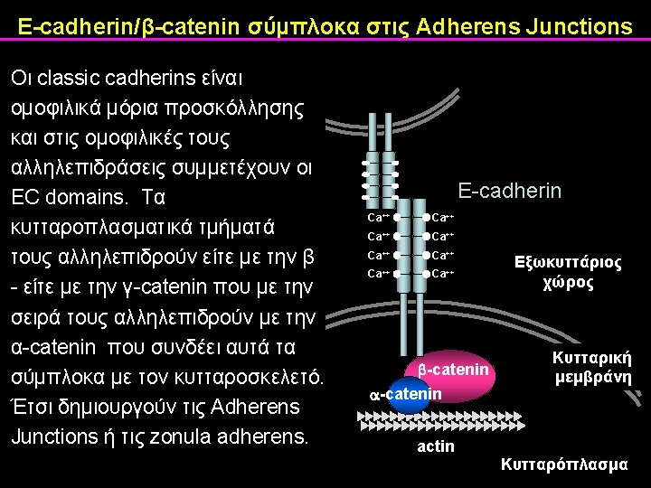
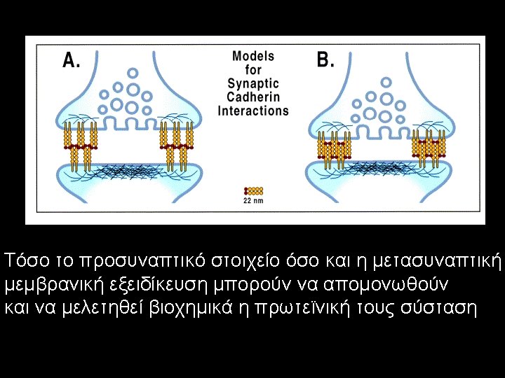
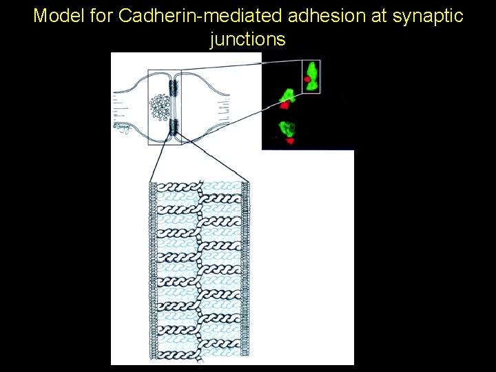
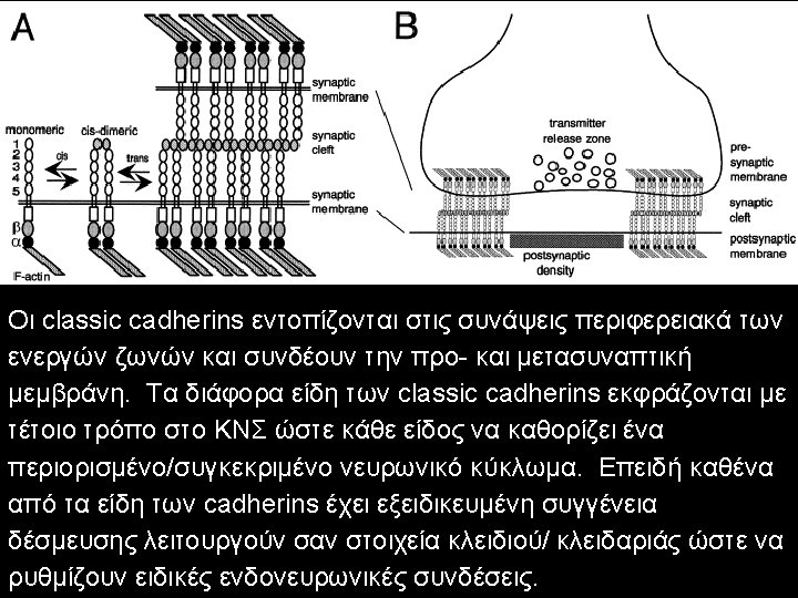
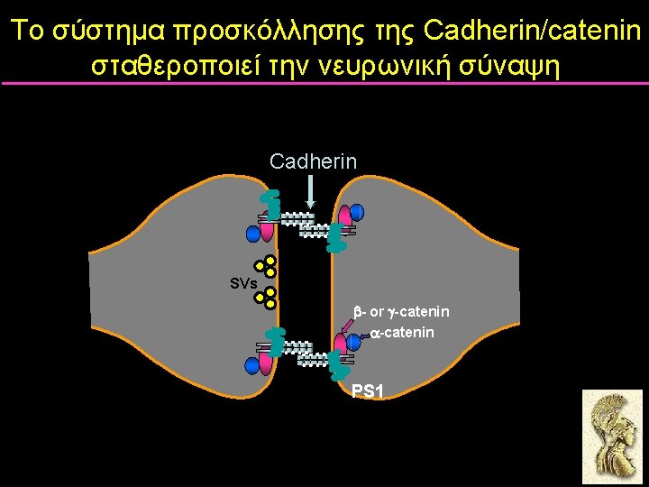
Model for Cadherin-mediated adhesion at synaptic junctions
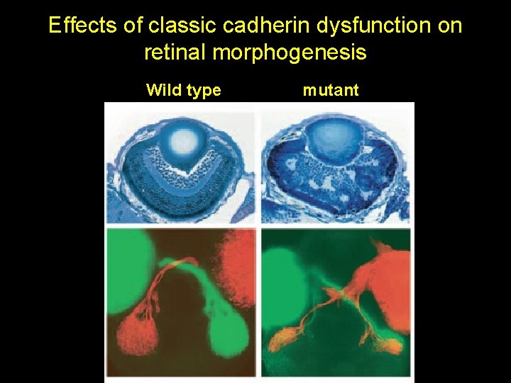
Effects of classic cadherin dysfunction on retinal morphogenesis Wild type mutant
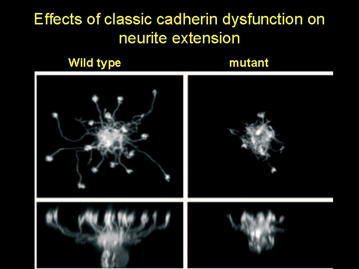
Effects of classic cadherin dysfunction on neurite extension Wild type mutant
N-Cadherin Redistribution during Synaptogenesis in Hippocampal Neurons Benson and Tanaka J. Neuroscience 18: 6892 -6904
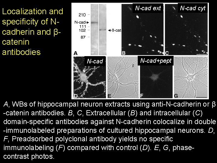
N-cad ext Localization and specificity of Ncadherin and βcatenin antibodies N-cad cyt N-cad+pept A, WΒs of hippocampal neuron extracts using anti-N-cadherin or β -catenin antibodies. B, C, Extracellular (B) and intracellular (C) domain-specific antibodies against N-cadherin colocalize in double -immunolabeled preparations of cultured hippocampal neurons. D, F, Preadsorbed polyclonal antibody yields no specific immunolabeling (F) compared with control (D). E, G, phasecontrast photos.
Hippocampal neurons cultured at low density are used to examine the localization of N-cadherin at the earliest stages of neuron development and synapse formation.
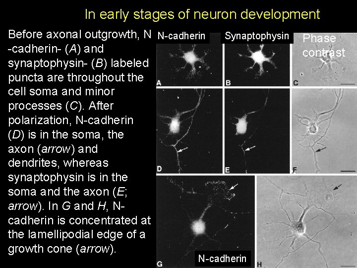
In early stages of neuron development Before axonal outgrowth, N N-cadherin- (A) and synaptophysin- (B) labeled puncta are throughout the cell soma and minor processes (C). After polarization, N-cadherin (D) is in the soma, the axon (arrow) and dendrites, whereas synaptophysin is in the soma and the axon (E; arrow). In G and H, Ncadherin is concentrated at the lamellipodial edge of a growth cone (arrow). Synaptophysin N-cadherin Phase contrast
N-cadherin is localized to developing synapses
Synaptic localization of N-Cadherin
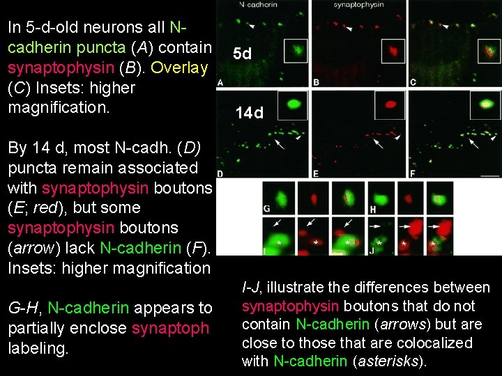
In 5 -d-old neurons all Ncadherin puncta (A) contain 5 d synaptophysin (B). Overlay (C) Insets: higher magnification. 14 d By 14 d, most N-cadh. (D) puncta remain associated with synaptophysin boutons (E; red), but some synaptophysin boutons (arrow) lack N-cadherin (F). Insets: higher magnification G-H, N-cadherin appears to partially enclose synaptoph labeling. I-J, illustrate the differences between synaptophysin boutons that do not contain N-cadherin (arrows) but are close to those that are colocalized with N-cadherin (asterisks).
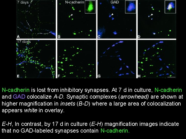
N-cadherin is lost from inhibitory synapses. At 7 d in culture, N-cadherin and GAD colocalize A-D. Synaptic complexes (arrowhead) are shown at higher magnification in insets (B-D) where a large area of colocalization appears white in overlay. E-H, In contrast, by 17 d in culture (E-H) magnification images indicate that no GAD-labeled synapses contain N-cadherin.
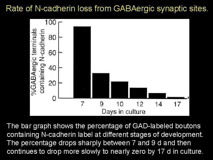
Rate of N-cadherin loss from GABAergic synaptic sites. The bar graph shows the percentage of GAD-labeled boutons containing N-cadherin label at different stages of development. The percentage drops sharply between 7 and 9 d and then continues to drop more slowly to nearly zero by 17 d in culture.
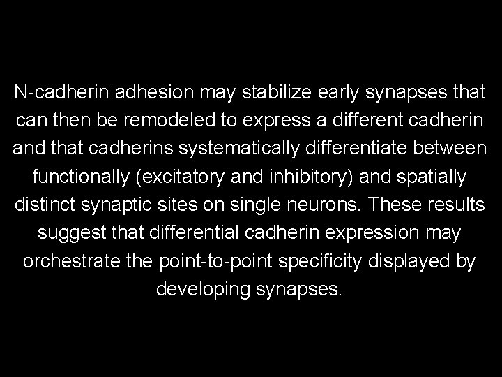
N-cadherin adhesion may stabilize early synapses that can then be remodeled to express a different cadherin and that cadherins systematically differentiate between functionally (excitatory and inhibitory) and spatially distinct synaptic sites on single neurons. These results suggest that differential cadherin expression may orchestrate the point-to-point specificity displayed by developing synapses.
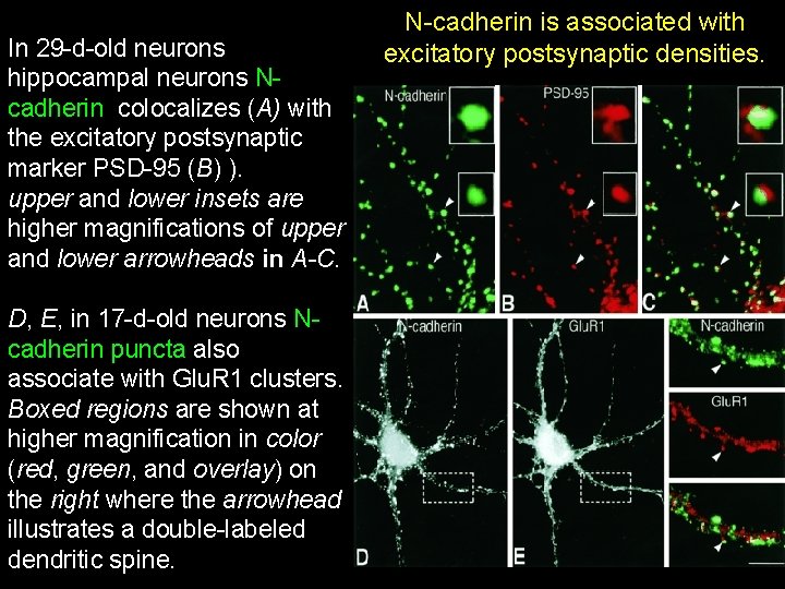
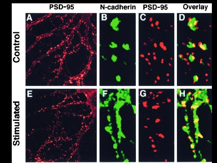
In 29 -d-old neurons hippocampal neurons Ncadherin colocalizes (A) with the excitatory postsynaptic marker PSD-95 (B) ). upper and lower insets are higher magnifications of upper and lower arrowheads in A-C. D, E, in 17 -d-old neurons Ncadherin puncta also associate with Glu. R 1 clusters. Boxed regions are shown at higher magnification in color (red, green, and overlay) on the right where the arrowhead illustrates a double-labeled dendritic spine. N-cadherin is associated with excitatory postsynaptic densities.
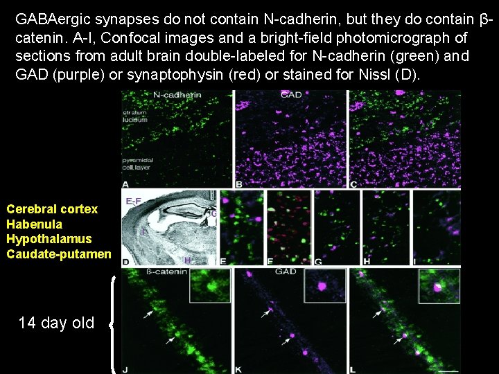
GABAergic synapses do not contain N-cadherin, but they do contain βcatenin. A-I, Confocal images and a bright-field photomicrograph of sections from adult brain double-labeled for N-cadherin (green) and GAD (purple) or synaptophysin (red) or stained for Nissl (D). Cerebral cortex Habenula Hypothalamus Caudate-putamen 14 day old
N-cadherin is present in axons and dendrites before synapse formation and then clusters at neuronal synapses. N-cadherin is expressed initially at all synaptic sites but rapidly becomes restricted to a subpopulation of excitatory synaptic sites. GABAergic, inhibitory synapses in mature cultures lack N-cadherin but are associated with clusters of βcatenin, implying that they contain a different classic cadherin.
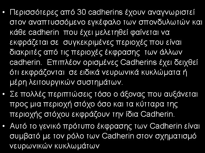
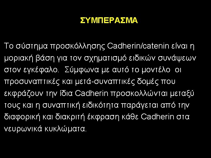
The differential distribution of cadherins and the specificity of the interactions between various cadherins is the basis for the specificity of neuronal interactions and synapse formation
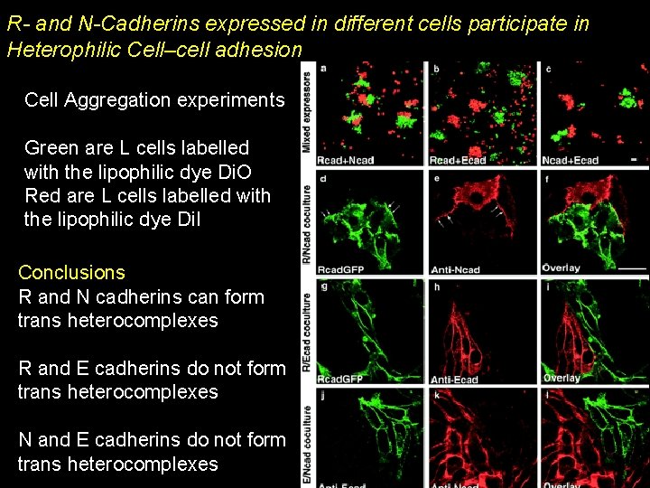
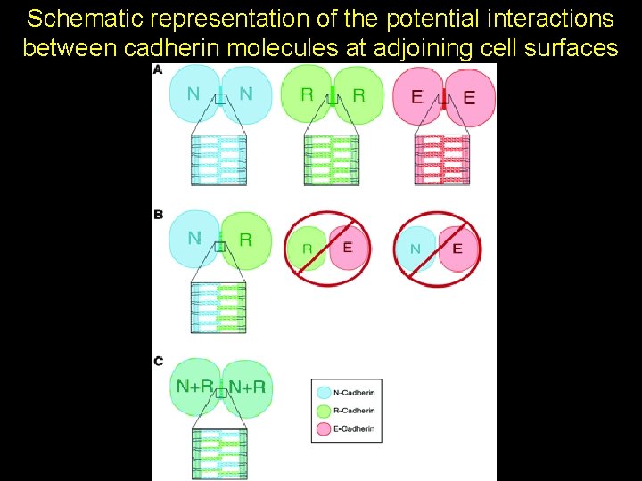
R- and N-Cadherins expressed in different cells participate in Heterophilic Cell–cell adhesion Cell Aggregation experiments Green are L cells labelled with the lipophilic dye Di. O Red are L cells labelled with the lipophilic dye Di. I Conclusions R and N cadherins can form trans heterocomplexes R and E cadherins do not form trans heterocomplexes N and E cadherins do not form trans heterocomplexes
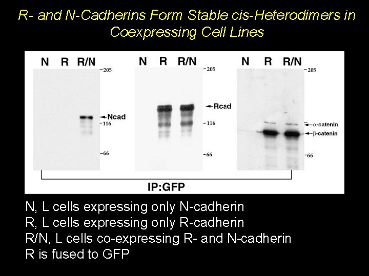
R- and N-Cadherins Form Stable cis-Heterodimers in Coexpressing Cell Lines N, L cells expressing only N-cadherin R, L cells expressing only R-cadherin R/N, L cells co-expressing R- and N-cadherin R is fused to GFP
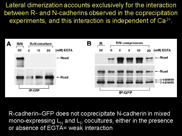
The heterophilic interaction between LR and LN cells is calcium-dependent. Therefore, to determine whether the R-/N-cadherin complexes obtained by immunoprecipitation originate from the same or opposing cells the calcium chelator EGTA was used to disrupt the adhesive interaction.
Lateral dimerization accounts exclusively for the interaction between R- and N-cadherins observed in the coprecipitation experiments, and this interaction is independent of Ca 2+. R-cadherin–GFP does not coprecipitate N-cadherin in mixed mono-expressing LN and LR cocultures, either in the presence or absence of EGTA= weak interaction
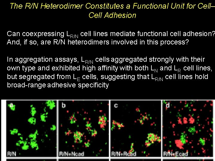
The R/N Heterodimer Constitutes a Functional Unit for Cell– Cell Adhesion Can coexpressing LR/N cell lines mediate functional cell adhesion? And, if so, are R/N heterodimers involved in this process? In aggregation assays, LR/N cells aggregated strongly with their own type and exhibited high affinity with both LN and LR cell lines, but segregated from LE cells, suggesting that LR/N cell lines hold broad-range adhesive specificity
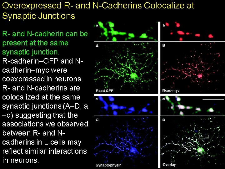
Overexpressed R- and N-Cadherins Colocalize at Synaptic Junctions R- and N-cadherin can be present at the same synaptic junction. R-cadherin–GFP and Ncadherin–myc were coexpressed in neurons. R- and N-cadherins are colocalized at the same synaptic junctions (A–D, a –d) suggesting that the associations we observed between R- and Ncadherins in L cells may reflect similar interactions in neurons.
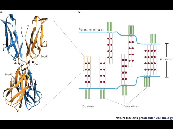
Schematic representation of the potential interactions between cadherin molecules at adjoining cell surfaces
Molecular Modification of N-Cadherin in Response to Synaptic Activity
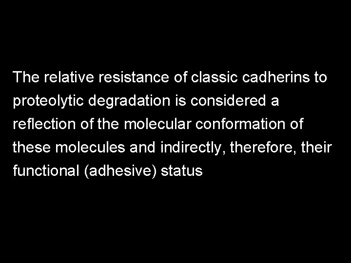
The relative resistance of classic cadherins to proteolytic degradation is considered a reflection of the molecular conformation of these molecules and indirectly, therefore, their functional (adhesive) status
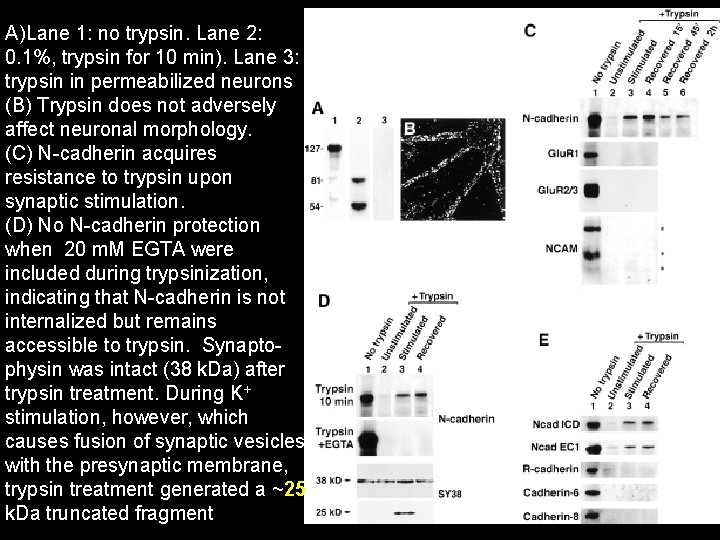
A)Lane 1: no trypsin. Lane 2: 0. 1%, trypsin for 10 min). Lane 3: trypsin in permeabilized neurons (B) Trypsin does not adversely affect neuronal morphology. (C) N-cadherin acquires resistance to trypsin upon synaptic stimulation. (D) No N-cadherin protection when 20 m. M EGTA were included during trypsinization, indicating that N-cadherin is not internalized but remains accessible to trypsin. Synaptophysin was intact (38 k. Da) after trypsin treatment. During K+ stimulation, however, which causes fusion of synaptic vesicles with the presynaptic membrane, trypsin treatment generated a ~25 k. Da truncated fragment
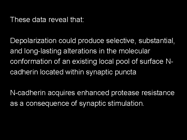
These data reveal that: Depolarization could produce selective, substantial, and long-lasting alterations in the molecular conformation of an existing local pool of surface Ncadherin located within synaptic puncta N-cadherin acquires enhanced protease resistance as a consequence of synaptic stimulation.
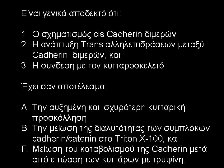
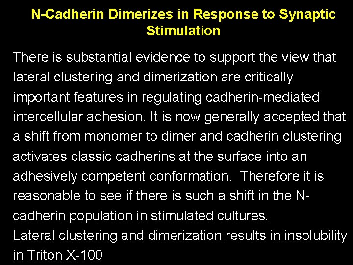
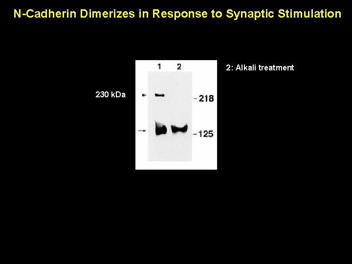
N-Cadherin Dimerizes in Response to Synaptic Stimulation There is substantial evidence to support the view that lateral clustering and dimerization are critically important features in regulating cadherin-mediated intercellular adhesion. It is now generally accepted that a shift from monomer to dimer and cadherin clustering activates classic cadherins at the surface into an adhesively competent conformation. Therefore it is reasonable to see if there is such a shift in the Ncadherin population in stimulated cultures. Lateral clustering and dimerization results in insolubility in Triton X-100
N-Cadherin Dimerizes in Response to Synaptic Stimulation 2: Alkali treatment 230 k. Da
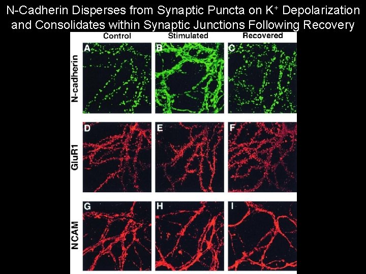
N-Cadherin Disperses from Synaptic Puncta on K+ Depolarization and Consolidates within Synaptic Junctions Following Recovery
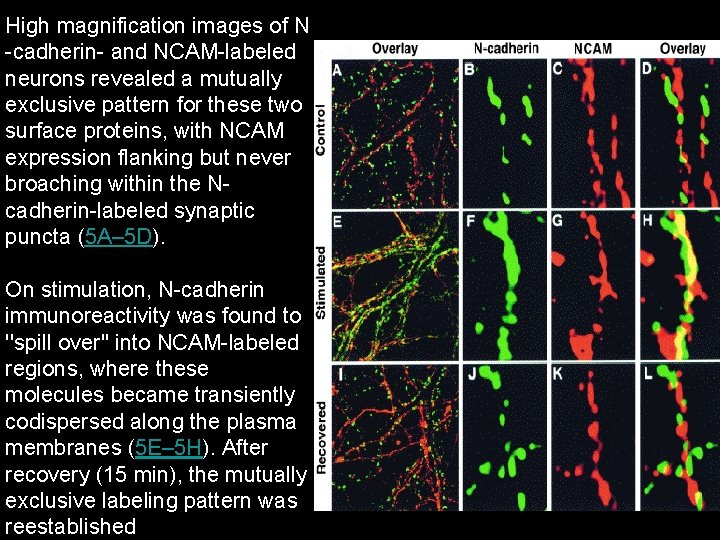
High magnification images of N -cadherin- and NCAM-labeled neurons revealed a mutually exclusive pattern for these two surface proteins, with NCAM expression flanking but never broaching within the Ncadherin-labeled synaptic puncta (5 A– 5 D). On stimulation, N-cadherin immunoreactivity was found to ''spill over'' into NCAM-labeled regions, where these molecules became transiently codispersed along the plasma membranes (5 E– 5 H). After recovery (15 min), the mutually exclusive labeling pattern was reestablished
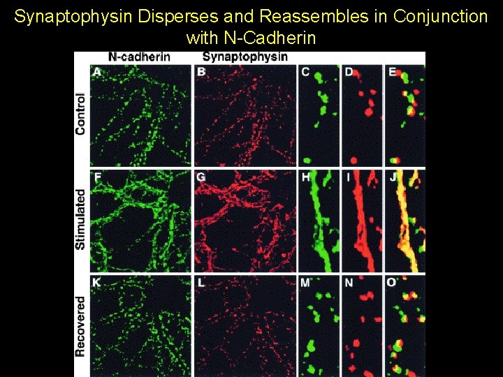
Synaptophysin Disperses and Reassembles in Conjunction with N-Cadherin
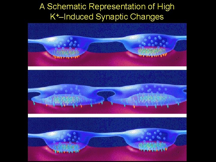
A Schematic Representation of High K+–Induced Synaptic Changes
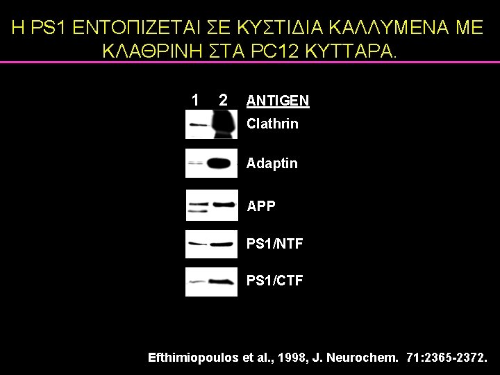
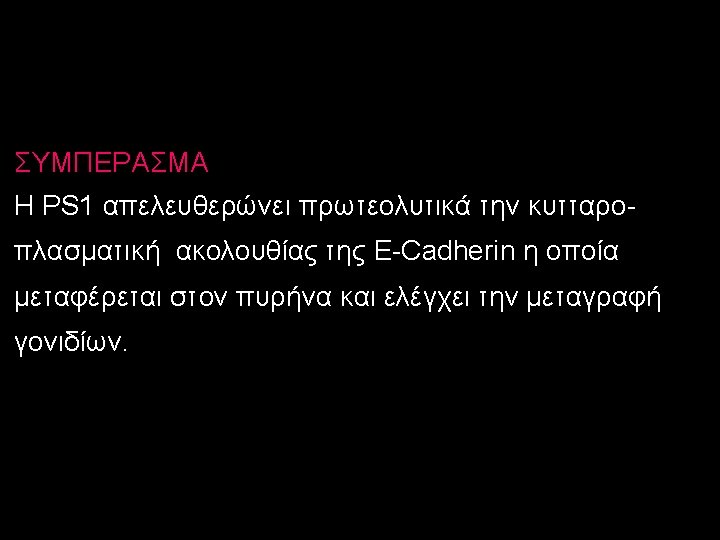
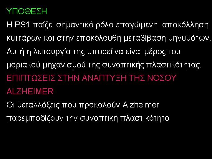
Η PS 1 ΕΝΤΟΠΙΖΕΤΑΙ ΣΕ ΚΥΣΤΙΔΙΑ ΚΑΛΛΥΜΕΝΑ ΜΕ ΚΛΑΘΡΙΝΗ ΣΤΑ PC 12 ΚΥΤΤΑΡΑ. 1 2 ANTIGEN Clathrin Adaptin APP PS 1/NTF PS 1/CTF Efthimiopoulos et al. , 1998, J. Neurochem. 71: 2365 -2372.
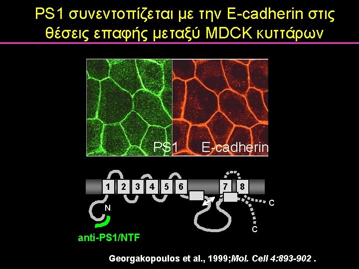
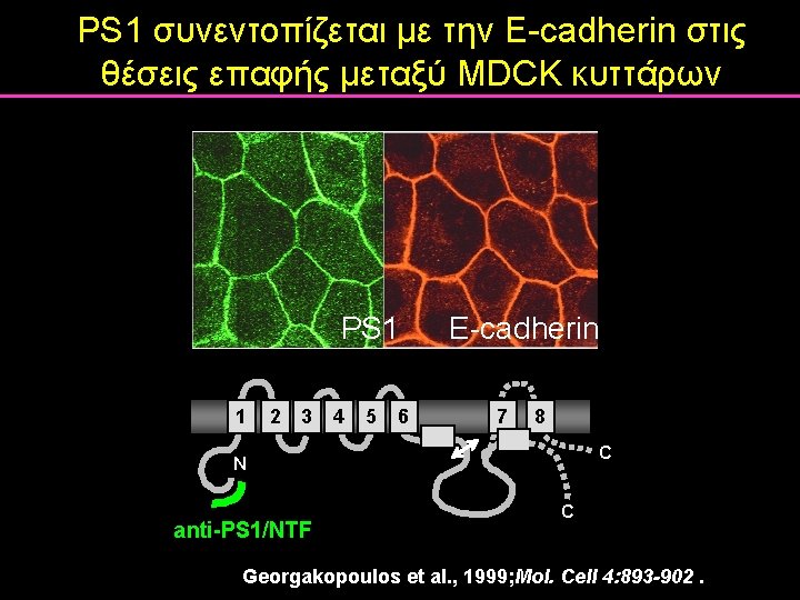
PS 1 συνεντοπίζεται με την E-cadherin στις θέσεις επαφής μεταξύ MDCK κυττάρων PS 1 1 2 3 4 5 E-cadherin 6 7 8 C N anti-PS 1/NTF C Georgakopoulos et al. , 1999; Mol. Cell 4: 893 -902.
PS 1 συνεντοπίζεται με την E-cadherin στις θέσεις επαφής μεταξύ MDCK κυττάρων PS 1 1 2 3 4 5 E-cadherin 6 7 8 C N anti-PS 1/NTF C Georgakopoulos et al. , 1999; Mol. Cell 4: 893 -902.
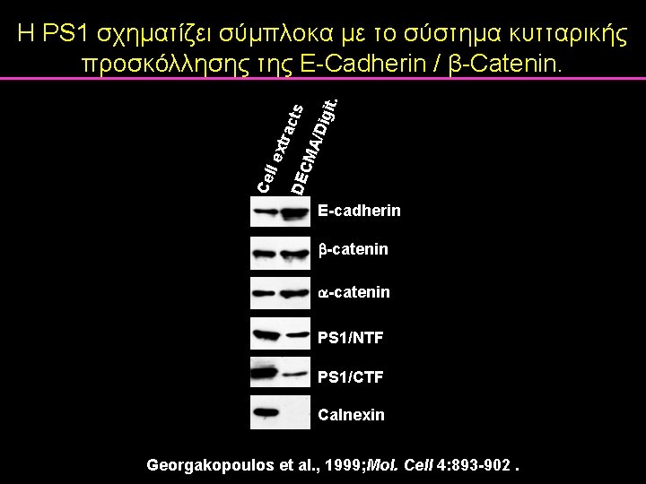
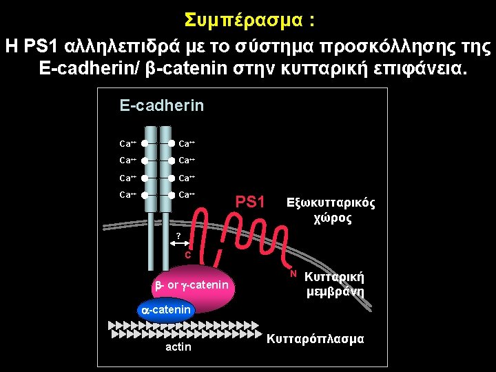
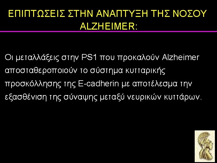
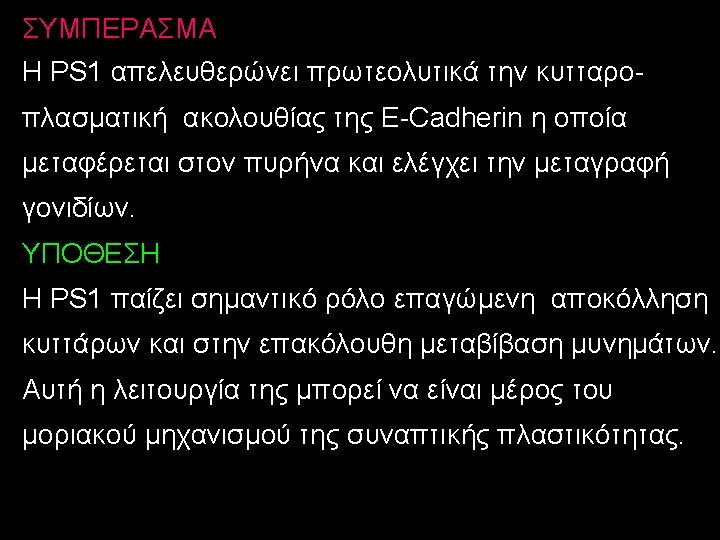
Cel l ex trac ts DEC MA /Dig it. Η PS 1 σχηματίζει σύμπλοκα με το σύστημα κυτταρικής προσκόλλησης της Ε-Cadherin / β-Catenin. E-cadherin -catenin PS 1/NTF PS 1/CTF Calnexin Georgakopoulos et al. , 1999; Mol. Cell 4: 893 -902.
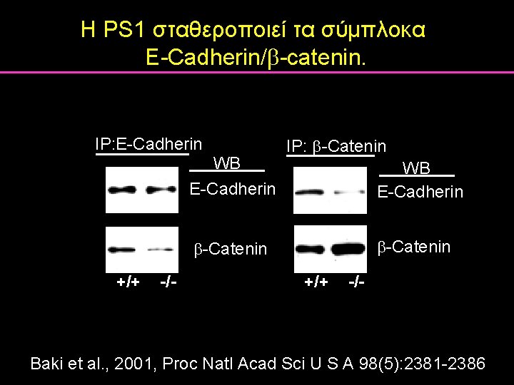
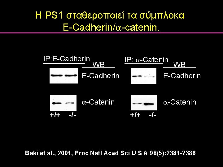
Η PS 1 σταθεροποιεί τα σύμπλοκα E-Cadherin/ -catenin. IP: E-Cadherin IP: -Catenin WB E-Cadherin -Catenin +/+ -/- Baki et al. , 2001, Proc Natl Acad Sci U S A 98(5): 2381 -2386
Η PS 1 σταθεροποιεί τα σύμπλοκα E-Cadherin/ -catenin. IP: E-Cadherin IP: -Catenin WB E-Cadherin -Catenin +/+ -/- Baki et al. , 2001, Proc Natl Acad Sci U S A 98(5): 2381 -2386
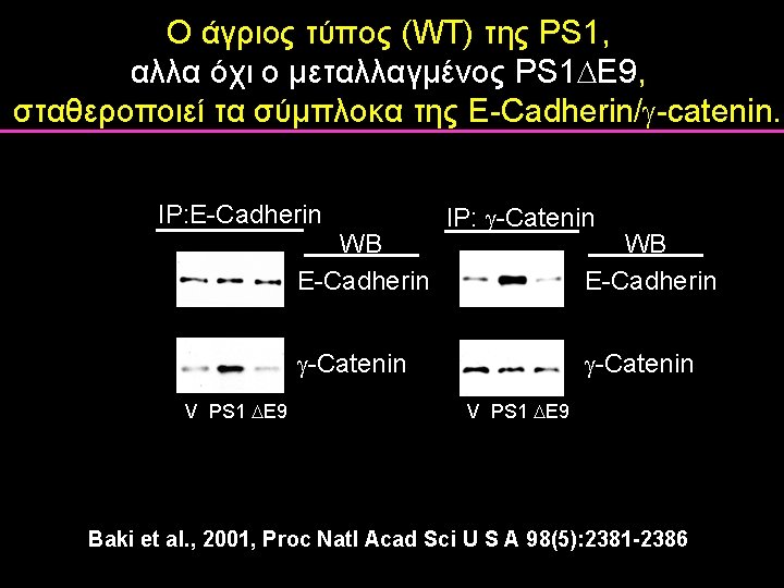
Ο άγριος τύπος (WT) της PS 1, αλλα όχι ο μεταλλαγμένος PS 1 E 9, σταθεροποιεί τα σύμπλοκα της E-Cadherin/ -catenin. IP: E-Cadherin IP: -Catenin WB E-Cadherin -Catenin V PS 1 E 9 Baki et al. , 2001, Proc Natl Acad Sci U S A 98(5): 2381 -2386
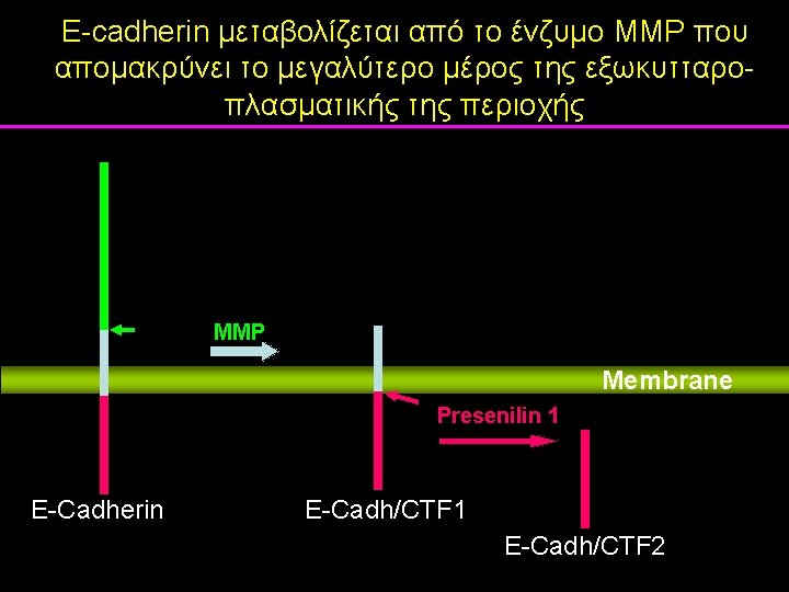
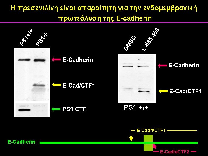
5, 4 58 68 DM S L- O -/PS 1+ /+ Η πρεσενιλίνη είναι απαραίτητη για την ενδομεμβρανική πρωτεόλυση της E-cadherin E-Cadherin E-Cad/CTF 1 PS 1 CTF E-Cad/CTF 1 PS 1 +/+ E-Cadh/CTF 1 E-Cadherin E-Cadh/CTF 2
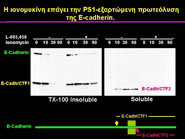
Η ιονομυκίνη επάγει την PS 1 -εξαρτώμενη πρωτεόλυση της E-cadherin. L-685, 458 - + - + . Ionomycin 0 10 30 90 0 10 30 90 0 10 30 90 0 10 30 90 E-Cadherin E-Cadh/CTF 1 E-Cadh/CTF 2 TX-100 Insoluble Soluble E-Cadh/CTF 1 E-Cadherin E-Cadh/CTF 2