In the name of GOD Neuro Anatomy Session










- Slides: 60
In the name of GOD Neuro. Anatomy- Session: 3 Session: 2 The Neurobiology of the Neuron and the a Nerve fibers & peripheral innervation Dr. Maria Zahiri
A 45 -year-old was recovering from a mild upper respiratory tract infection when he suddenly noticed weakness in both legs while walking up stairs He also developed a numb sensation over the lower part of both legs and the feet Two days later, while shaving, he noticed a weakness of the muscles on the right side of his face 2/28/2021 DR. MARIA ZAHIRI 2
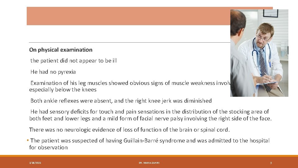
On physical examination the patient did not appear to be ill He had no pyrexia Examination of his leg muscles showed obvious signs of muscle weakness involving both legs, especially below the knees Both ankle reflexes were absent, and the right knee jerk was diminished He had sensory deficits for touch and pain sensations in the distribution of the stocking area of both feet and lower legs and a mild form of facial nerve palsy involving the right side of the face. There was no neurologic evidence of loss of function of the brain or spinal cord. • The patient was suspected of having Guillain-Barré syndrome and was admitted to the hospital for observation 2/28/2021 DR. MARIA ZAHIRI 3
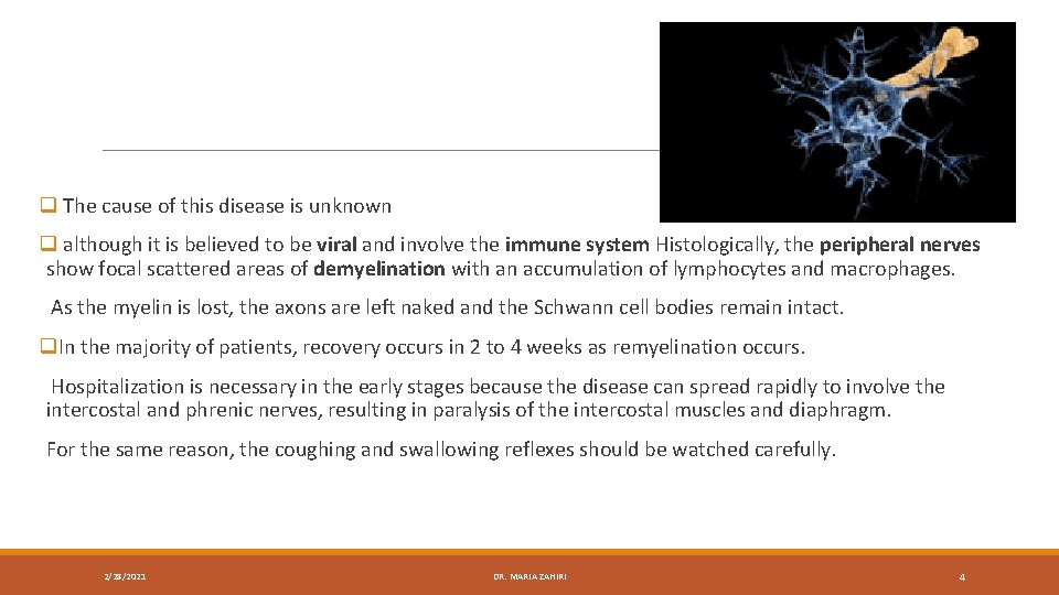
q The cause of this disease is unknown q although it is believed to be viral and involve the immune system Histologically, the peripheral nerves show focal scattered areas of demyelination with an accumulation of lymphocytes and macrophages. As the myelin is lost, the axons are left naked and the Schwann cell bodies remain intact. q. In the majority of patients, recovery occurs in 2 to 4 weeks as remyelination occurs. Hospitalization is necessary in the early stages because the disease can spread rapidly to involve the intercostal and phrenic nerves, resulting in paralysis of the intercostal muscles and diaphragm. For the same reason, the coughing and swallowing reflexes should be watched carefully. 2/28/2021 DR. MARIA ZAHIRI 4
ØTo consider the basic structure and function of nerve fibers Ø To understand the process of nerve degeneration and regeneration Ø To review the special organs that lie at the ends of sensory and motor nerves Ø To examine the different sensory modalities ØTo learn the terms used in assessing skin sensory loss and abnormal muscle activity ØTo review the special organs that lie at the ends of sensory and motor nerves 2/28/2021 DR. MARIA ZAHIRI 5
Nerve Fibers q Nerve fiber is the name given to an axon (or a dendrite) of a nerve cell. q Bundles of nerve fibers found in the CNS are often referred to as nerve tracts qbundles of nerve fibers found in the PNS are called peripheral nerves q 2 types of nerve fibers are present in the central and peripheral parts of the nervous system: myelinated and nonmyelinated. 2/28/2021 DR. MARIA ZAHIRI 6
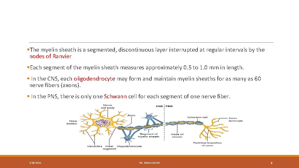
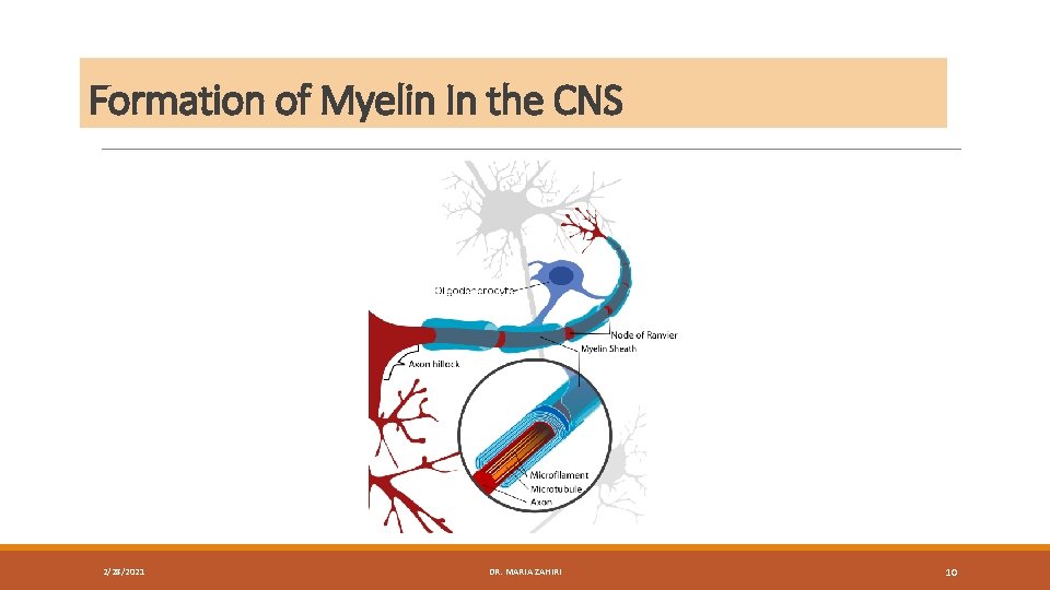
Myelinated Nerve Fibers q A myelinated nerve fiber is one that is surrounded by a myelin sheath. q The myelin sheath is not part of the neuron but is formed by a supporting cell q In the CNS, the supporting cell is called the oligodendrocyte q in the PNS, it is called the Schwann cell. 2/28/2021 DR. MARIA ZAHIRI 7
§The myelin sheath is a segmented, discontinuous layer interrupted at regular intervals by the nodes of Ranvier §Each segment of the myelin sheath measures approximately 0. 5 to 1. 0 mm in length. § In the CNS, each oligodendrocyte may form and maintain myelin sheaths for as many as 60 nerve fibers (axons). § In the PNS, there is only one Schwann cell for each segment of one nerve fiber. 2/28/2021 DR. MARIA ZAHIRI 8
Formation of Myelin In the PNS 2/28/2021 DR. MARIA ZAHIRI 9
Formation of Myelin In the CNS 2/28/2021 DR. MARIA ZAHIRI 10
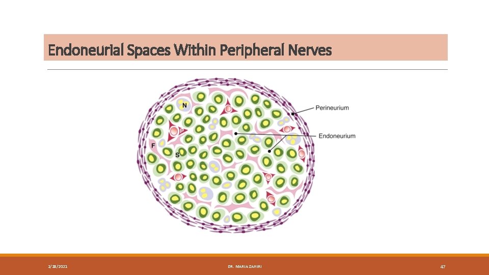
Peripheral Transvers. Nerve section of peripheral nerve Longitudinal section of peripheral nerve Endoneurium: surrond individual nerve fiber -is a loose connective tissue -The endoneurium is in contact with basal lamina of the Schwann cells Perineurium: cover each bundle of nerve fibers(fascicle) is composed of dense irregular CT. Epineurium: the outermost layer covering of the nerve -composed of dense irregular CT -containing some thick elastic fibers -is thick near the CNS where continues with dura matter DR. MARIA ZAHIRI
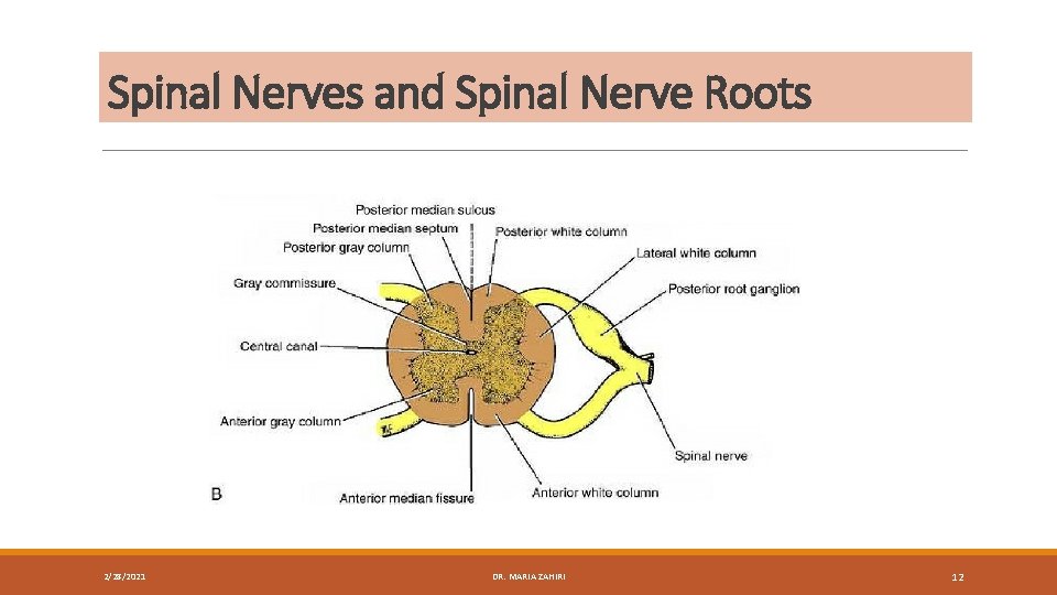
Spinal Nerves and Spinal Nerve Roots 2/28/2021 DR. MARIA ZAHIRI 12
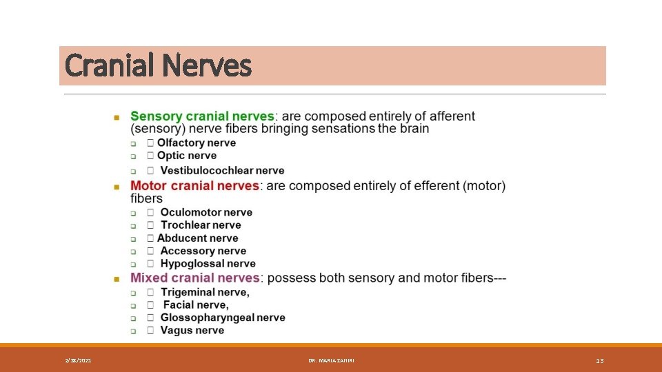
Cranial Nerves 2/28/2021 DR. MARIA ZAHIRI 13
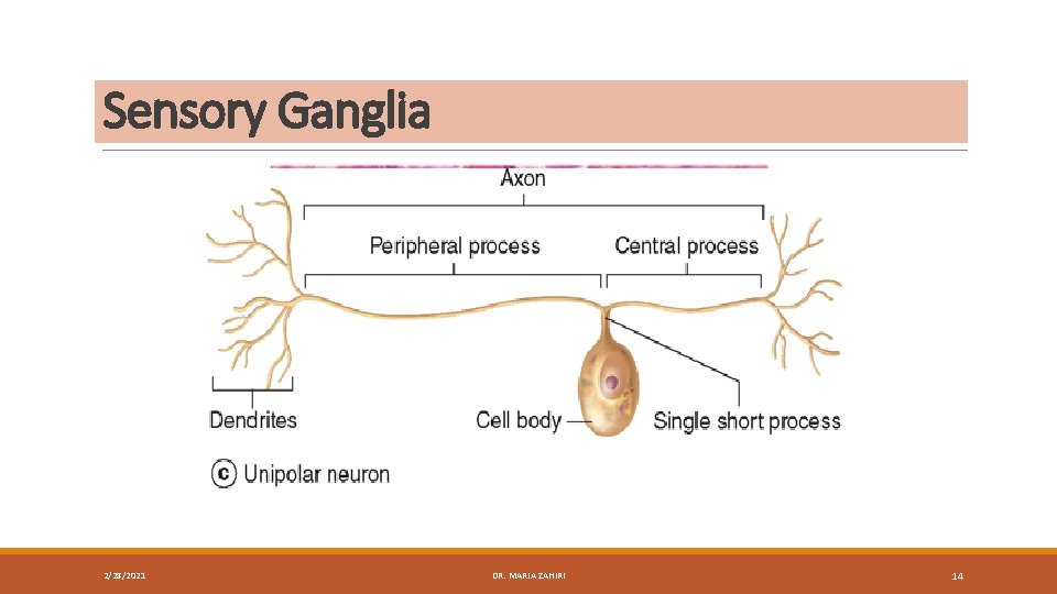
Sensory Ganglia 2/28/2021 DR. MARIA ZAHIRI 14
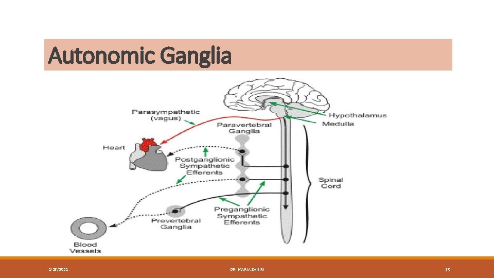
Autonomic Ganglia 2/28/2021 DR. MARIA ZAHIRI 15
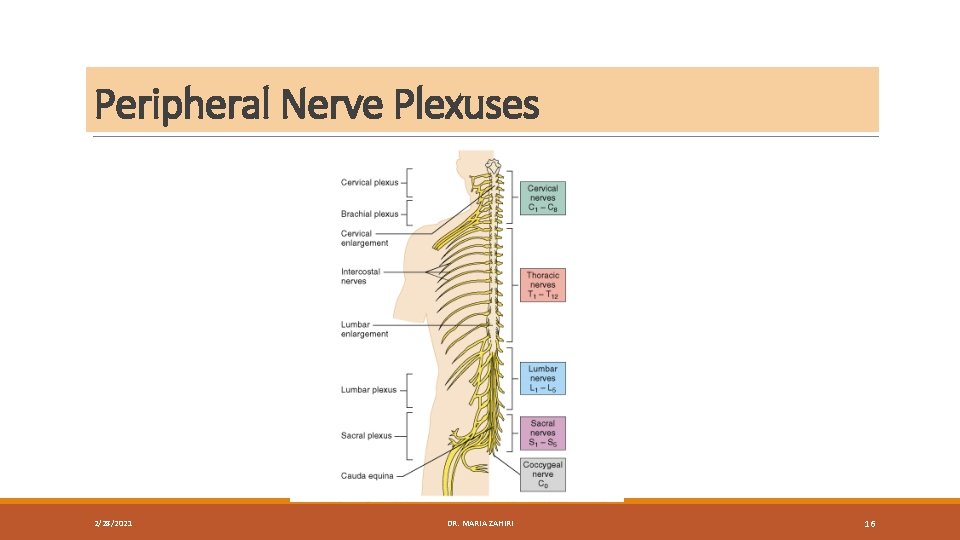
Peripheral Nerve Plexuses 2/28/2021 DR. MARIA ZAHIRI 16
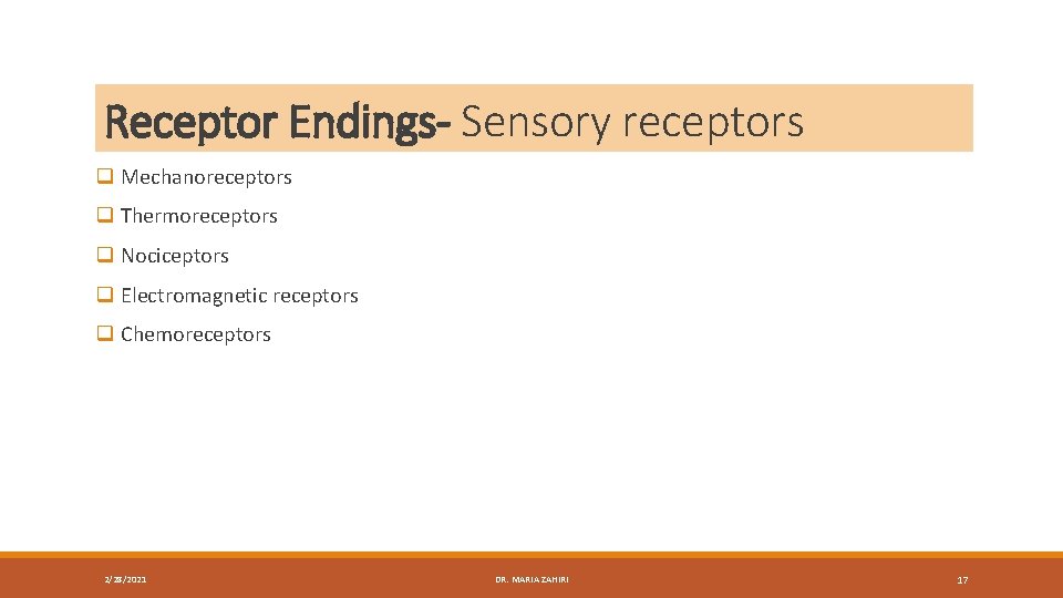
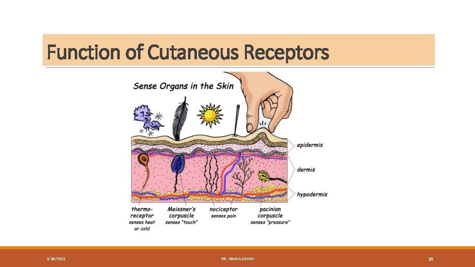
Receptor Endings- Sensory receptors q Mechanoreceptors q Thermoreceptors q Nociceptors q Electromagnetic receptors q Chemoreceptors 2/28/2021 DR. MARIA ZAHIRI 17
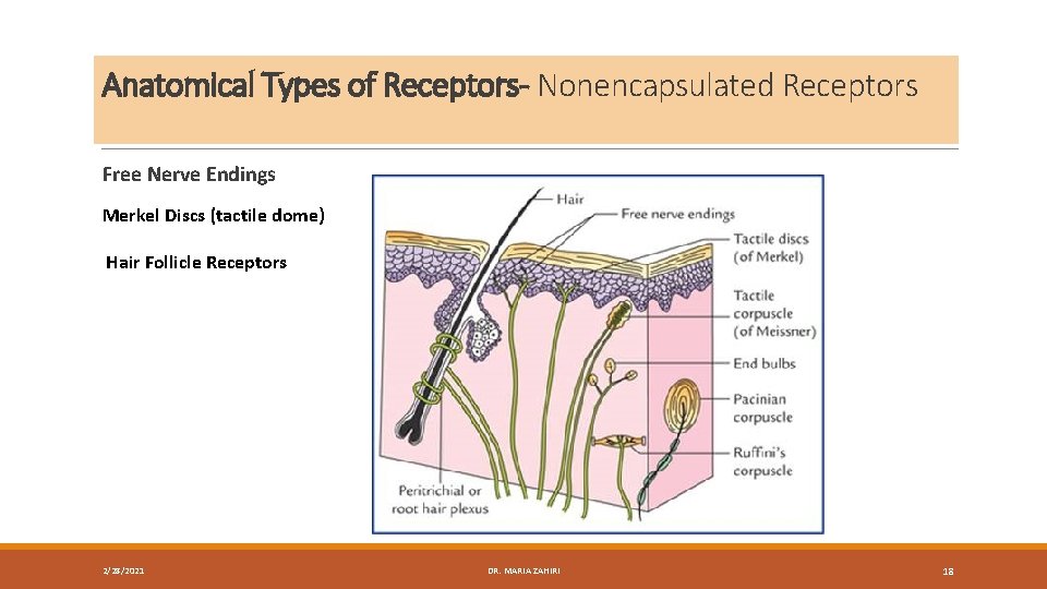
Anatomical Types of Receptors- Nonencapsulated Receptors Free Nerve Endings Merkel Discs (tactile dome) Hair Follicle Receptors 2/28/2021 DR. MARIA ZAHIRI 18
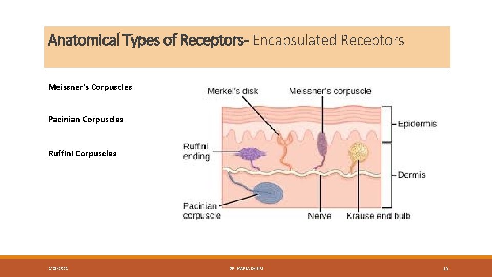
Anatomical Types of Receptors- Encapsulated Receptors Meissner's Corpuscles Pacinian Corpuscles Ruffini Corpuscles 2/28/2021 DR. MARIA ZAHIRI 19
Function of Cutaneous Receptors 2/28/2021 DR. MARIA ZAHIRI 20
Transduction of Sensory Stimuli Into Nerve Impulses 2/28/2021 DR. MARIA ZAHIRI 21
Joint Receptors q 4 types of sensory endings can be located in the capsule and ligaments of synovial joints: q 3 of these endings are encapsulated and resemble pacinian, Ruffini, and tendon stretch receptors. q A fourth type of ending is nonencapsulated and is thought to be sensitive to excessive movements and to transmit pain sensations. 2/28/2021 DR. MARIA ZAHIRI 22
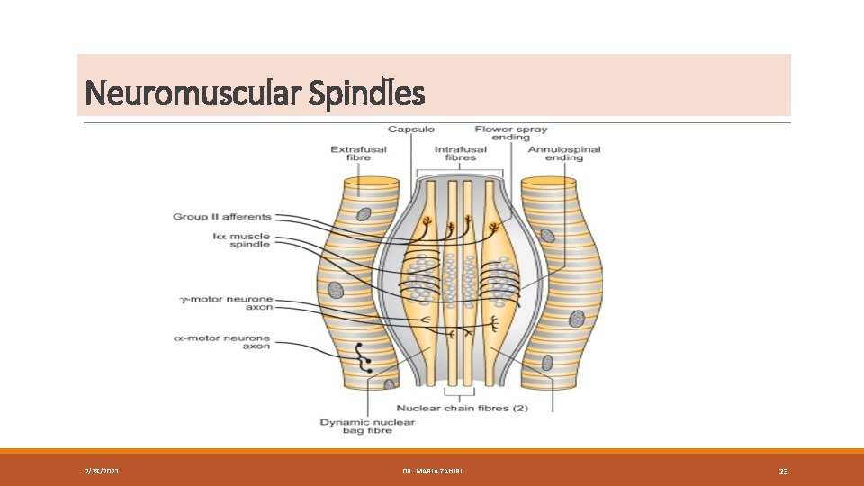
Neuromuscular Spindles 2/28/2021 DR. MARIA ZAHIRI 23
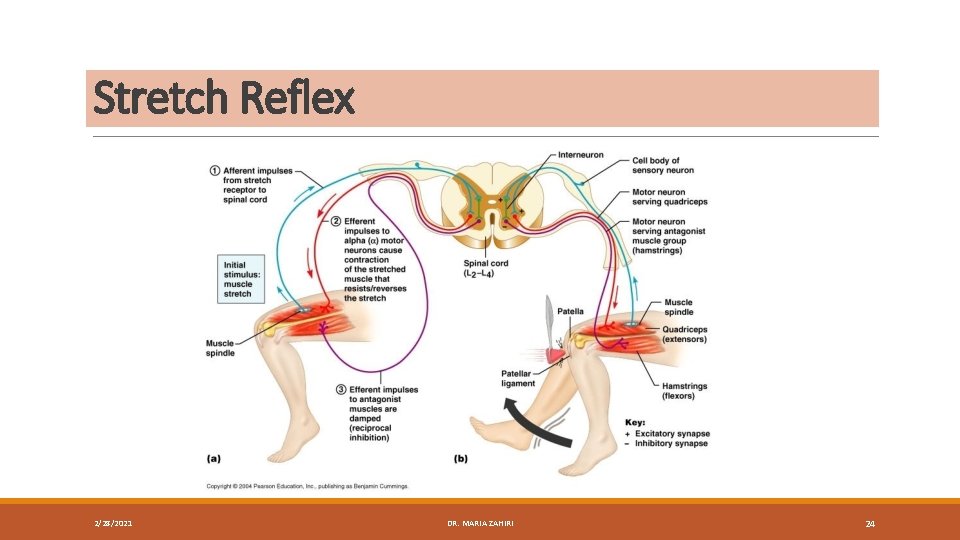
Stretch Reflex 2/28/2021 DR. MARIA ZAHIRI 24
Neurotendinous Spindles(Golgi tendon organs) 2/28/2021 DR. MARIA ZAHIRI 25
Effector Endings- Innervation of Skeletal Muscle q the neurovascular hilus The nerve to a muscle contains motor and sensory fibers. q The motor fibers are of three types: (1) large alpha myelinated fibers, (2) small gamma myelinated fibers, and (3) fine unmyelinated fibers. q The sensory fibers are of three main types: (1) the myelinated fibers, which originate in the annulospiral and flower-spray endings of the neuromuscular spindles (2) the myelinated fibers, which originate in the neurotendinous spindles (3) the myelinated and nonmyelinated fibers, which originate from a variety of sensory endings in the connective tissue of the muscle. 2/28/2021 DR. MARIA ZAHIRI 26
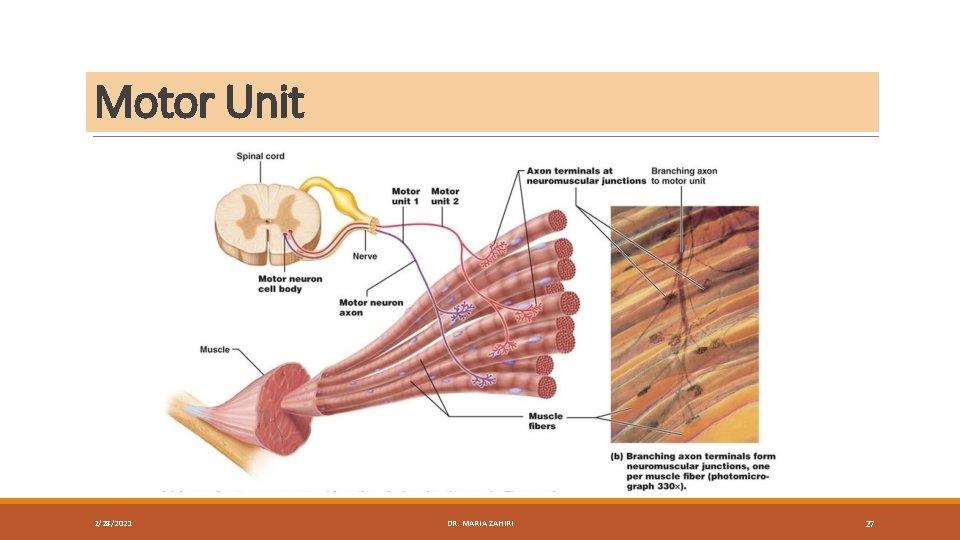
Motor Unit 2/28/2021 DR. MARIA ZAHIRI 27
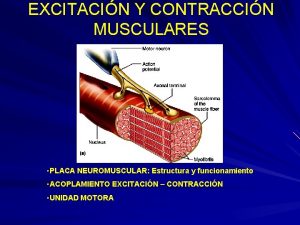
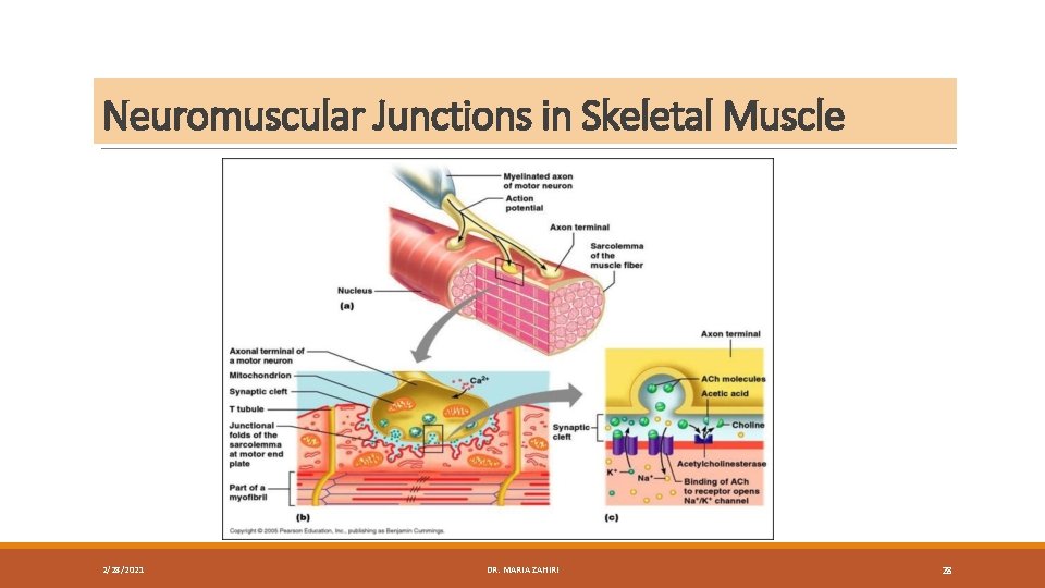
Neuromuscular Junctions in Skeletal Muscle 2/28/2021 DR. MARIA ZAHIRI 28
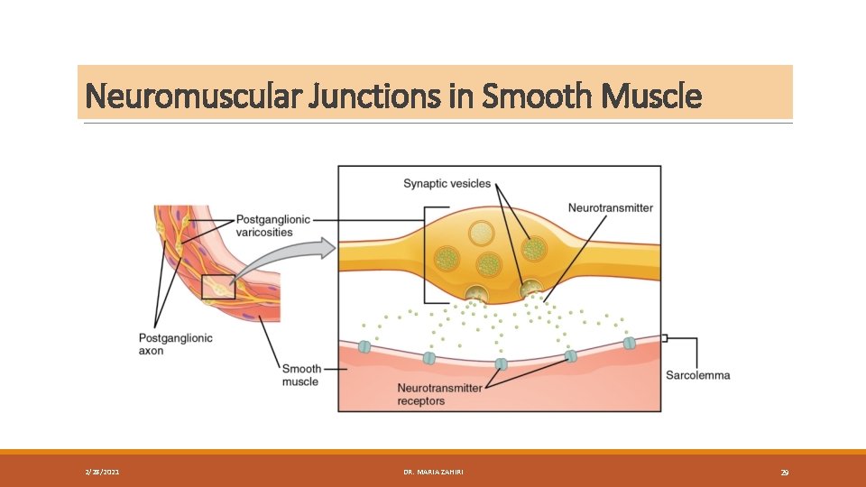
Neuromuscular Junctions in Smooth Muscle 2/28/2021 DR. MARIA ZAHIRI 29
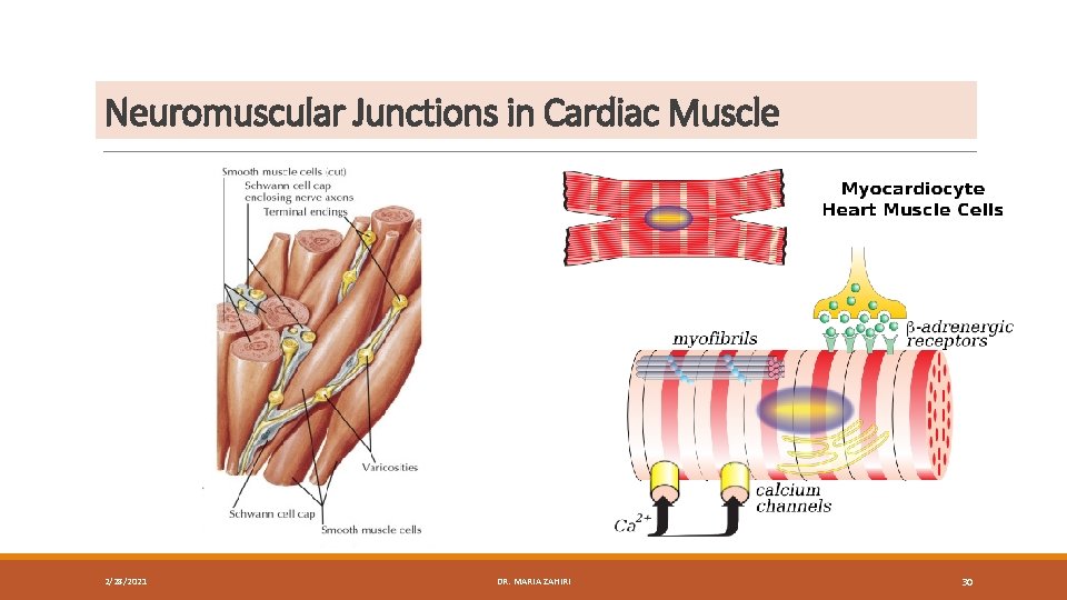
Neuromuscular Junctions in Cardiac Muscle 2/28/2021 DR. MARIA ZAHIRI 30
Neuromuscular Junctions in glands 2/28/2021 DR. MARIA ZAHIRI 31
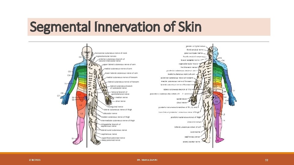
Segmental Innervation of Skin 2/28/2021 DR. MARIA ZAHIRI 32
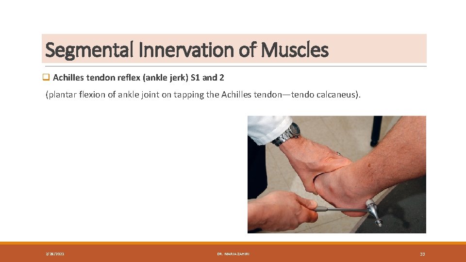
Segmental Innervation of Muscles q Achilles tendon reflex (ankle jerk) S 1 and 2 Biceps brachii tendon reflex (C 5 -6 ) Triceps tendon reflex C 6 -7 and C 8 Brachioradialis tendon reflex C 5 -6 and C 7 Abdominal superficial reflexes Patellar tendon reflex (knee jerk) L 2, L 3, and L 4 (plantar flexion of ankle joint on tapping the Achilles tendon—tendo calcaneus). (flexion of the elbow joint by tapping the biceps tendon). (extension of the elbow joint by tapping the triceps tendon). (supination of the radioulnar joints by tapping the insertion of the brachioradialis tendon). (contraction of underlying abdominal muscles by stroking the skin). Upper abdominal skin T 6 -7; (extension of knee joint on tapping the patellar tendon). middle abdominal skin T 8 -9; lower abdominal skin T 10 -12. 2/28/2021 DR. MARIA ZAHIRI 33
Muscle Tone and Muscle Action q A motor unit consists of a motor neuron in the anterior gray column (horn) of the spinal cord and all the muscle fibers it supplies q muscle tone q Summation of Motor Units q Muscle Fatigue q Posture 2/28/2021 DR. MARIA ZAHIRI 34
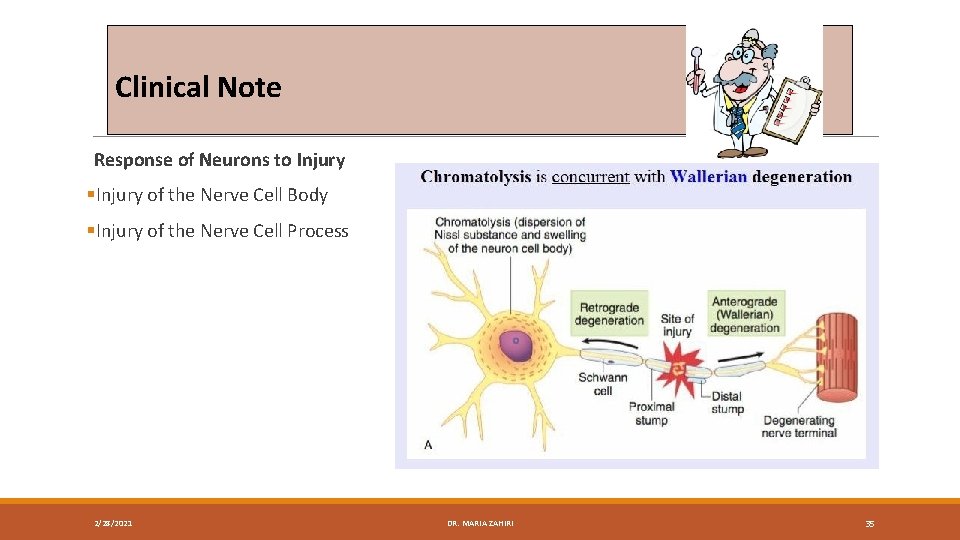
Clinical Note Response of Neurons to Injury §Injury of the Nerve Cell Body §Injury of the Nerve Cell Process 2/28/2021 DR. MARIA ZAHIRI 35
Recovery of Neurons The following mechanisms are thought to be involved: (1) the axons are attracted by chemotropic factors secreted by the Schwann cells in the distal stump (2) growth-stimulating factors exist within the distal stump (3) inhibitory factors are present in the perineurium to inhibit the axons from leaving the nerve. 2/28/2021 DR. MARIA ZAHIRI 36
Recovery of Neurons The satisfactory regeneration of axons and the return of normal function depend on the following factors: 1. intact endoneurial sheaths 2. severity of injury 3. distance between the proximal and distal stumps of the completely severed 4. Injury of mixed nerves (those containing sensory, motor, and autonomic fibers 5. Inadequate physiotherapy to the paralyzed muscles 6. The presence of infection at the site of the wound 2/28/2021 DR. MARIA ZAHIRI 37
Recovery of Neurons 2/28/2021 DR. MARIA ZAHIRI 38
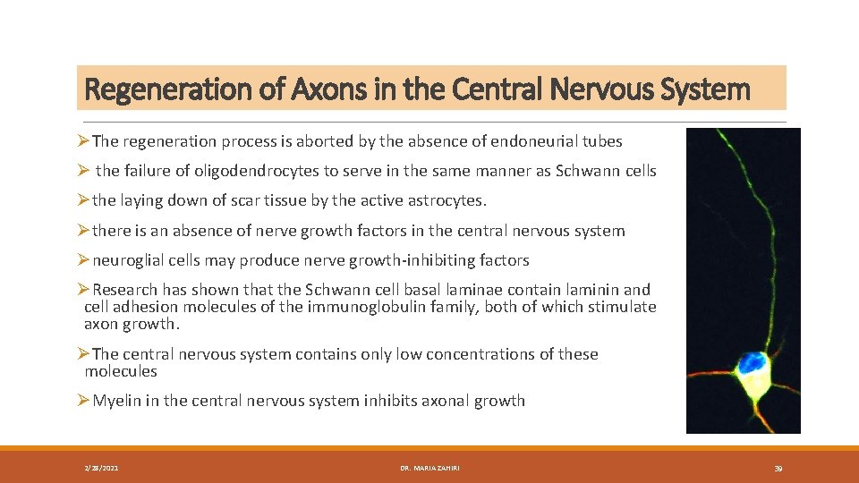
Regeneration of Axons in the Central Nervous System ØThe regeneration process is aborted by the absence of endoneurial tubes Ø the failure of oligodendrocytes to serve in the same manner as Schwann cells Øthe laying down of scar tissue by the active astrocytes. Øthere is an absence of nerve growth factors in the central nervous system Øneuroglial cells may produce nerve growth-inhibiting factors ØResearch has shown that the Schwann cell basal laminae contain laminin and cell adhesion molecules of the immunoglobulin family, both of which stimulate axon growth. ØThe central nervous system contains only low concentrations of these molecules ØMyelin in the central nervous system inhibits axonal growth 2/28/2021 DR. MARIA ZAHIRI 39
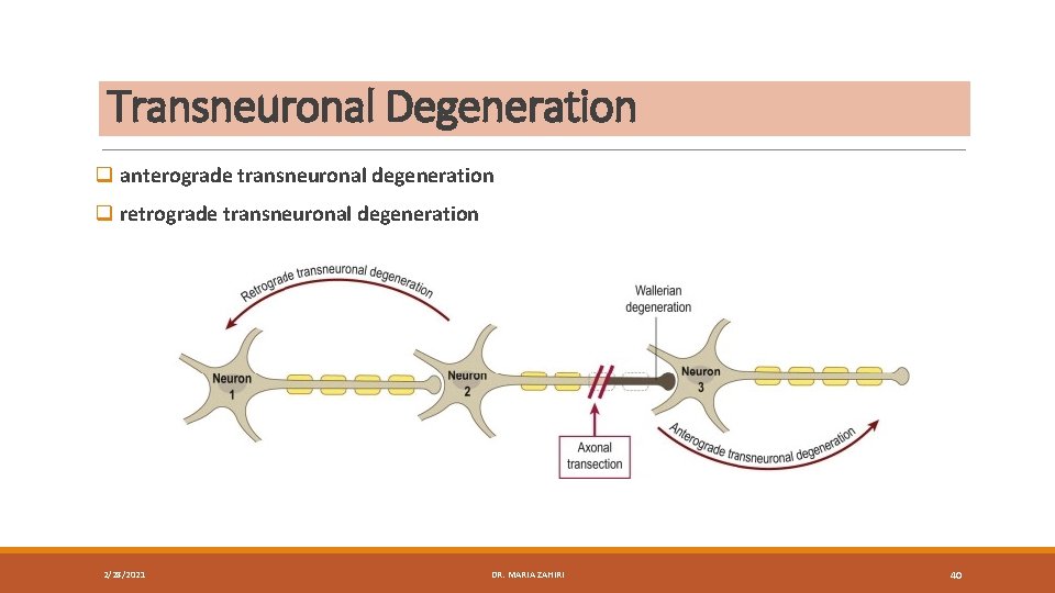
Transneuronal Degeneration q anterograde transneuronal degeneration q retrograde transneuronal degeneration 2/28/2021 DR. MARIA ZAHIRI 40
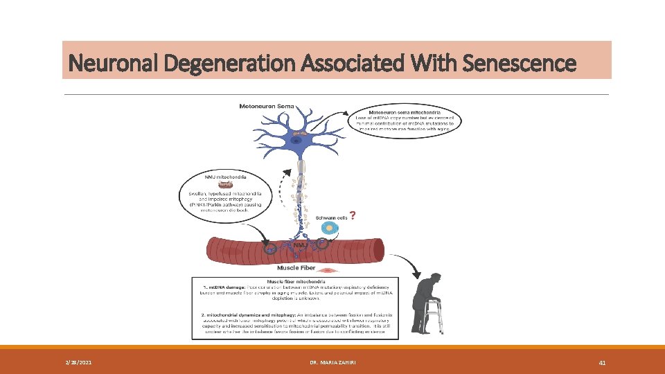
Neuronal Degeneration Associated With Senescence 2/28/2021 DR. MARIA ZAHIRI 41
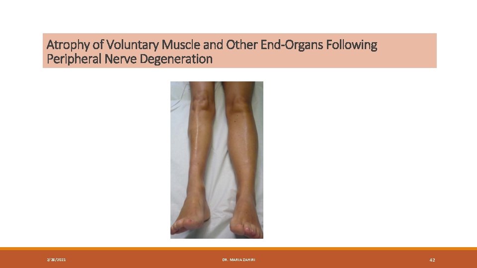
Atrophy of Voluntary Muscle and Other End-Organs Following Peripheral Nerve Degeneration 2/28/2021 DR. MARIA ZAHIRI 42
Traumatic Lesions of Peripheral Nerve Seddon's classification 2/28/2021 DR. MARIA ZAHIRI 43
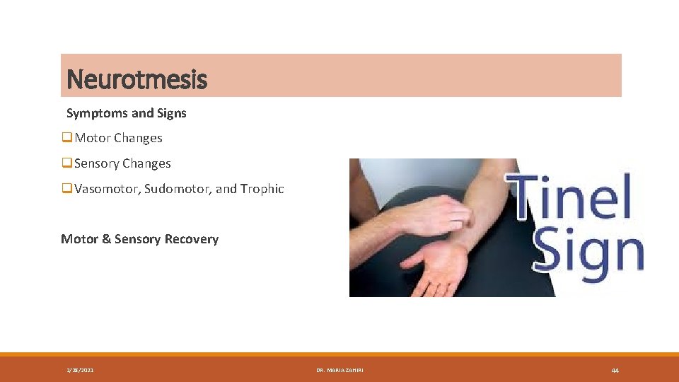
Neurotmesis Symptoms and Signs q. Motor Changes q. Sensory Changes q. Vasomotor, Sudomotor, and Trophic Motor & Sensory Recovery 2/28/2021 DR. MARIA ZAHIRI 44
Nerve Transplantation 2/28/2021 DR. MARIA ZAHIRI 45
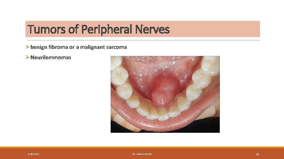
Tumors of Peripheral Nerves Øbenign fibroma or a malignant sarcoma ØNeurilemmomas 2/28/2021 DR. MARIA ZAHIRI 46
Endoneurial Spaces Within Peripheral Nerves 2/28/2021 DR. MARIA ZAHIRI 47
Action of Local Anesthetics on Nerve Conduction 2/28/2021 DR. MARIA ZAHIRI 48
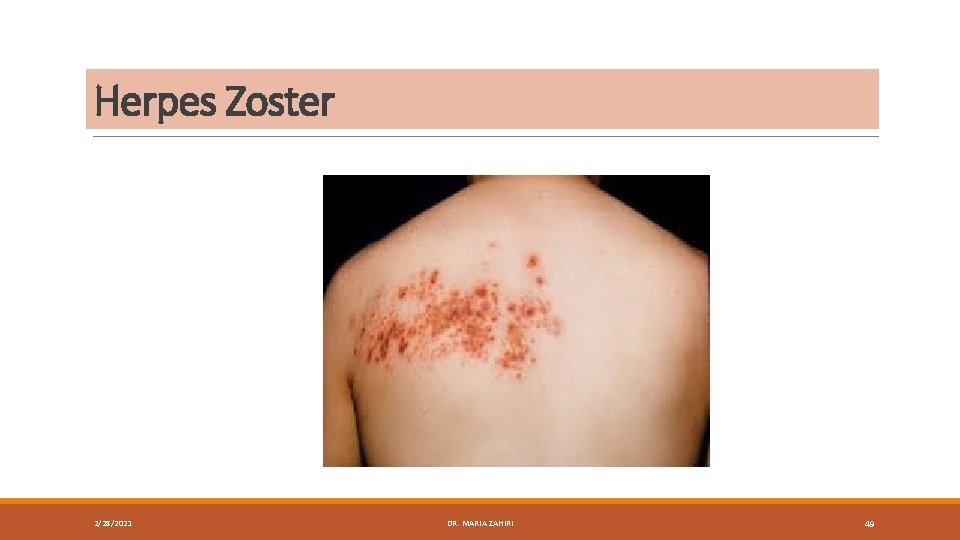
Herpes Zoster 2/28/2021 DR. MARIA ZAHIRI 49
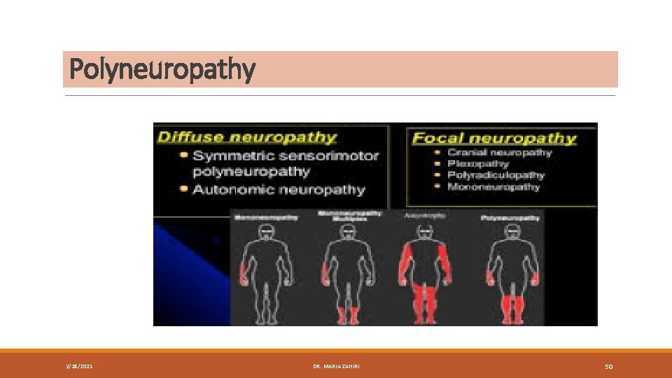
Polyneuropathy 2/28/2021 DR. MARIA ZAHIRI 50
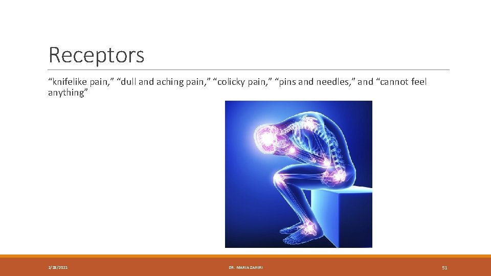
Receptors “knifelike pain, ” “dull and aching pain, ” “colicky pain, ” “pins and needles, ” and “cannot feel anything” 2/28/2021 DR. MARIA ZAHIRI 51
Sensory Receptors and Age 2/28/2021 DR. MARIA ZAHIRI 52
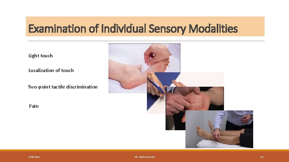
Examination of Individual Sensory Modalities Light touch Localization of touch Two-point tactile discrimination Pain 2/28/2021 DR. MARIA ZAHIRI 53
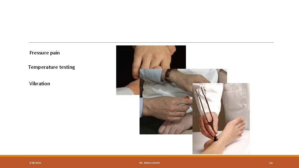
Pressure pain Temperature testing Vibration 2/28/2021 DR. MARIA ZAHIRI 54
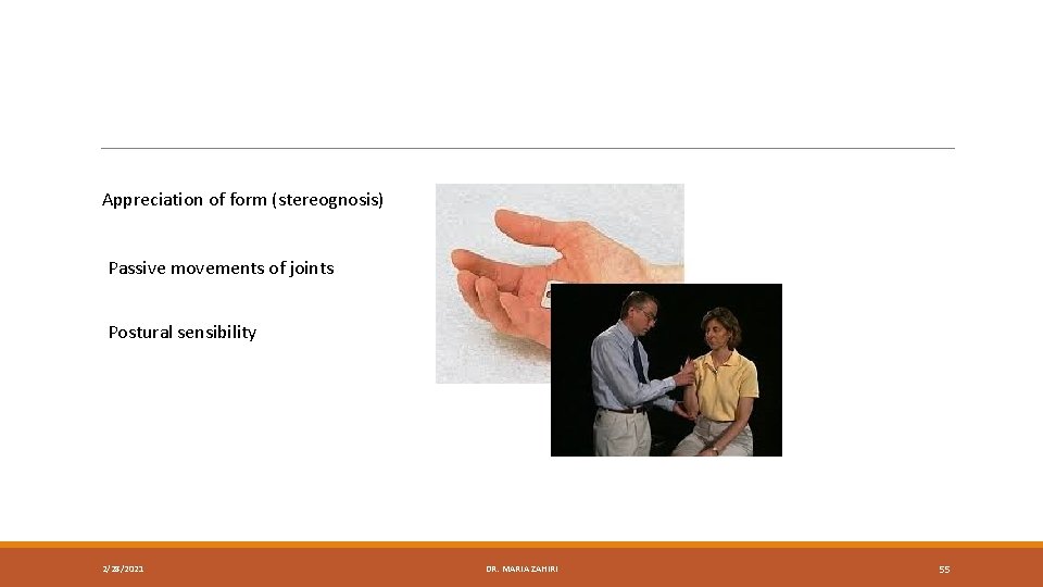
Appreciation of form (stereognosis) Passive movements of joints Postural sensibility 2/28/2021 DR. MARIA ZAHIRI 55
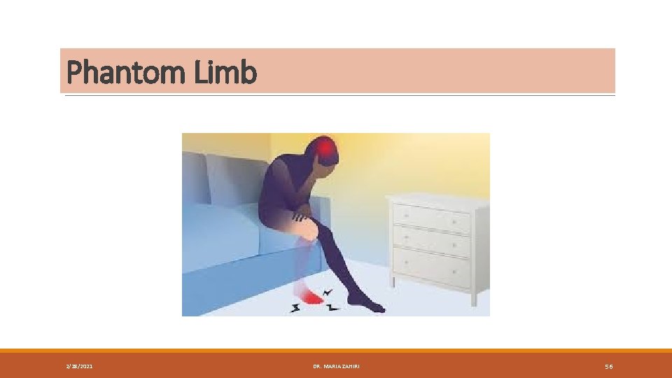
Phantom Limb 2/28/2021 DR. MARIA ZAHIRI 56
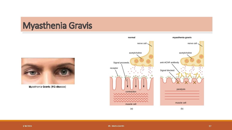
Myasthenia Gravis 2/28/2021 DR. MARIA ZAHIRI 57
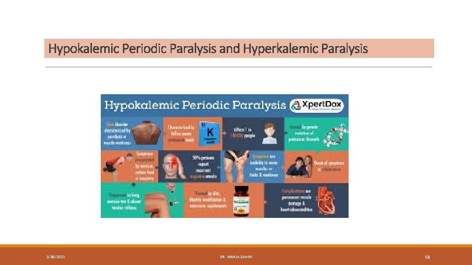
Hypokalemic Periodic Paralysis and Hyperkalemic Paralysis 2/28/2021 DR. MARIA ZAHIRI 58