
DIFFERENTIATING HEADACHES TESTING AND MANAGEMENT Dr Bhadresh B
DIFFERENTIATING HEADACHES , TESTING AND MANAGEMENT Dr Bhadresh B Mangukiya D. M. Neurology (Clinic-504, Param doc House, Station road , surat)
TALK…………………. . • • Basic stuctures and pathophysiology Primary or secondary headache? Differ primary Headache? What is natural History disease? EEG , MRI , CT SCAN and OTHERS Role When to start prophylaxis? Drug of Choice?

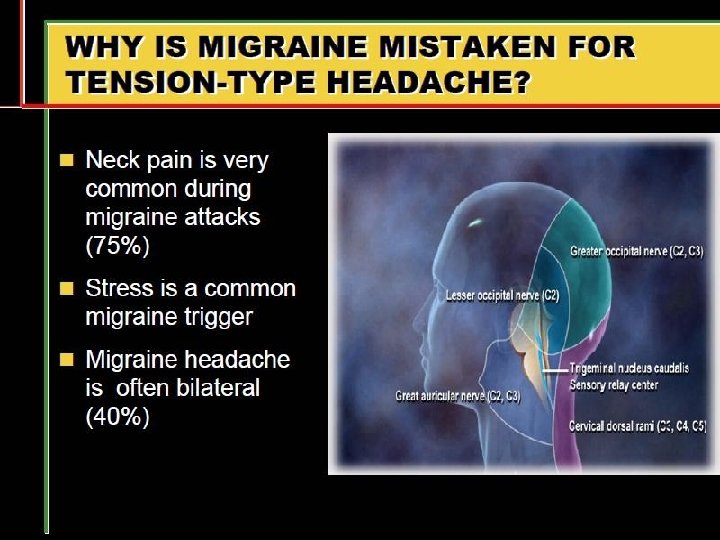
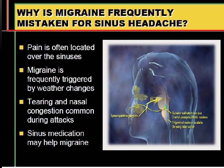
PAIN-SENSITIVE STRUCTURES IN THE HEAD AND NECK Extra-cranial Intracranial • • • • Scalp muscles Skull Carotid and vertebral arteries Paranasal sinuses Eyes and orbits Mouth, teeth, and pharynx Ears Cervical spine and ligaments Cervical muscles • • Periosteum Cranial nerves Meningeal arteries and dural sinuses Proximal intracranial arteries Sphenoid sinus Thalamic nuclei Brainstem pain-modulating centers

WHAT CAUSES HEDACHE?
WHAT ARE COMMON CAUSES?

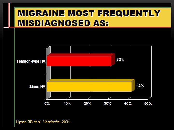
DIFFERENTIAL DIAGNOSIS OF 906 PATIENTS WHO PRESENTED TO A GENERAL NEUROLOGY CLINIC WITH HEADACHE OR FACIAL PAIN AS THE MAJOR OR ONLY SYMPTOM Diagnosis Number % Tension headache Migraine Headache ? Cause Post-traumatic Facial pain ? cause Depression Trigeminal neuralgia Cluster headache Malignant IC Tumour Benign IC Tumour Temporal arteritis Post-herpetic neuralgia Benign IC hypertension Cough headache Subdural haematoma Sinus infection 296 241 139 71 38 29 29 19 14 9 6 32 27 15 8 4 3 3 2 1. 5 5 4 3 2 1
HEADACHE DIAGNOSIS AND TESTING


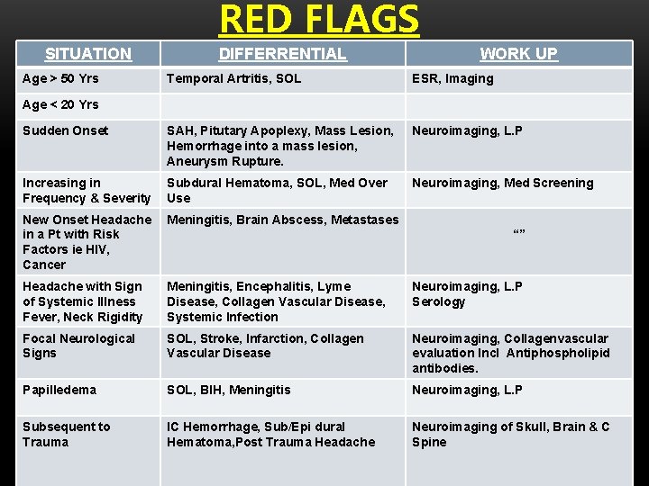
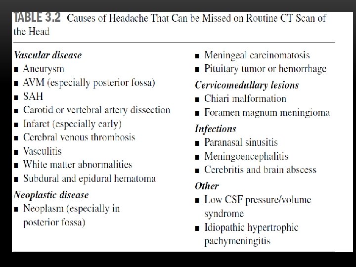
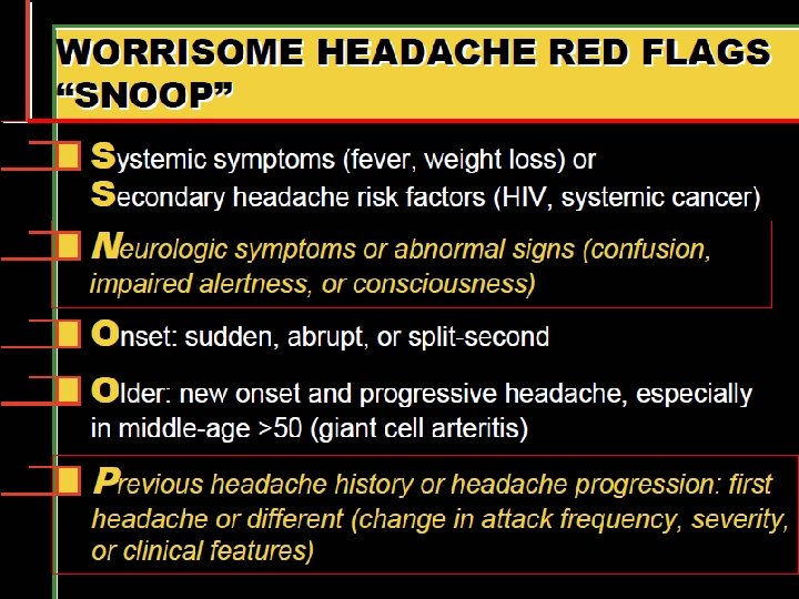
RED FLAGS SITUATION Age > 50 Yrs DIFFERRENTIAL WORK UP Temporal Artritis, SOL ESR, Imaging Sudden Onset SAH, Pitutary Apoplexy, Mass Lesion, Hemorrhage into a mass lesion, Aneurysm Rupture. Neuroimaging, L. P Increasing in Frequency & Severity Subdural Hematoma, SOL, Med Over Use Neuroimaging, Med Screening New Onset Headache in a Pt with Risk Factors ie HIV, Cancer Meningitis, Brain Abscess, Metastases Headache with Sign of Systemic Illness Fever, Neck Rigidity Meningitis, Encephalitis, Lyme Disease, Collagen Vascular Disease, Systemic Infection Neuroimaging, L. P Serology Focal Neurological Signs SOL, Stroke, Infarction, Collagen Vascular Disease Neuroimaging, Collagenvascular evaluation Incl Antiphospholipid antibodies. Papilledema SOL, BIH, Meningitis Neuroimaging, L. P Subsequent to Trauma IC Hemorrhage, Sub/Epi dural Hematoma, Post Trauma Headache Neuroimaging of Skull, Brain & C Spine Age < 20 Yrs “”
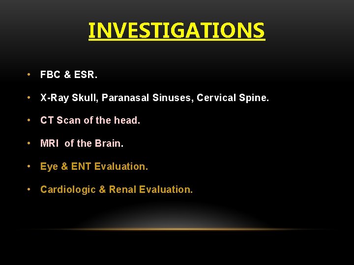
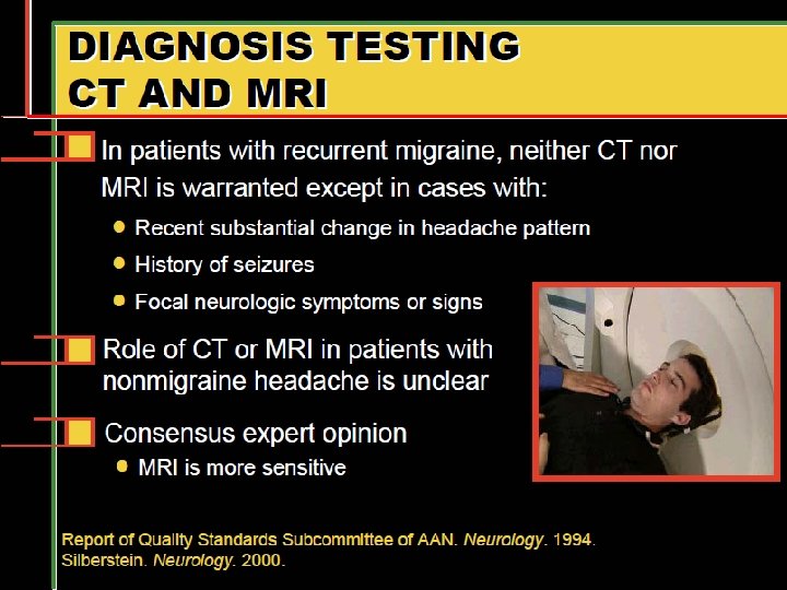
INVESTIGATIONS • FBC & ESR. • X-Ray Skull, Paranasal Sinuses, Cervical Spine. • CT Scan of the head. • MRI of the Brain. • Eye & ENT Evaluation. • Cardiologic & Renal Evaluation.
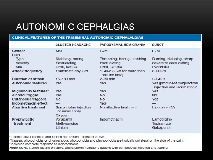
AUTONOMI C CEPHALGIAS
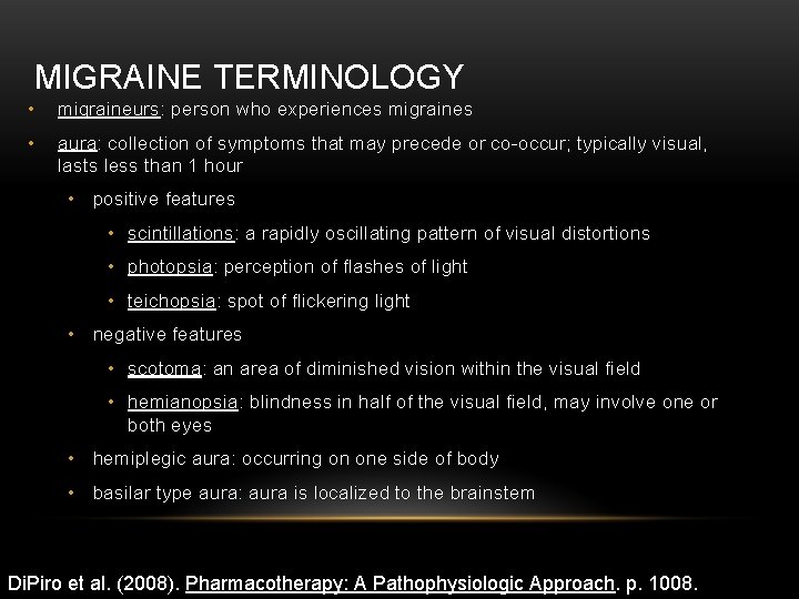
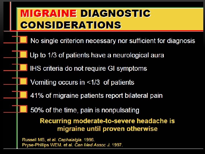
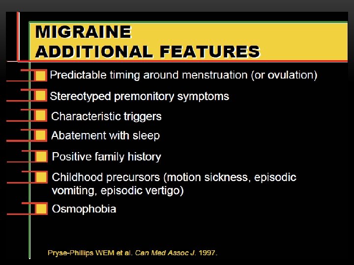
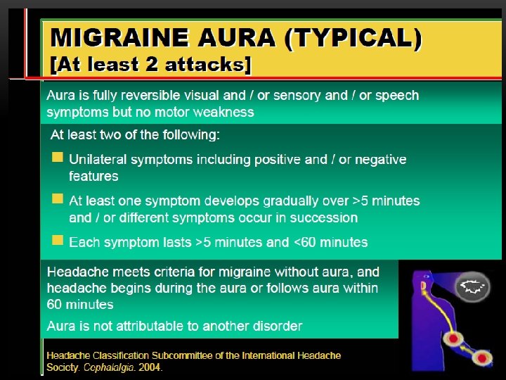
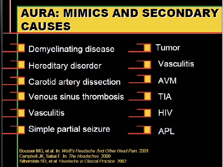
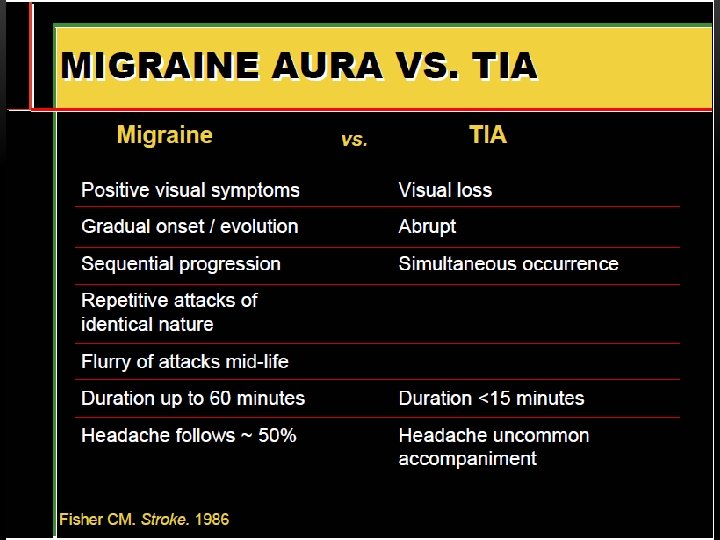
MIGRAINE TERMINOLOGY • migraineurs: person who experiences migraines • aura: collection of symptoms that may precede or co-occur; typically visual, lasts less than 1 hour • positive features • scintillations: a rapidly oscillating pattern of visual distortions • photopsia: perception of flashes of light • teichopsia: spot of flickering light • negative features • scotoma: an area of diminished vision within the visual field • hemianopsia: blindness in half of the visual field, may involve one or both eyes • hemiplegic aura: occurring on one side of body • basilar type aura: aura is localized to the brainstem Di. Piro et al. (2008). Pharmacotherapy: A Pathophysiologic Approach. p. 1008.
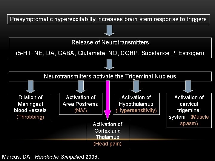
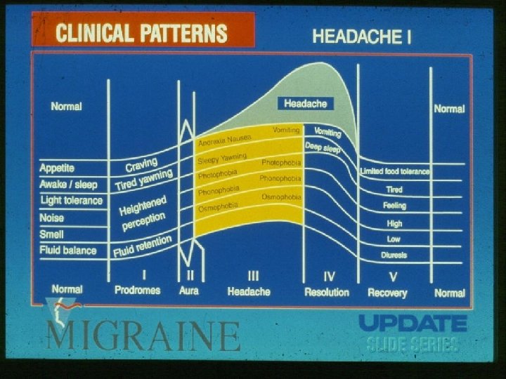
Presymptomatic hyperexcitabilty increases brain stem response to triggers Release of Neurotransmitters (5 -HT, NE, DA, GABA, Glutamate, NO, CGRP, Substance P, Estrogen) Neurotransmitters activate the Trigeminal Nucleus Dilation of Meningeal blood vessels (Throbbing) Activation of Area Postrema (N/V) Activation of Hypothalamus (Hypersensitivity) Activation of Cortex and Thalamus (Head pain) Marcus, DA. Headache Simplified 2008. Activation of cervical trigeminal system (Muscle spasm)
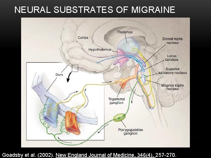
NEURAL SUBSTRATES OF MIGRAINE Goadsby et al. (2002). New England Journal of Medicine, 346(4), 257 -270.
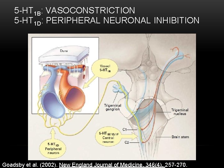
5 -HT 1 B: VASOCONSTRICTION 5 -HT 1 D: PERIPHERAL NEURONAL INHIBITION Goadsby et al. (2002). New England Journal of Medicine, 346(4), 257 -270.
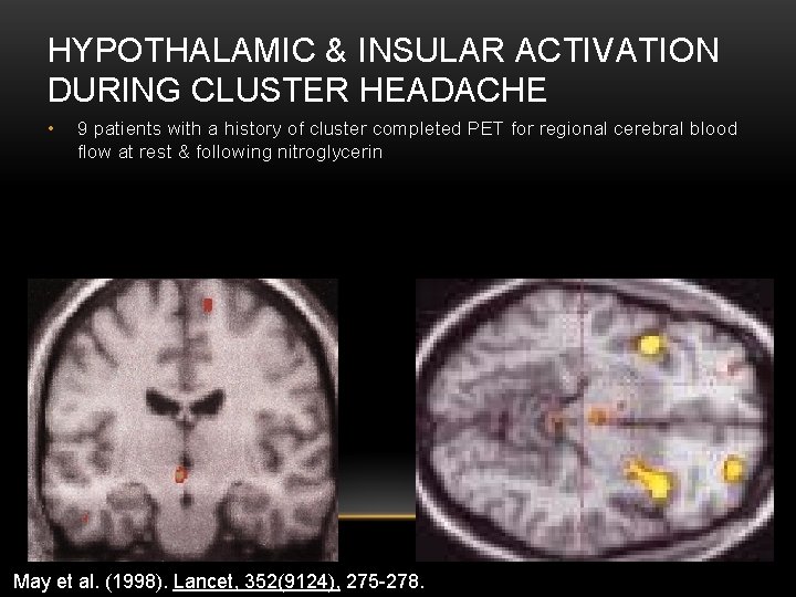
HYPOTHALAMIC & INSULAR ACTIVATION DURING CLUSTER HEADACHE • 9 patients with a history of cluster completed PET for regional cerebral blood flow at rest & following nitroglycerin May et al. (1998). Lancet, 352(9124), 275 -278.
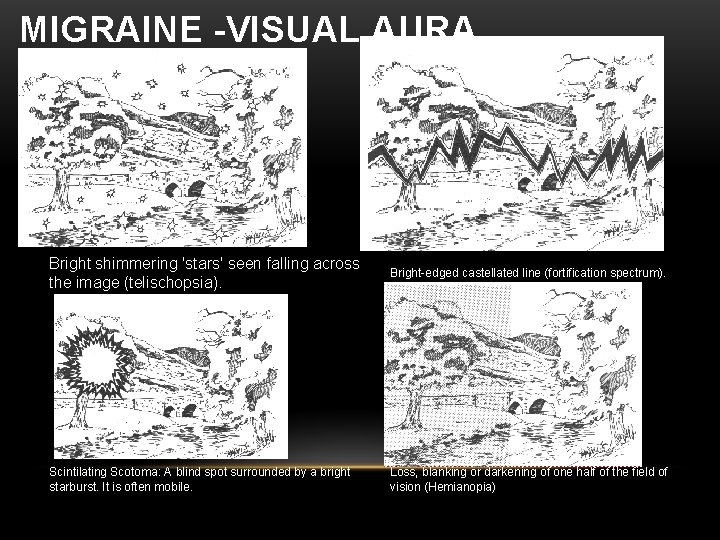
MIGRAINE -VISUAL AURA Bright shimmering 'stars' seen falling across the image (telischopsia). Scintilating Scotoma: A blind spot surrounded by a bright starburst. It is often mobile. Bright-edged castellated line (fortification spectrum). Loss, blanking or darkening of one half of the field of vision (Hemianopia)
MIGRAINE -VISUAL AURA

- Slides: 50