Children with Surgical Disorders Dr S Nishan Silva



- Slides: 75
Children with Surgical Disorders Dr. S. Nishan Silva (MBBS)
Congenital Abnormalities • Malformation of the Central Nervous System ▫ Myelomeningocele ▫ Congenital Hydrocephalus • Malformations of the Gastrointestinal Tract ▫ Cleft Lip and Cleft Palate ▫ Anorectal Malformations Imperforated Anus Pyloric Stenosis Intersusception Hirschsprung’s Disease
Other… • Urinary system related ( covered later) ▫ ▫ Hypospedias Phimosis Upper urinary track Hydrocoele • Other
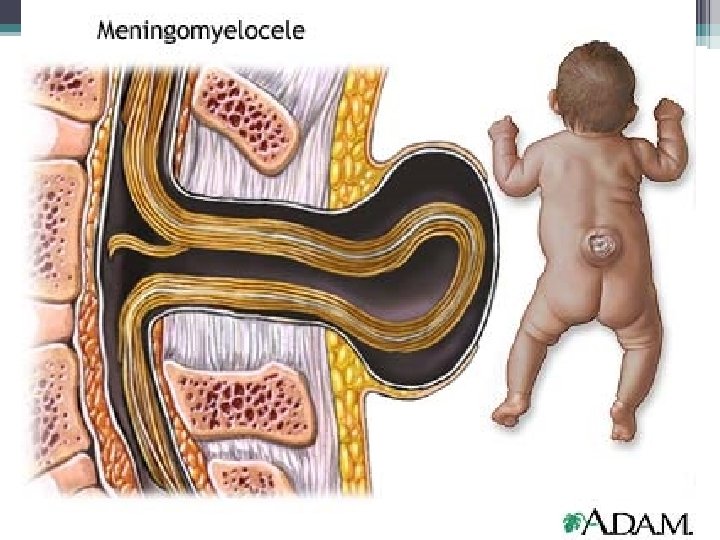
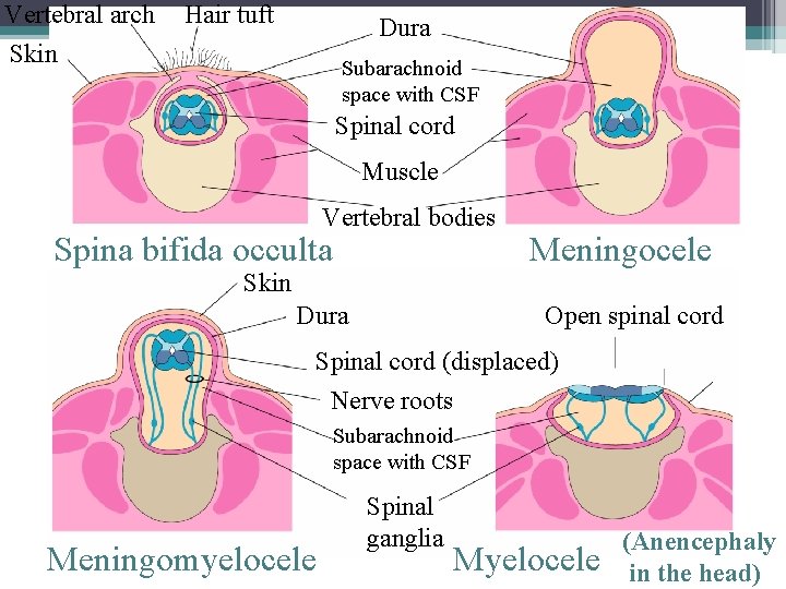
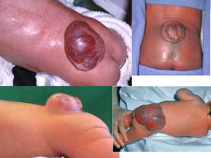
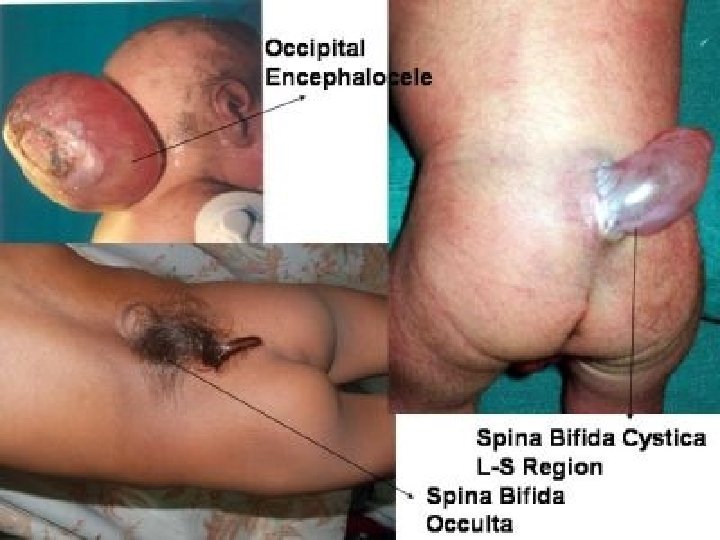
Myelomeningocele • 1 st 28 days of pregnancy • Associated problems ▫ ▫ Hydrocephalus Other spinal cord / M/S system disorders Syringomyelia Hip dislocation • Causes ▫ Folic Acid ▫ Heriditory?
Clinical Manifestations • Can be seen after birth • Defect is related to the anatomical level ▫ Bowel bladder incontinance ▫ Sensory deficit ▫ Paralysis • Infection risk
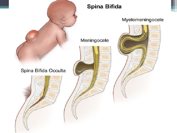
Vertebral arch Skin Hair tuft Dura Subarachnoid space with CSF Spinal cord Muscle Vertebral bodies Spina bifida occulta Skin Dura Meningocele Open spinal cord Spinal cord (displaced) Nerve roots Subarachnoid space with CSF Meningomyelocele Spinal ganglia Myelocele (Anencephaly in the head)
Diagnosis (tests) • • • MRI USS CT Myelography Perinatal diagnosis ▫ Anomaly scan ▫ Alpha feto protein in maternal blood and amniotic fluid
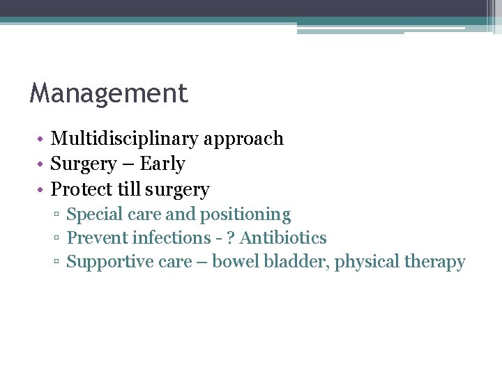
Management • Multidisciplinary approach • Surgery – Early • Protect till surgery ▫ Special care and positioning ▫ Prevent infections - ? Antibiotics ▫ Supportive care – bowel bladder, physical therapy
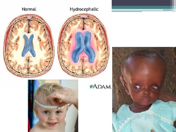
Hydrocephalus • CSF block ▫ SOL ▫ Infection ▫ Injury • Or CSF overproduction • Fluid accumulating in the cerebral ventricals • Skull expands
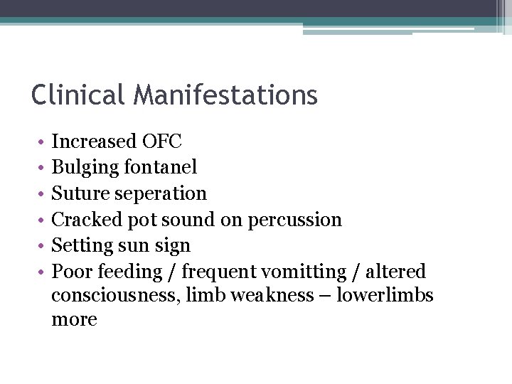
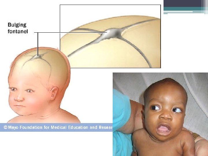
Clinical Manifestations • • • Increased OFC Bulging fontanel Suture seperation Cracked pot sound on percussion Setting sun sign Poor feeding / frequent vomitting / altered consciousness, limb weakness – lowerlimbs more
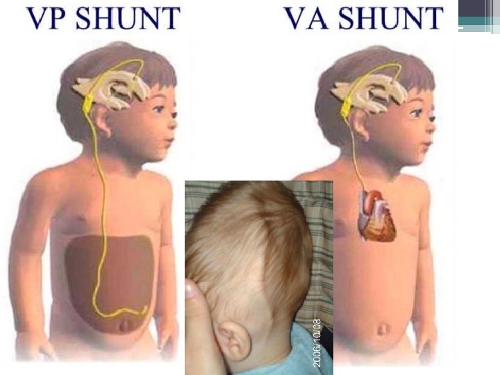
Management • The relief of hydrocephalus ▫ Ventriculo-peritoneal shunt - younger ▫ Ventriculo- atrial shunt - older ▫ Lumbo – peritoneal shunt • Treatment of complications ▫ ▫ Infections Mechanical obstructions Displacement with growth Perforations • Ensure proper development
Nursing care • Perioperative ▫ ▫ Check for raised ICP daily OFC daily Observe for behavioral change Assist with nutritional needs • Postoperative ▫ ▫ ▫ Positioning – prevent pressure on valve Raised ICP – pupil dilatation, hypertension Peritonitis ? IP/OP chart Family support
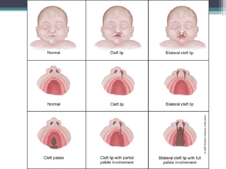
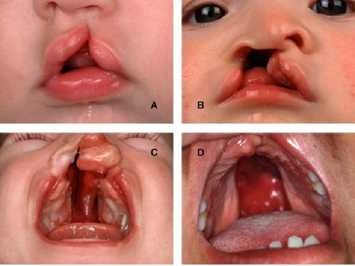
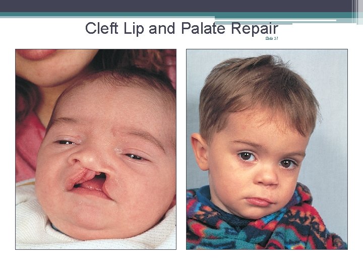
Cleft Lip / Cleft Palate • Cleft lip , uni/bilateral • May be associated with cleft palate, nasal maldevelopments, maxillary anomalies • CL can see clearly. CP not. So, check carefully. Use a gloved finger in baby’s mouth. • CL surgery – early • CP delayed till 12 -18 months to use natural shaping of the palate. • When CP is associated there may be a speech defect. • Feeding done in an upright position.
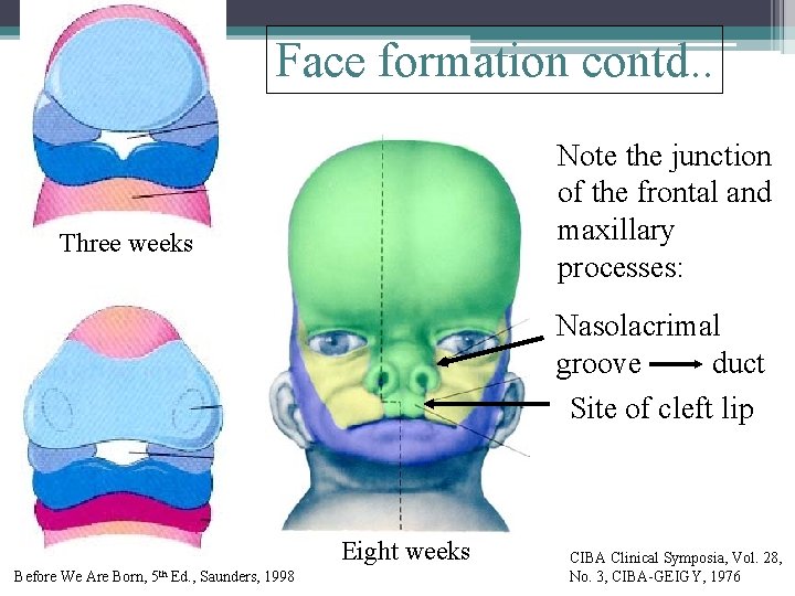
Face formation contd. . Note the junction of the frontal and maxillary processes: Three weeks Nasolacrimal groove duct Site of cleft lip Eight weeks Before We Are Born, 5 th Ed. , Saunders, 1998 CIBA Clinical Symposia, Vol. 28, No. 3, CIBA-GEIGY, 1976
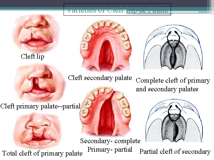
Varieties of Cleft Lip & Palate Cleft lip Cleft secondary palate Complete cleft of primary and secondary palates Cleft primary palate--partial Secondary- complete Primary- partial Partial cleft of secondary Total cleft of primary palate
Post operative • Lip protection device, arm restrainer, check suture line carefully. • Continue to feed upright • CP repair – keep the child on abdomen initially. Packing will be removed in 2 -3 days. • Care to feed only soft foods. • Educate on mouth guards, mouth care • Guidance for speech therapy
Cleft Lip and Palate Repair Slide 27
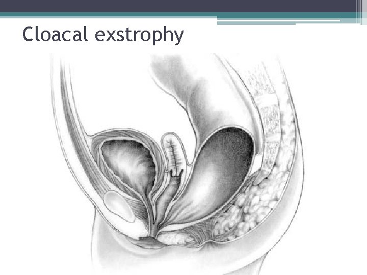
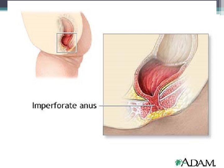
Anorectal malformations • Forms of Imperforated anus • Genitourinary anomalies • Cloacal exstrophy
Cloacal exstrophy
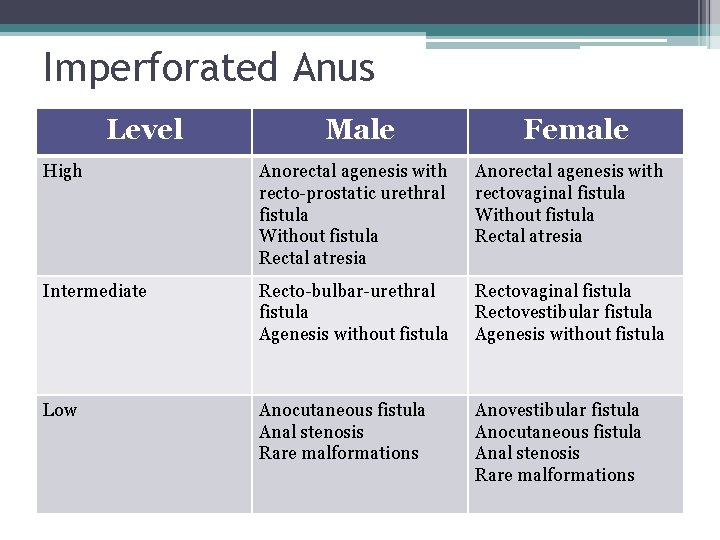
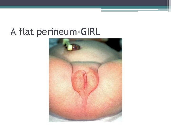
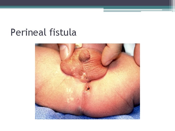
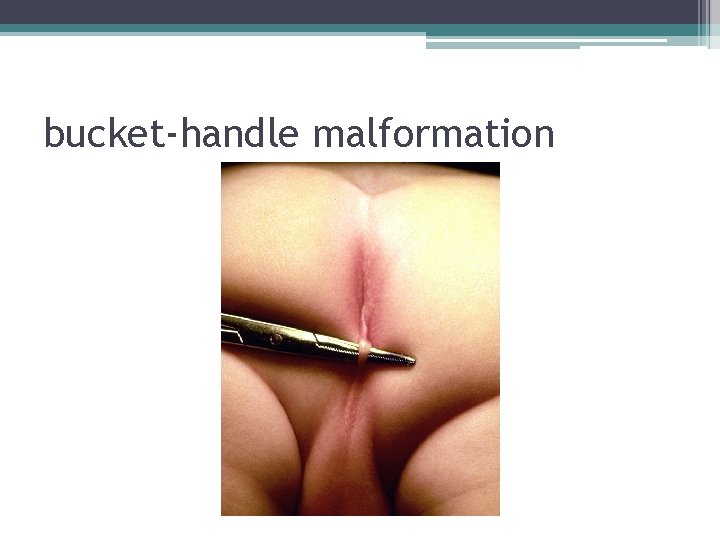
Imperforated Anus Level Male Female High Anorectal agenesis with recto-prostatic urethral fistula Without fistula Rectal atresia Anorectal agenesis with rectovaginal fistula Without fistula Rectal atresia Intermediate Recto-bulbar-urethral fistula Agenesis without fistula Rectovaginal fistula Rectovestibular fistula Agenesis without fistula Low Anocutaneous fistula Anal stenosis Rare malformations Anovestibular fistula Anocutaneous fistula Anal stenosis Rare malformations
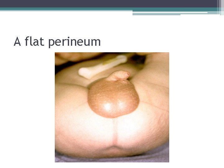
A flat perineum
A flat perineum-GIRL
Perineal fistula
bucket-handle malformation
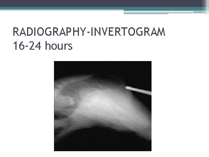
Diagnosis ▫ External physical findings Patency Fistula Meconium track ▫ Radiography USS
RADIOGRAPHY-INVERTOGRAM 16 -24 hours
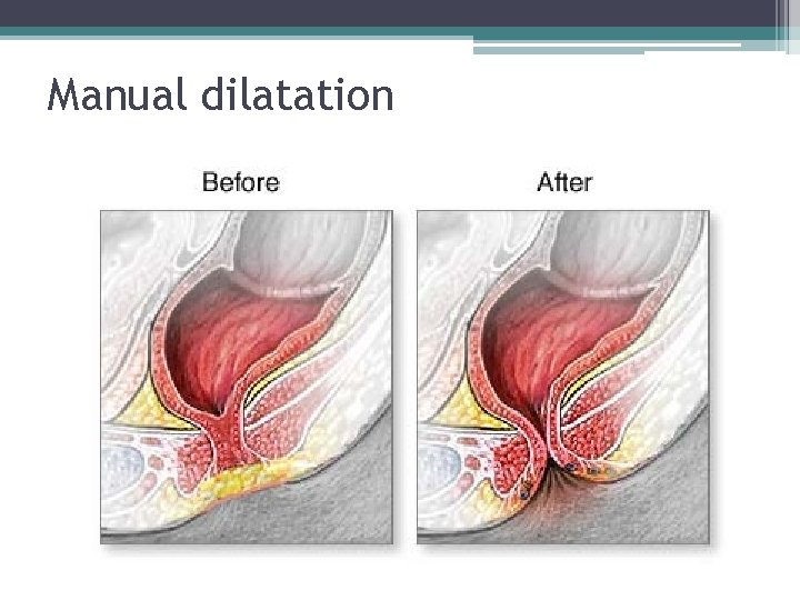
Management • Low category, with good muscles, well innervated – Manual dilatation • High or Intermediate categories – Elective repair with sigmoid colostomy – at 6 -12 months
Manual dilatation
Nursing Considerations • Suspicion – No passage of stools within first 24 hours • Pre-operative care ▫ Gastric decompression ▫ IV fluids • Post-operative care – usual ▫ Skin care – Zinc oxide, hydrocolloid dressing ▫ Cleansing skin ▫ Position for minimal contamination • Family support
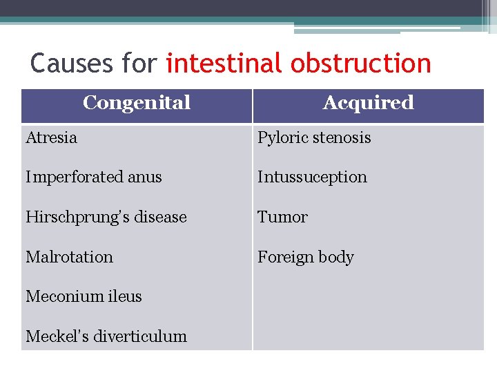
Causes for intestinal obstruction Congenital Acquired Atresia Pyloric stenosis Imperforated anus Intussuception Hirschprung’s disease Tumor Malrotation Foreign body Meconium ileus Meckel’s diverticulum
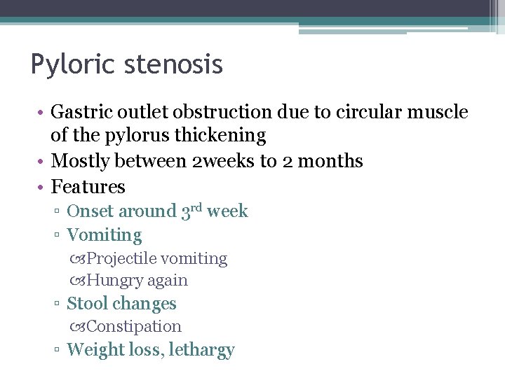
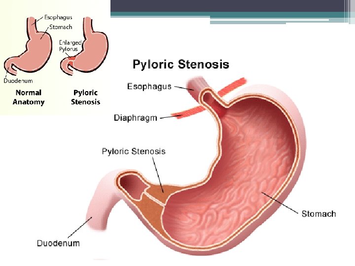
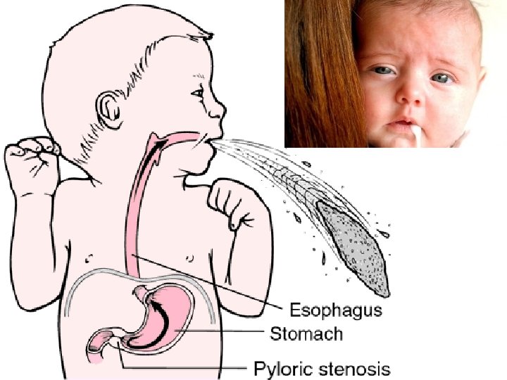
Pyloric stenosis • Gastric outlet obstruction due to circular muscle of the pylorus thickening • Mostly between 2 weeks to 2 months • Features ▫ Onset around 3 rd week ▫ Vomiting Projectile vomiting Hungry again ▫ Stool changes Constipation ▫ Weight loss, lethargy
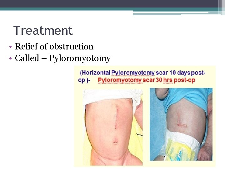
Treatment • Relief of obstruction • Called – Pyloromyotomy
Nursing considerations • Perioperative ▫ Hydration – IP/OP ▫ Electrolyte correction ▫ Stomach decompression through NG - amount • Perioperative ▫ ▫ IP/OP Vital signs NG – amount Feeding – after 24 hours – start with clear fluid
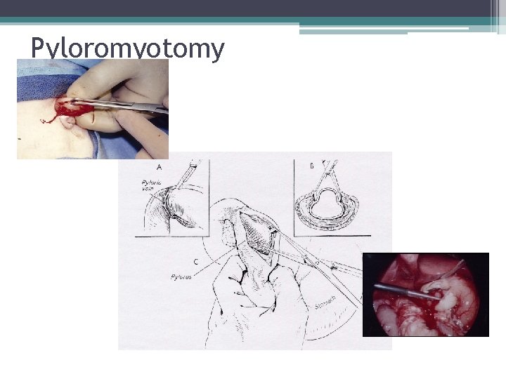
Pyloromyotomy
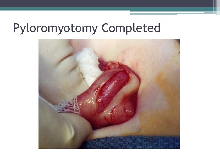
Pyloromyotomy Completed
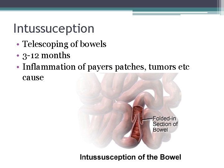
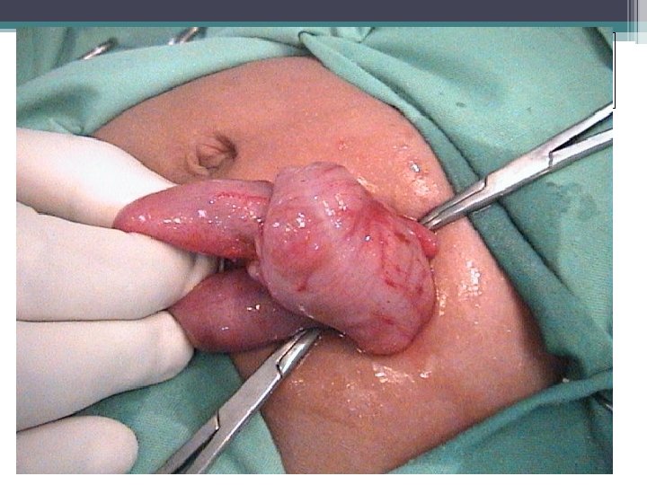
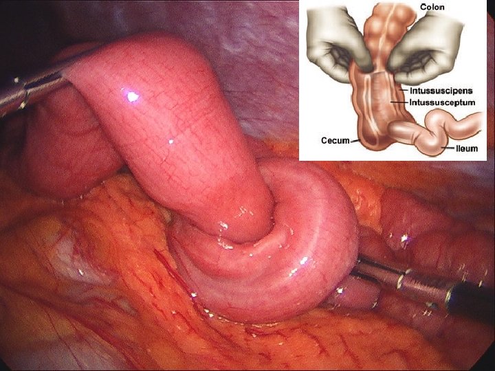
Intussuception • Telescoping of bowels • 3 -12 months • Inflammation of payers patches, tumors etc cause
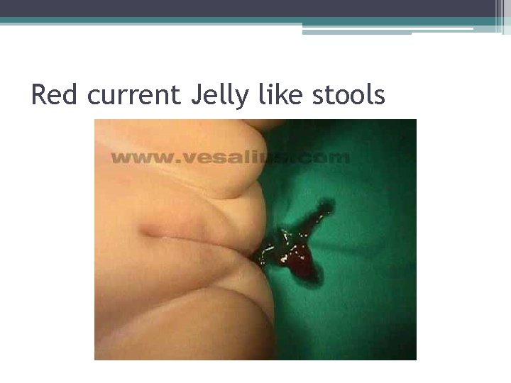
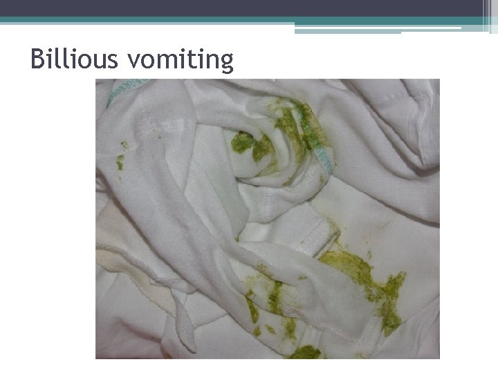
Clinical manifestations • • Sudden severe pain – crying Abdominal distension Bloody stools – Red current jelly like Billious vomiting Lethargy Shallow breathing Grunting Disgnosis ▫ USS – sausage shape appearance of a bowel segment
Red current Jelly like stools
Billious vomiting
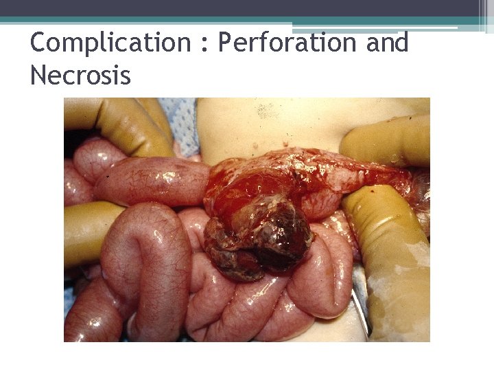
Complication : Perforation and Necrosis
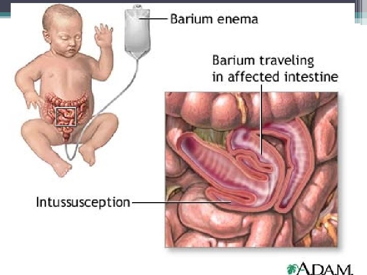
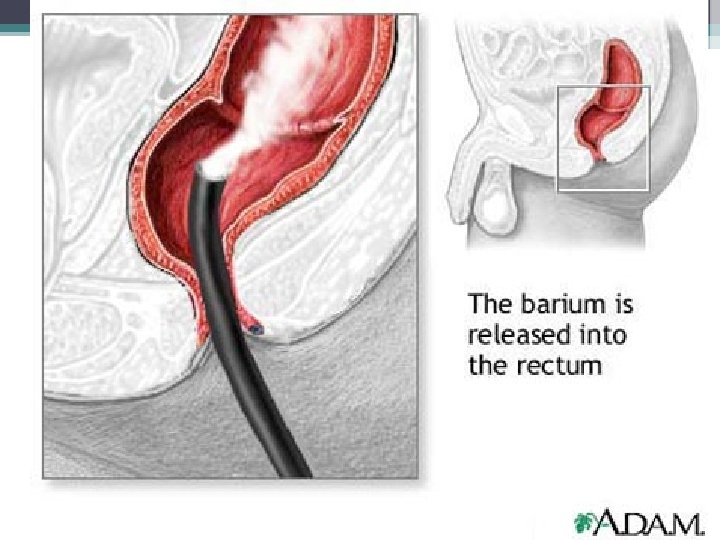
Management • Hydrostatic reduction or barium enema • Surgical release • Or resection of bowel if non-viable
Intussusception
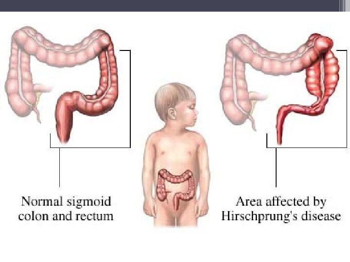
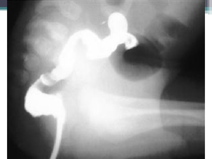
Hirschsprung’s Disease • Congenital aganglionic megacolon • Anus upwards – aganglionic segment • Affected length – usually less than a foot
Clinical Features • Newborn – no meconium, vomitting, abdominal distension, intestinal obstruction • Infant – Inadequate weight gain, constipation, abdominal distension, episodic diarrhoea, vomiting. • Childhood – Constipation, streaky/ heavily smelly stools, abdominal distension, palpable fecal mass • Diagnosis – Rectal biopsy
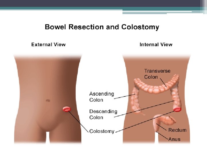
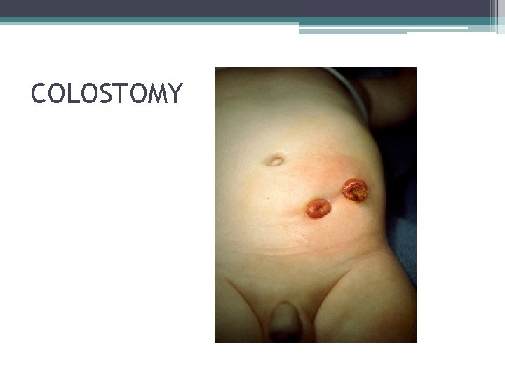
Treatment • Surgical ▫ 1 st Stage – Colostomy ▫ 2 nd stage –Pull through surgery once the weight is right • Nursing ▫ Nutrition • Homecare ▫ - Colostomy care Skin care Bad changing Stump cleaning
COLOSTOMY
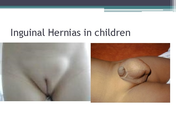
Inguinal Hernias in children
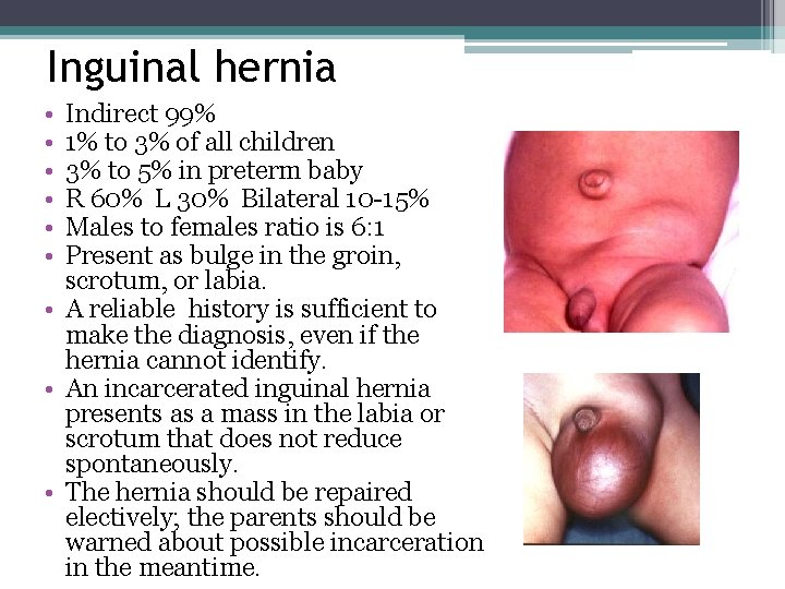
Inguinal hernia • • • Indirect 99% 1% to 3% of all children 3% to 5% in preterm baby R 60% L 30% Bilateral 10 -15% Males to females ratio is 6: 1 Present as bulge in the groin, scrotum, or labia. • A reliable history is sufficient to make the diagnosis, even if the hernia cannot identify. • An incarcerated inguinal hernia presents as a mass in the labia or scrotum that does not reduce spontaneously. • The hernia should be repaired electively; the parents should be warned about possible incarceration in the meantime.
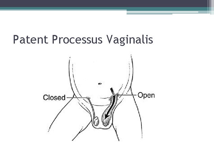
Patent Processus Vaginalis
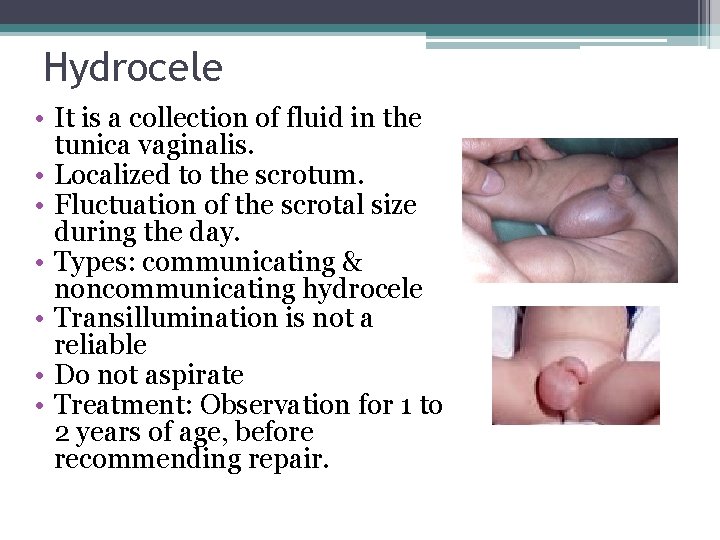
Hydrocele • It is a collection of fluid in the tunica vaginalis. • Localized to the scrotum. • Fluctuation of the scrotal size during the day. • Types: communicating & noncommunicating hydrocele • Transillumination is not a reliable • Do not aspirate • Treatment: Observation for 1 to 2 years of age, before recommending repair.
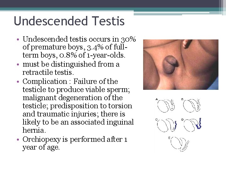
Undescended Testis • Undescended testis occurs in 30% of premature boys, 3. 4% of fullterm boys, 0. 8% of 1 -year-olds. • must be distinguished from a retractile testis. • Complication : Failure of the testicle to produce viable sperm; malignant degeneration of the testicle; predisposition to torsion and traumatic injuries; there is likely to be an associated inguinal hernia. • Orchiopexy is performed after 1 year of age.
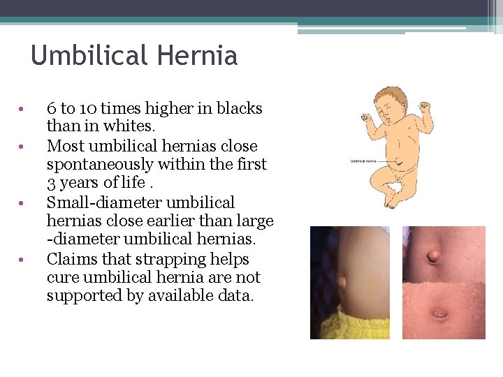
Umbilical Hernia • • 6 to 10 times higher in blacks than in whites. Most umbilical hernias close spontaneously within the first 3 years of life. Small-diameter umbilical hernias close earlier than large -diameter umbilical hernias. Claims that strapping helps cure umbilical hernia are not supported by available data.
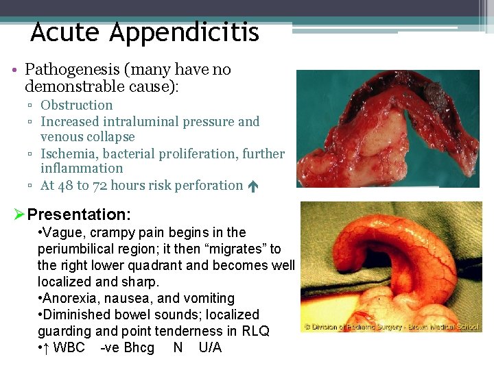
Acute Appendicitis • Pathogenesis (many have no demonstrable cause): ▫ Obstruction ▫ Increased intraluminal pressure and venous collapse ▫ Ischemia, bacterial proliferation, further inflammation ▫ At 48 to 72 hours risk perforation ØPresentation: • Vague, crampy pain begins in the periumbilical region; it then “migrates” to the right lower quadrant and becomes well localized and sharp. • Anorexia, nausea, and vomiting • Diminished bowel sounds; localized guarding and point tenderness in RLQ • ↑ WBC -ve Bhcg N U/A
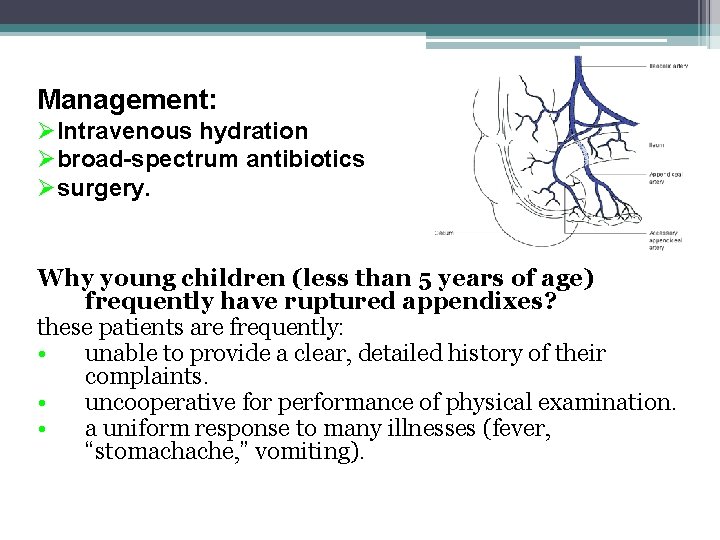
Management: ØIntravenous hydration Øbroad-spectrum antibiotics Øsurgery. Why young children (less than 5 years of age) frequently have ruptured appendixes? these patients are frequently: • unable to provide a clear, detailed history of their complaints. • uncooperative for performance of physical examination. • a uniform response to many illnesses (fever, “stomachache, ” vomiting).