Chapter 7 Acute Coronary Syndrome Imposters Copyright 2012

- Slides: 59
Chapter 7 Acute Coronary Syndrome Imposters Copyright © 2012, 2006, 1996 by Mosby, Inc. , an affiliate of Elsevier Inc. All rights reserved. 1
Objectives Recognize that many conditions other than infarction may produce ST-segment elevation. Identify the electrocardiographic (ECG) criteria for left ventricular hypertrophy, bundle branch block (BBB), ventricular rhythms, benign early repolarization, and pericarditis. Copyright © 2012, 2006, 1996 by Mosby, Inc. , an affiliate of Elsevier Inc. All rights reserved. 2
Objectives Recognize the manner in which the STsegment is displaced by left ventricular hypertrophy, left bundle branch block (LBBB), and ventricular rhythms. Review the anatomy of the intraventricular conduction system. Understand the clinical significance of infarctinduced BBB. Copyright © 2012, 2006, 1996 by Mosby, Inc. , an affiliate of Elsevier Inc. All rights reserved. 3
Objectives Gain familiarity with the various causes of BBB. Develop an approach to differentiating right bundle branch block (RBBB) from LBBB. Describe the clinical presentation of pericarditis. Copyright © 2012, 2006, 1996 by Mosby, Inc. , an affiliate of Elsevier Inc. All rights reserved. 4
Introduction to Acute Coronary Syndromes Imposters 1. 2. 3. 4. 5. Left ventricular hypertrophy Bundle branch block Ventricular rhythms Benign early repolarization Pericarditis Copyright © 2012, 2006, 1996 by Mosby, Inc. , an affiliate of Elsevier Inc. All rights reserved. 5
Left Ventricular Hypertrophy Copyright © 2012, 2006, 1996 by Mosby, Inc. , an affiliate of Elsevier Inc. All rights reserved. 6
Left Ventricular Hypertrophy (LVH) Dilatation Ø Hypertrophy Ø Increase in diameter of a heart chamber caused by volume overload Increase in thickness of a heart chamber because of chronic pressure overload Enlargement Ø Implies presence of dilatation or hypertrophy or both Copyright © 2012, 2006, 1996 by Mosby, Inc. , an affiliate of Elsevier Inc. All rights reserved. 7
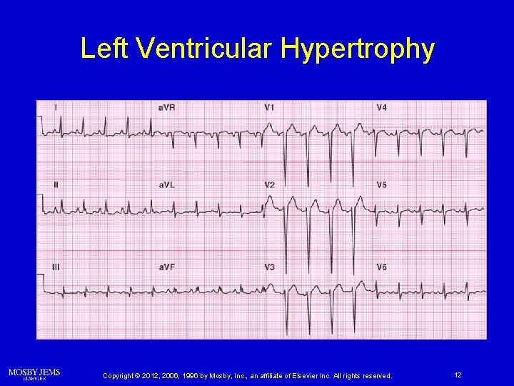
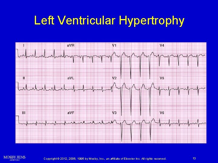
Left Ventricular Hypertrophy Ø Increases QRS amplitude Ø Often associated with ST depression and asymmetric T-wave inversion Copyright © 2012, 2006, 1996 by Mosby, Inc. , an affiliate of Elsevier Inc. All rights reserved. 8
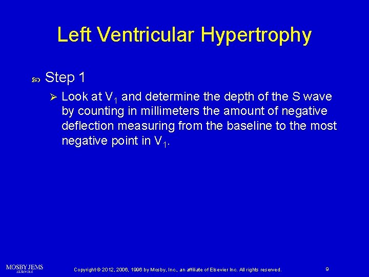
Left Ventricular Hypertrophy Step 1 Ø Look at V 1 and determine the depth of the S wave by counting in millimeters the amount of negative deflection measuring from the baseline to the most negative point in V 1. Copyright © 2012, 2006, 1996 by Mosby, Inc. , an affiliate of Elsevier Inc. All rights reserved. 9
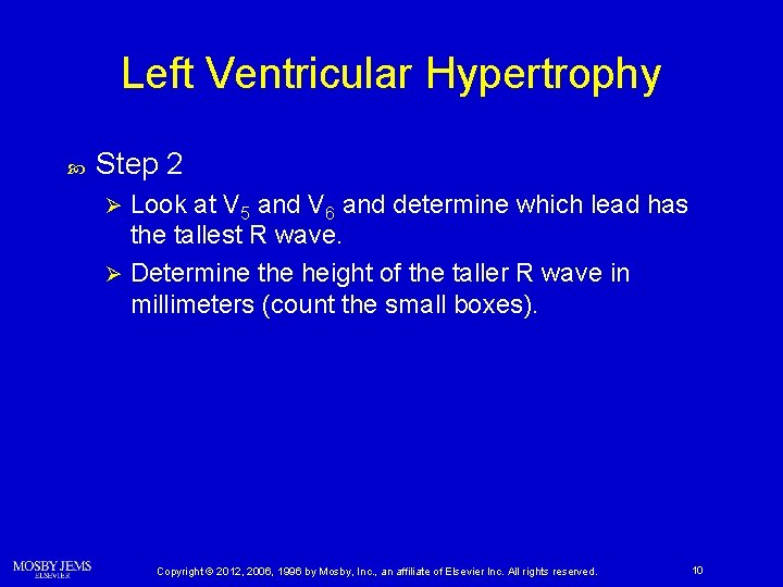
Left Ventricular Hypertrophy Step 2 Look at V 5 and V 6 and determine which lead has the tallest R wave. Ø Determine the height of the taller R wave in millimeters (count the small boxes). Ø Copyright © 2012, 2006, 1996 by Mosby, Inc. , an affiliate of Elsevier Inc. All rights reserved. 10
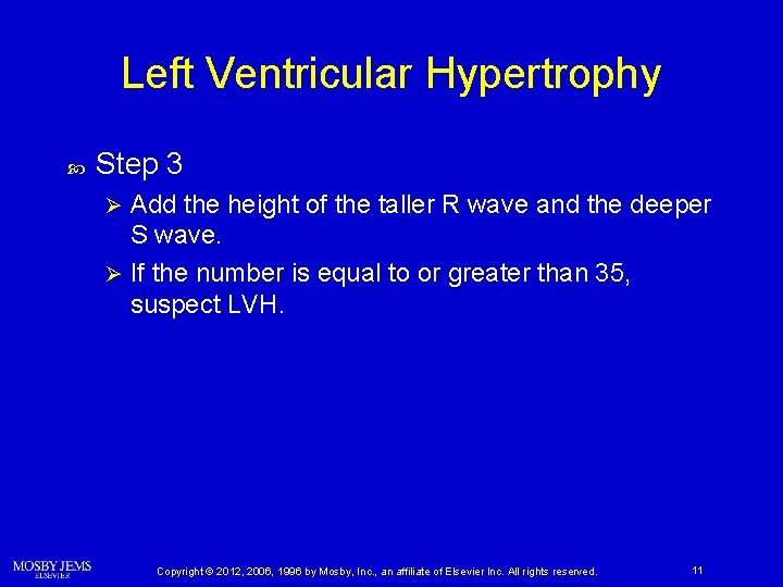
Left Ventricular Hypertrophy Step 3 Add the height of the taller R wave and the deeper S wave. Ø If the number is equal to or greater than 35, suspect LVH. Ø Copyright © 2012, 2006, 1996 by Mosby, Inc. , an affiliate of Elsevier Inc. All rights reserved. 11
Left Ventricular Hypertrophy Copyright © 2012, 2006, 1996 by Mosby, Inc. , an affiliate of Elsevier Inc. All rights reserved. 12
Left Ventricular Hypertrophy Copyright © 2012, 2006, 1996 by Mosby, Inc. , an affiliate of Elsevier Inc. All rights reserved. 13
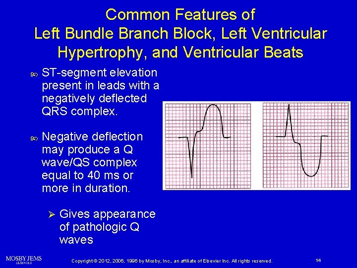
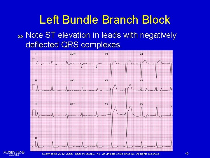
Common Features of Left Bundle Branch Block, Left Ventricular Hypertrophy, and Ventricular Beats ST-segment elevation present in leads with a negatively deflected QRS complex. Negative deflection may produce a Q wave/QS complex equal to 40 ms or more in duration. Ø Gives appearance of pathologic Q waves Copyright © 2012, 2006, 1996 by Mosby, Inc. , an affiliate of Elsevier Inc. All rights reserved. 14
Left Ventricular Hypertrophy 12 -lead machine checks for the presence of LVH “Meets voltage criteria for left ventricular hypertrophy” Ø “Possible criteria for left ventricular hypertrophy with repolarization abnormality” Ø Copyright © 2012, 2006, 1996 by Mosby, Inc. , an affiliate of Elsevier Inc. All rights reserved. 15
Bundle Branch Blocks Copyright © 2012, 2006, 1996 by Mosby, Inc. , an affiliate of Elsevier Inc. All rights reserved. 16
Structures of the Intraventricular Conduction System After passing through the AV node, the electrical impulse enters the bundle of His. Ø Normally only electrical connection between atria and ventricles Ø Connects AV node with two bundle branches Ø Conducts impulse to right and left bundle branches Copyright © 2012, 2006, 1996 by Mosby, Inc. , an affiliate of Elsevier Inc. All rights reserved. 17
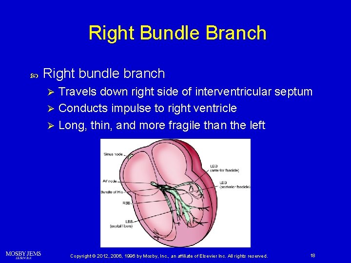
Right Bundle Branch Right bundle branch Travels down right side of interventricular septum Ø Conducts impulse to right ventricle Ø Long, thin, and more fragile than the left Ø Copyright © 2012, 2006, 1996 by Mosby, Inc. , an affiliate of Elsevier Inc. All rights reserved. 18
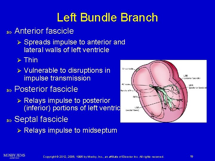
Left Bundle Branch Anterior fascicle Spreads impulse to anterior and lateral walls of left ventricle Ø Thin Ø Vulnerable to disruptions in impulse transmission Ø Posterior fascicle Ø Relays impulse to posterior (inferior) portions of left ventricle Septal fascicle Ø Relays impulse to midseptum Copyright © 2012, 2006, 1996 by Mosby, Inc. , an affiliate of Elsevier Inc. All rights reserved. 19
Bundle Branch Activation Normally, left side of the interventricular septum is stimulated first. Electrical impulse then crosses the septum to stimulate the right side. Left and right ventricles are then depolarized simultaneously. Copyright © 2012, 2006, 1996 by Mosby, Inc. , an affiliate of Elsevier Inc. All rights reserved. 20
Bundle Branch Activation If a delay or block occurs in one of the bundle branches, the ventricles will be depolarized asynchronously. Ø Impulse travels first down the unblocked branch Ø Stimulates that ventricle Ø Impulse must then travel from cell to cell through myocardium to stimulate other ventricle Copyright © 2012, 2006, 1996 by Mosby, Inc. , an affiliate of Elsevier Inc. All rights reserved. 21
Significance of Bundle Branch Blocks Infarct-induced BBB Ø Increased mortality rate Ø Rate of cardiogenic shock increases BBB itself does not cause these outcomes. New-onset BBB indicates extensive infarction. Copyright © 2012, 2006, 1996 by Mosby, Inc. , an affiliate of Elsevier Inc. All rights reserved. 22
Significance of Bundle Branch Blocks Patients experiencing septal and anteroseptal infarctions are most likely to develop BBB. Patients with BBB in the setting of infarction are at increased risk of developing complete AV block. Ø Standby pacing indicated when BBB complicates infarction LBBB can produce ST-segment elevation and wide Q waves that look similar to infarction. Copyright © 2012, 2006, 1996 by Mosby, Inc. , an affiliate of Elsevier Inc. All rights reserved. 23
Causes of Right Bundle Branch Blocks Can occur in individuals with no underlying heart disease. Ø RBBB present in 3% to 7% of acute MIs. Ø More commonly occurs in presence of coronary artery disease May require pacemaker Progression of RBBB to complete AV block occurs twice as often as that of LBBB. Copyright © 2012, 2006, 1996 by Mosby, Inc. , an affiliate of Elsevier Inc. All rights reserved. 24
Causes of Left Bundle Branch Blocks Acute LBBB may occur secondary to: Ø Anteroseptal MI (more common) Ø Inferior MI Ø Acute congestive heart failure Ø Acute pericarditis Ø Myocarditis Copyright © 2012, 2006, 1996 by Mosby, Inc. , an affiliate of Elsevier Inc. All rights reserved. 25
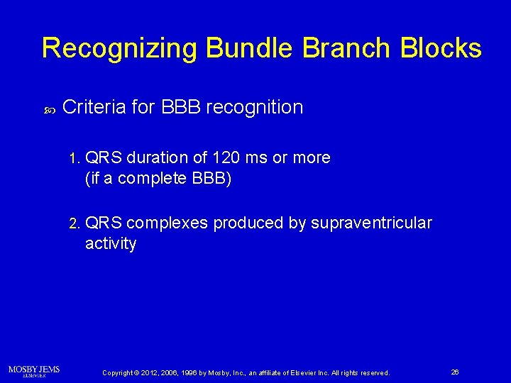
Recognizing Bundle Branch Blocks Criteria for BBB recognition 1. QRS duration of 120 ms or more (if a complete BBB) 2. QRS complexes produced by supraventricular activity Copyright © 2012, 2006, 1996 by Mosby, Inc. , an affiliate of Elsevier Inc. All rights reserved. 26
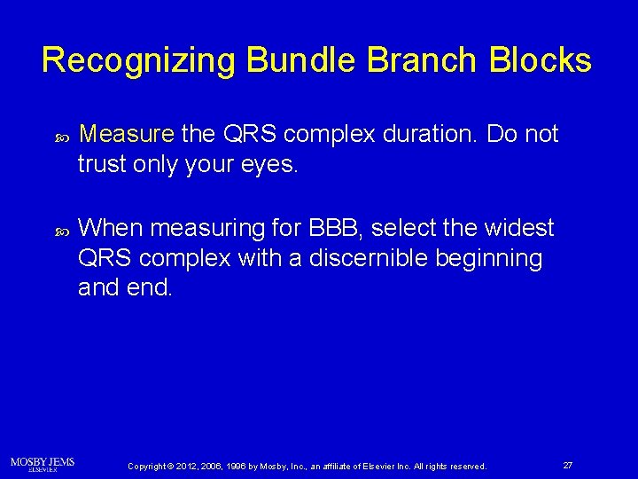
Recognizing Bundle Branch Blocks Measure the QRS complex duration. Do not trust only your eyes. When measuring for BBB, select the widest QRS complex with a discernible beginning and end. Copyright © 2012, 2006, 1996 by Mosby, Inc. , an affiliate of Elsevier Inc. All rights reserved. 27
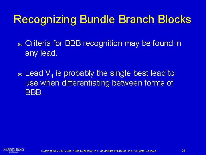
Recognizing Bundle Branch Blocks Criteria for BBB recognition may be found in any lead. Lead V 1 is probably the single best lead to use when differentiating between forms of BBB. Copyright © 2012, 2006, 1996 by Mosby, Inc. , an affiliate of Elsevier Inc. All rights reserved. 28
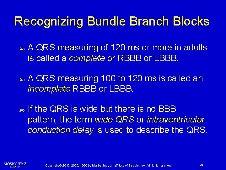
Recognizing Bundle Branch Blocks A QRS measuring of 120 ms or more in adults is called a complete or RBBB or LBBB. A QRS measuring 100 to 120 ms is called an incomplete RBBB or LBBB. If the QRS is wide but there is no BBB pattern, the term wide QRS or intraventricular conduction delay is used to describe the QRS. Copyright © 2012, 2006, 1996 by Mosby, Inc. , an affiliate of Elsevier Inc. All rights reserved. 29
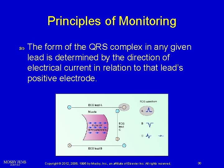
Principles of Monitoring The form of the QRS complex in any given lead is determined by the direction of electrical current in relation to that lead’s positive electrode. Copyright © 2012, 2006, 1996 by Mosby, Inc. , an affiliate of Elsevier Inc. All rights reserved. 30
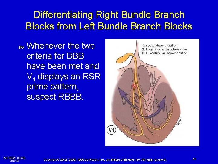
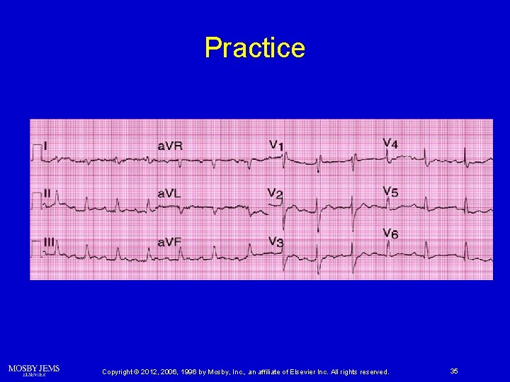
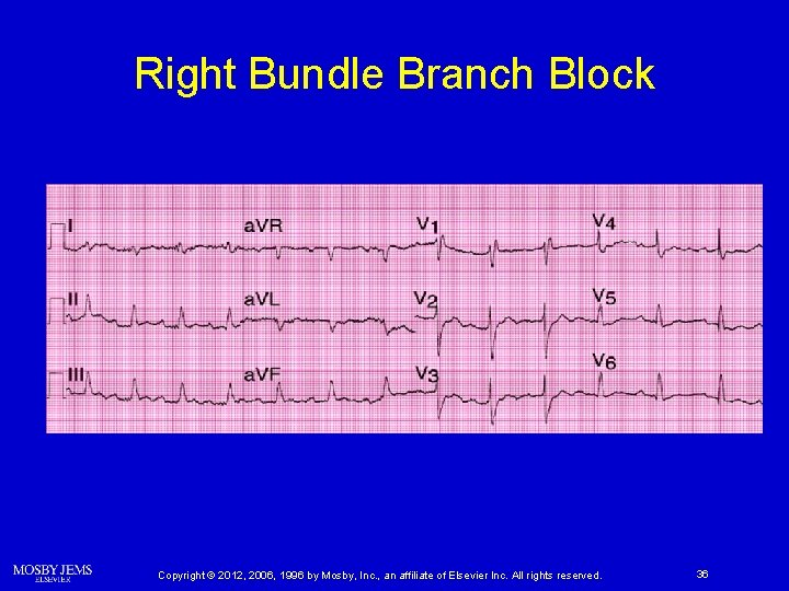
Differentiating Right Bundle Branch Blocks from Left Bundle Branch Blocks Whenever the two criteria for BBB have been met and V 1 displays an RSR prime pattern, suspect RBBB. Copyright © 2012, 2006, 1996 by Mosby, Inc. , an affiliate of Elsevier Inc. All rights reserved. 31
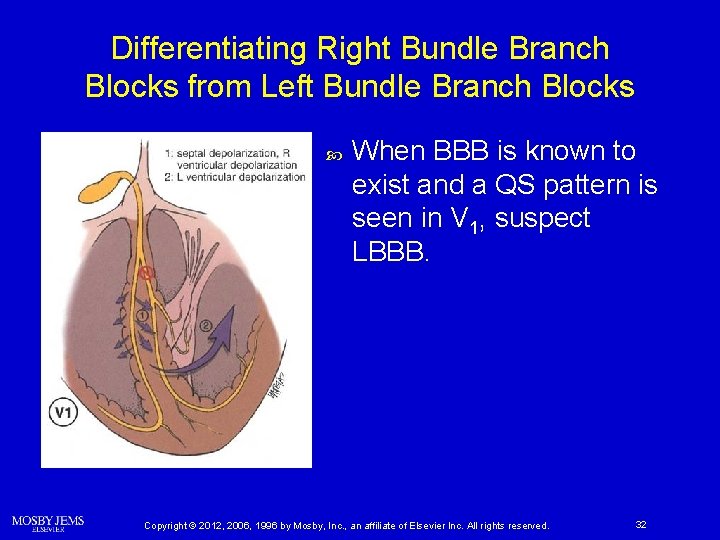
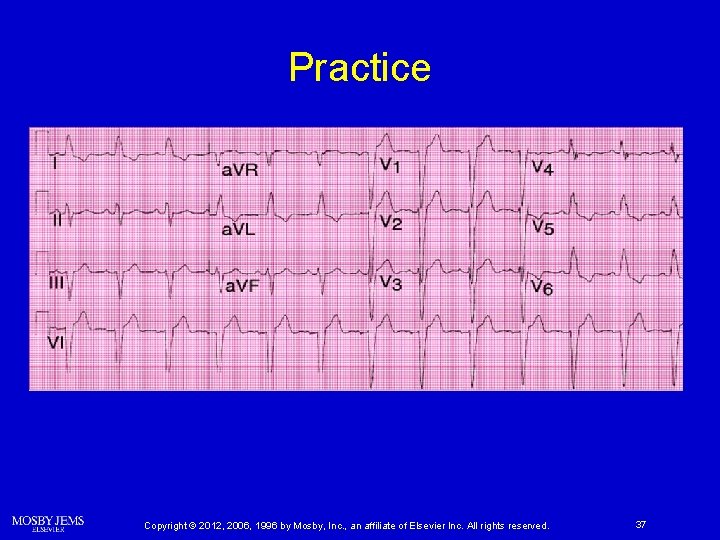
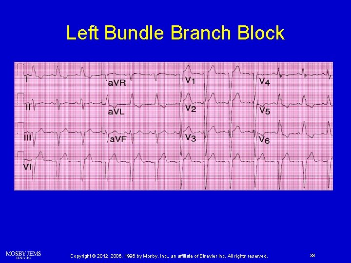
Differentiating Right Bundle Branch Blocks from Left Bundle Branch Blocks When BBB is known to exist and a QS pattern is seen in V 1, suspect LBBB. Copyright © 2012, 2006, 1996 by Mosby, Inc. , an affiliate of Elsevier Inc. All rights reserved. 32
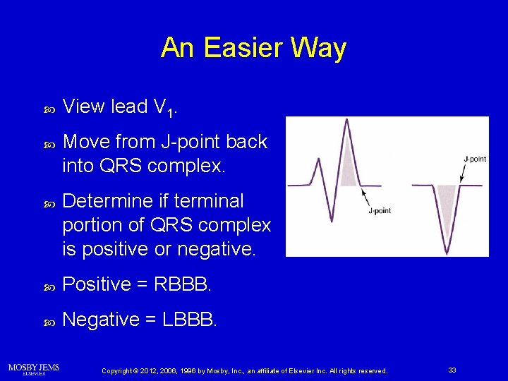
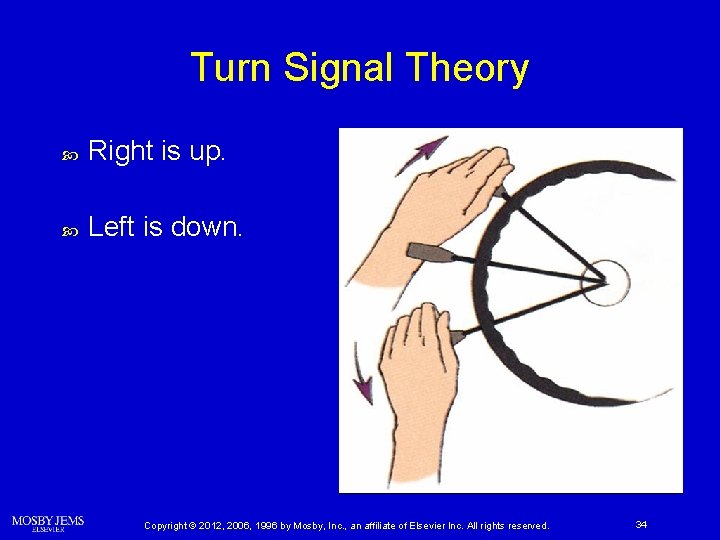
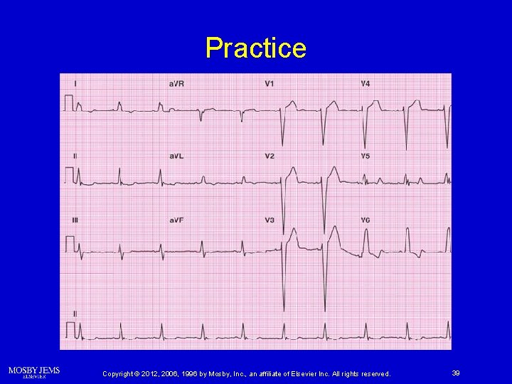
An Easier Way View lead V 1. Move from J-point back into QRS complex. Determine if terminal portion of QRS complex is positive or negative. Positive = RBBB. Negative = LBBB. Copyright © 2012, 2006, 1996 by Mosby, Inc. , an affiliate of Elsevier Inc. All rights reserved. 33
Turn Signal Theory Right is up. Left is down. Copyright © 2012, 2006, 1996 by Mosby, Inc. , an affiliate of Elsevier Inc. All rights reserved. 34
Practice Copyright © 2012, 2006, 1996 by Mosby, Inc. , an affiliate of Elsevier Inc. All rights reserved. 35
Right Bundle Branch Block Copyright © 2012, 2006, 1996 by Mosby, Inc. , an affiliate of Elsevier Inc. All rights reserved. 36
Practice Copyright © 2012, 2006, 1996 by Mosby, Inc. , an affiliate of Elsevier Inc. All rights reserved. 37
Left Bundle Branch Block Copyright © 2012, 2006, 1996 by Mosby, Inc. , an affiliate of Elsevier Inc. All rights reserved. 38
Practice Copyright © 2012, 2006, 1996 by Mosby, Inc. , an affiliate of Elsevier Inc. All rights reserved. 39
Left Bundle Branch Block Note ST elevation in leads with negatively deflected QRS complexes. Copyright © 2012, 2006, 1996 by Mosby, Inc. , an affiliate of Elsevier Inc. All rights reserved. 40
Exceptions Junctional rhythms Wolff-Parkinson-White (WPW) syndrome Copyright © 2012, 2006, 1996 by Mosby, Inc. , an affiliate of Elsevier Inc. All rights reserved. 44
Exceptions Nonspecific intraventricular conduction delay (NSIVCD) Ø Do not display the typical V 1 morphologies generally produced by BBB. Ø Origin may not be due to a complete BBB. Ø Atypical patterns of BBB can be attributed to NSIVCD. Copyright © 2012, 2006, 1996 by Mosby, Inc. , an affiliate of Elsevier Inc. All rights reserved. 45
Ventricular Rhythms Copyright © 2012, 2006, 1996 by Mosby, Inc. , an affiliate of Elsevier Inc. All rights reserved. 46
Ventricular Rhythms Impulses from the ventricles may be the result of: Natural pacemaker sites Ø Implanted pacemakers Ø May show ST elevation not due to MI Copyright © 2012, 2006, 1996 by Mosby, Inc. , an affiliate of Elsevier Inc. All rights reserved. 47
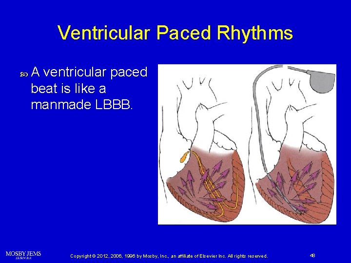
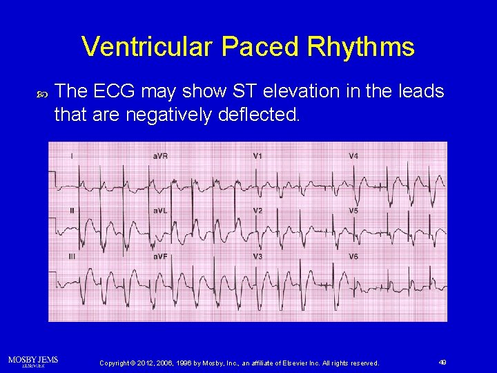
Ventricular Paced Rhythms A ventricular paced beat is like a manmade LBBB. Copyright © 2012, 2006, 1996 by Mosby, Inc. , an affiliate of Elsevier Inc. All rights reserved. 48
Ventricular Paced Rhythms The ECG may show ST elevation in the leads that are negatively deflected. Copyright © 2012, 2006, 1996 by Mosby, Inc. , an affiliate of Elsevier Inc. All rights reserved. 49
Benign Early Repolarization Copyright © 2012, 2006, 1996 by Mosby, Inc. , an affiliate of Elsevier Inc. All rights reserved. 50
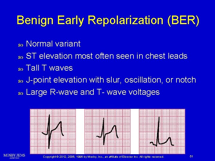
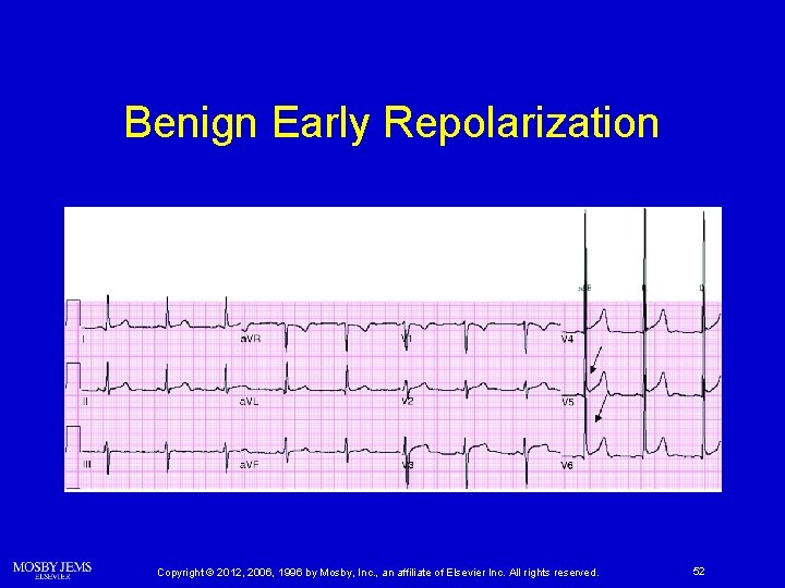
Benign Early Repolarization (BER) Normal variant ST elevation most often seen in chest leads Tall T waves J-point elevation with slur, oscillation, or notch Large R-wave and T- wave voltages Copyright © 2012, 2006, 1996 by Mosby, Inc. , an affiliate of Elsevier Inc. All rights reserved. 51
Benign Early Repolarization Copyright © 2012, 2006, 1996 by Mosby, Inc. , an affiliate of Elsevier Inc. All rights reserved. 52
Pericarditis Copyright © 2012, 2006, 1996 by Mosby, Inc. , an affiliate of Elsevier Inc. All rights reserved. 53
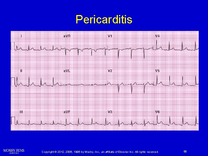
Pericarditis ST elevation occurs because of inflammation. Anyone can develop pericarditis. Ø Post-MI and postcardiac surgery patients are especially prone. Copyright © 2012, 2006, 1996 by Mosby, Inc. , an affiliate of Elsevier Inc. All rights reserved. 54
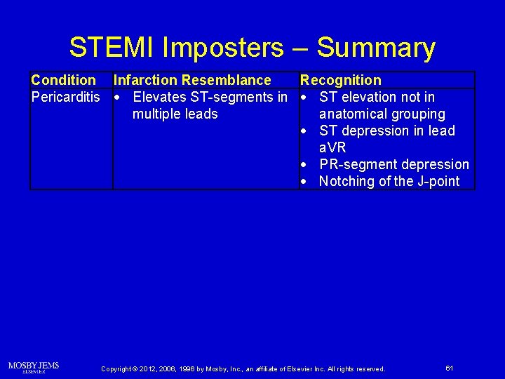
Pericarditis ST elevation occurs frequently. Ø Usually diffuse and not strictly grouped into leads that are anatomically contiguous. PR-segment depression. Notching of the J-point. Copyright © 2012, 2006, 1996 by Mosby, Inc. , an affiliate of Elsevier Inc. All rights reserved. 55
Pericarditis Copyright © 2012, 2006, 1996 by Mosby, Inc. , an affiliate of Elsevier Inc. All rights reserved. 56
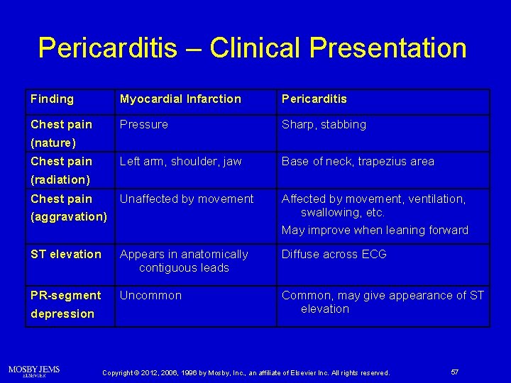
Pericarditis – Clinical Presentation Finding Myocardial Infarction Pericarditis Chest pain (nature) Pressure Sharp, stabbing Chest pain (radiation) Left arm, shoulder, jaw Base of neck, trapezius area Chest pain (aggravation) Unaffected by movement Affected by movement, ventilation, swallowing, etc. May improve when leaning forward ST elevation Appears in anatomically contiguous leads Diffuse across ECG PR-segment depression Uncommon Common, may give appearance of ST elevation Copyright © 2012, 2006, 1996 by Mosby, Inc. , an affiliate of Elsevier Inc. All rights reserved. 57
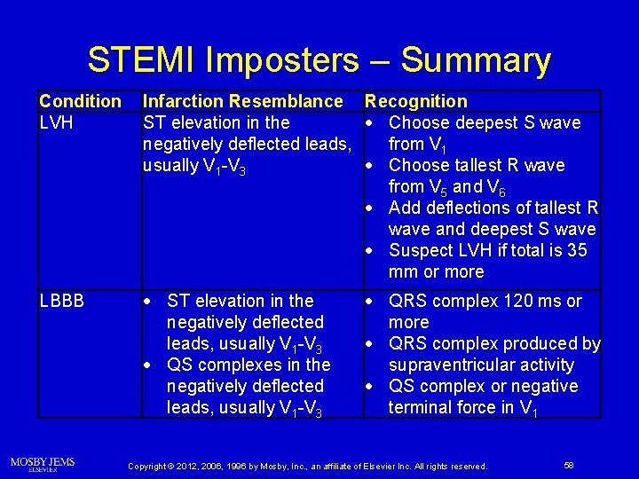
STEMI Imposters – Summary Condition LVH Infarction Resemblance Recognition ST elevation in the Choose deepest S wave negatively deflected leads, from V 1 usually V 1 -V 3 Choose tallest R wave from V 5 and V 6 Add deflections of tallest R wave and deepest S wave Suspect LVH if total is 35 mm or more LBBB ST elevation in the negatively deflected leads, usually V 1 -V 3 QS complexes in the negatively deflected leads, usually V 1 -V 3 QRS complex 120 ms or more QRS complex produced by supraventricular activity QS complex or negative terminal force in V 1 Copyright © 2012, 2006, 1996 by Mosby, Inc. , an affiliate of Elsevier Inc. All rights reserved. 58
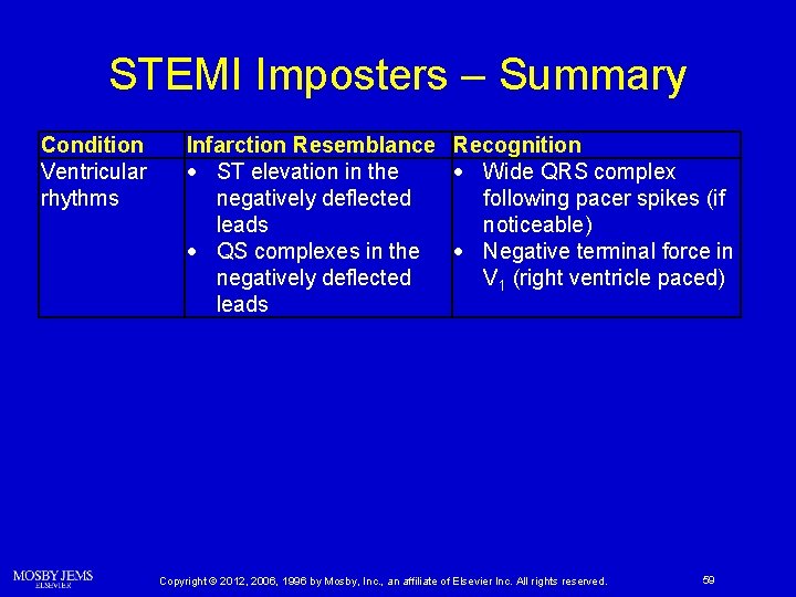
STEMI Imposters – Summary Condition Ventricular rhythms Infarction Resemblance Recognition ST elevation in the Wide QRS complex negatively deflected following pacer spikes (if leads noticeable) QS complexes in the Negative terminal force in negatively deflected V 1 (right ventricle paced) leads Copyright © 2012, 2006, 1996 by Mosby, Inc. , an affiliate of Elsevier Inc. All rights reserved. 59
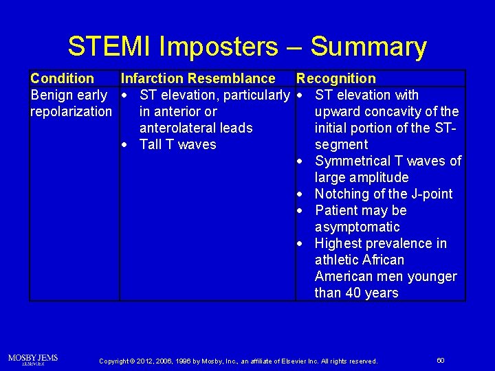
STEMI Imposters – Summary Condition Infarction Resemblance Recognition Benign early ST elevation, particularly ST elevation with repolarization in anterior or upward concavity of the anterolateral leads initial portion of the ST Tall T waves segment Symmetrical T waves of large amplitude Notching of the J-point Patient may be asymptomatic Highest prevalence in athletic African American men younger than 40 years Copyright © 2012, 2006, 1996 by Mosby, Inc. , an affiliate of Elsevier Inc. All rights reserved. 60
STEMI Imposters – Summary Condition Infarction Resemblance Recognition Pericarditis Elevates ST-segments in ST elevation not in multiple leads anatomical grouping ST depression in lead a. VR PR-segment depression Notching of the J-point Copyright © 2012, 2006, 1996 by Mosby, Inc. , an affiliate of Elsevier Inc. All rights reserved. 61
Questions? Copyright © 2012, 2006, 1996 by Mosby, Inc. , an affiliate of Elsevier Inc. All rights reserved. 62