RENAL DISEASE GLOMERULONEPHRITIS AND NEPHROTIC SYNDROME Pathophysiology of


- Slides: 8

RENAL DISEASE: GLOMERULONEPHRITIS AND NEPHROTIC SYNDROME Pathophysiology of Disease: Chapter 16 (398 -401) Jack De. Ruiter, Ph. D Department of Pharmacal Sciences April, 2000

GLOMERULONEPHRITIS AND NEPHROTIC SYNDROME (Pages 398 -400) • • • Acute Glomerulonephritis: Rapidly Progressive Glomerulonephritis Chronic Glomerulonephritis Nephrotic Syndrome Asymptomatic urinary Abnormalities

GLOMERULONEPHRITIS: Clinical manifestations (pages 400 -401) • Proteinuria and hematuria: Damage to capillary wall allows “leakage” • Decreased GFR: – Infiltration of glomerular capillaries with inflammatory cells, OR – Expansion of contractile mesangial cells • Edema and Hypertension: Fluid and salt overload from decreased GFR • Transient decrease in serum complement • Transient elevations antibody to streptococcal antigen

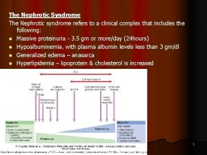
NEPHROTIC SYNDROME: Clinical manifestations (pages 400 -401) • Decreased oncotic pressure: loss of serum protein: – Intravascular volume depletion with syncope, shock and acute renal failure – Activation of renin-angiotensin-aldosterone system – Activation of sympathetic nervous system – Increased secretion of vasopressin – Hyperlipidemia: Increases hepatic VLDL production • Loss of other plasma proteins: – – Increased susceptibility to infection Hypercoagulability Vitamin D deficiency: loss of Vit D binding protein Altered thyroxine binding protein/thyroid tests

Acute Glomerulonephritis: (page 398 -400) • Clinical Presentation – Abrupt hematuria and proteinuria – reduced GFR, salt and water retention • Pathology & Pathogenesis – Infection: Immune response to pathogen (i. e. Streptococci) antigen resulting in deposition of immune complexes and complement in glomerular capillary bed (intrarenal!) – Onset: 7 -10 days after initial infection – Full recovery typically occurs within weeks on infection

Rapidly Progressive Glomerulonephritis (pages 398 & 400) • Clinical Presentation: “Intermediate” stage – Failure to recover from Acute Glomerulonephritis: Origin unknown – Worsening renal function with irreversible and complete renal failure a the outcome • Pathology & Pathogenesis – – Intermediate stage between “Acute” and “Chronic” Extracapillary cellular proliferation: 70% of glomeruli Basement membrane gap/discontinuities Deposition of anti-GBM antibodies or granular immunoglobulins

Chronic Glomerulonephritis (pages 398 & 400) • Clinical Presentation – Slow progression from acute disease to chronic renal failure (5 -20 years) • Pathology & Pathogenesis – Capillary or mesangial cellular proliferation, OR – Structural obliteration of glomeruli: sclerosing CG, OR – Subepithelial proteinaceous deposits: Membranous G

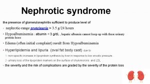
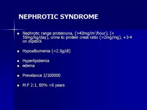
Nephrotic Syndrome (page 398 -400) • Clinical Presentation: See Table 16 -15 – Marked proteinuria (albuminuria) >3. 5 g/24 hr with hypoalbuminemia – Edema – Hyperlipidemia – Fat bodies in the urine • Pathology & Pathogenesis: See Table 16 -15 – Minimal cellular infiltration of glomeruli – Deposition of antigen-antibody complexes in the BM
 Ira pré renal renal e pós renal
 Ira pré renal renal e pós renal
 Acute glomerulonephritis pathophysiology
 Dr kriti mohan
 Primary and secondary causes of nephrotic syndrome
 Nephrotic syndrome differential diagnosis
 Why there is hyperlipidemia in nephrotic syndrome
 Nephrotic syndrome
 Polyclonal gammopathy electrophoresis pattern

