Arthritis OA versus RA Fractures Tendon Injuries RSDCRPS


 hypermobile")


 and")


? Purpose is to immobilize the")


 weeks: Remove")


 Shoulder hand syndrome (2) Reflex sympathetic dystrophy (3)")
")
 Pain (2) decreased ROM (3) changes in")

; 2. stress loading (1) Scrub")


- Slides: 76
Arthritis: OA versus RA Fractures Tendon Injuries RSD/CRPS
Major Issues re Arthritis Main goal is to prevent loss of ROM and avoid deforming forces and habits. Interventions aimed at this use splints, joint protection education, adaptive equipment, energy conservation education. Critical to differentiate osteo arthritis or OA (also called degenerative arthritis and deg. Jt. Disease or DJD) from rheumatoid arthritis or RA.
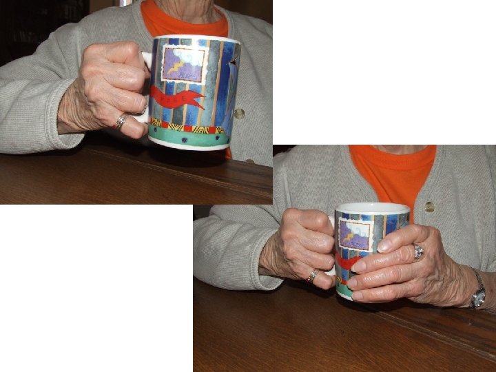
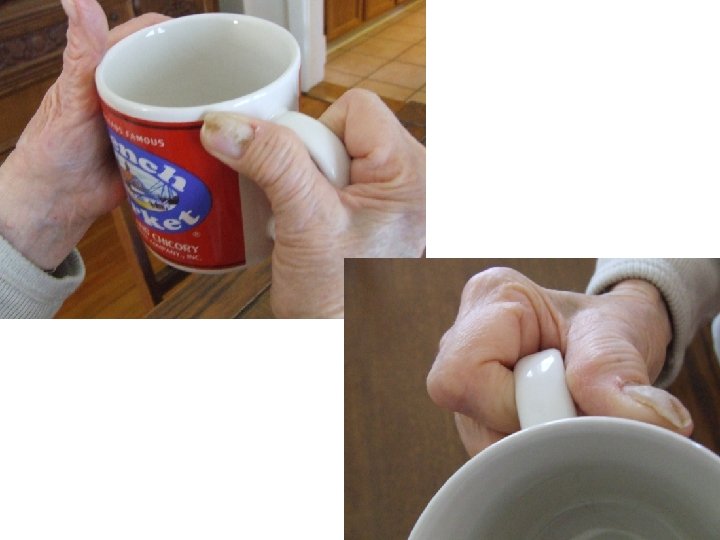
OA=Joint-specific RA=systemic disease Joint "end feel" is bony Joint "end feel" is soft Joints limited in ROM but are stiff and may be fused (ankylosed) with spurs and bony bridges across joint space. May involve only one joint. Joints have limited ROM but are hypermobile, may be subluxed or even dislocated; sign of RA is instability in joints. Joints involved may go through flare ups and get worse; more joints can become involved (polyarthritis).
OA RA Predisposed to from previous fractures involving the articular surface (fracture is said to "cross the joint line") - interrupted hyaline cartilage heals with fibrocartilage, which is rough and leads to degenerative changes over time. Occurs in multiple joints - bilateral involvement although not symmetrical. This is a systemic disease involving autoimmunity - Starts with production of auto antibodies against normally circulating antibodies in the synovial fluid. This results in inflammatory response in which the synovial tissue and then other joint structures (ligaments and bone) responsible for joint integrity and strength are damaged. Very common for OTs to work with individuals with severe involvement in MCPs, PIPs, wrists; sometimes shoulders and elbows.
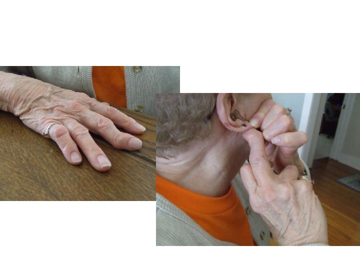
Evaluations ROM - goniometry - but look for different effects – RA (1) hypermobile joints early with full ROM and soft end feels and (2) later ROM limitations associated with subluxation and dislocation deformities like ulnar drift of MCPs, volar subluxation and dislocation of MCPs, boutonniere, swan neck, Types I – IV thumb deformities; also more likely to have multiple joint involvement (polyarthritis) as this is a systemic autoimmune disease OA with ROM limitations and hard or bony end feels; more likely to have isolated joint involvement
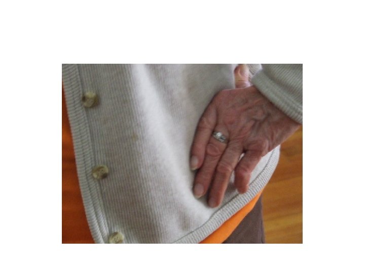
Evaluations Functional ROM within activities - occupational performance eval by observation - especially how limitations in grip and pinch motions affect performance of daily tasks Need for splints for support, adaptive equipment Need for joint protection strategies Need for energy conservation strategies Observation and documentation of specific "deformities“ Edema measurement No MMT or grip strength - assess strength through function
Joint Protection Principles • An example • Also good to communicate in pictures:
MCP Arthroplasty & Splinting
Review of Anatomy
Review of Anatomy
Review of Anatomy
Major Issues re Fractures Main goal is to protect healing bone (via immobilization) and avoid secondary problems: • (joint or muscle) contractures 2 o to immob • CRPS • tendon adhesion to fx site • re dislocation • Intrinsic muscle tightness (intrinsic “+” hand)
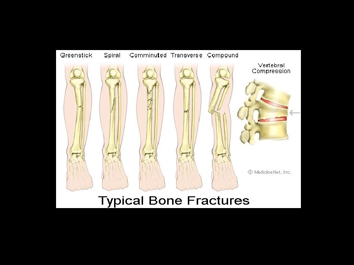
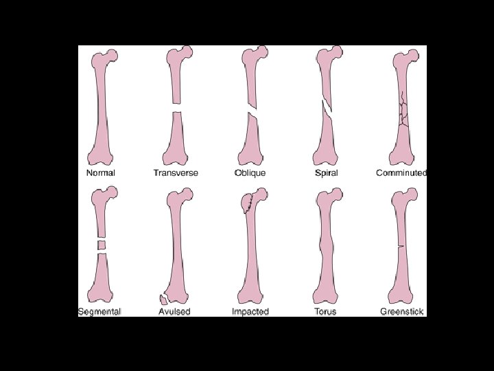
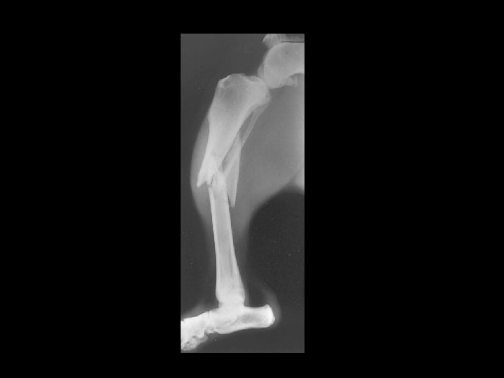
Types of fractures
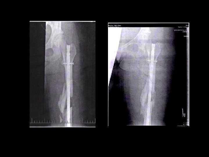
Internal Fixation
External Fixator
Ilizarov and external fixator
• Use of dorsiflexor splint or straps indicated for someone in external fixator secondary to tibfib fx to prevent foot-drop and eventual shortening of gastrocnemius-soleus when Common Peronneal nerve is damaged.
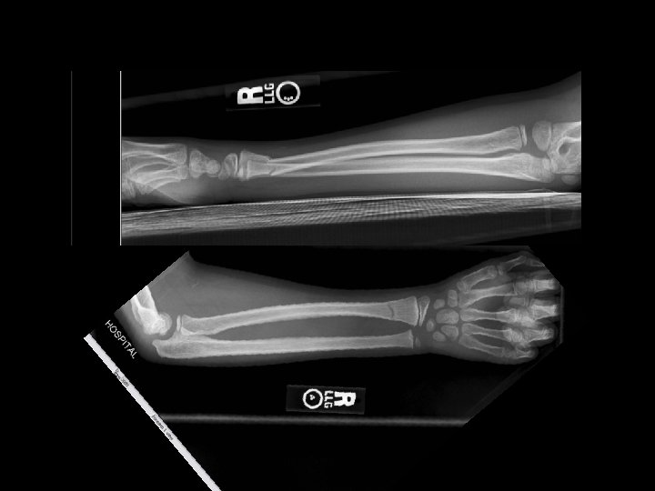
Closed Reduction and Casting Long arm cast primarily to limit: ? Short arm cast to limit: ?
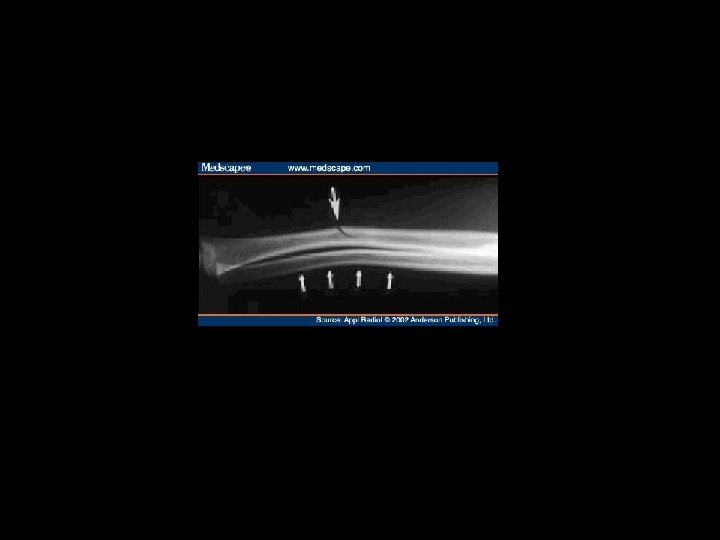
One more type of fracture is avulsion fracture. What injury typically occurs with (or causes) and avulsion fracture? What is avulsed and why? A fracture in which a piece of bone is pulled off with the tendon or ligament. Instead of the tendon or ligament tearing off of its attachment, it is so solidly attached it pulls off its attachment. It would be like pulling an electrical cord to unplug it (should never do, right? ? ) and instead of coming unplugged, the entire socket comes out of the wall. Occurs with joint sprains where the joint is forced in an extreme position and great stress is placed on the ligament (remember - holds bone to bone) or the tendon (holds muscle to bone).
Immobilization: Purpose is to protect the injury part and allow healing following: • Fracture • Tendon injury and repair • Ligament injury and dislocation • Nerve Injury
What is the purpose of immobilization: To protect the injury part and allow healing After fracture? Think: muscle tendons pull on bones and often have reflexive contractions when bone fractures; if there was a mid shaft fracture of the prox phalanx and the interosseus muscles pulled the proximal fragment into flexion at the MCP, what would the finger look like? Intrinsic muscle would pull proximal fragment of proximal phalanx into flexion but remaining part of finger would not flex. So after fracture, the normal pull of muscles across bones can displace the fragments - this can happen after the fracture has been reduced. So the fragments once realigned must be stabilized.
After ligament injury (e. g. , associated with dislocation)? Purpose is to immobilize the joint so the ligament is in slack position - this would be the loose-packed position. After tendon injury? Purpose is to immobilize the joint so the tendon is in slack position - this would NOT be the loose-packed position - as the tendon is not intimately associated with the joint like the ligament is. You could place a joint crossed by a tendon in the loose-packed position and the tendon tension would not change very much. To place tendons on slack, passively move (and immobilize) all the joints crossed by the tendon in the same direction as function of the muscle/tendon. For example, if the flexor dig profundus tendon is torn and repaired, place the wrist, MCP, PIP and DIP joints all in flexion.
After nerve injury? Again, we want slack so the reconnected nerve does not have tension pulling it apart. Slack position depends on the nerve's relationship to the joint: median and ulnar nerves are anterior to the wrist so wrist flexion will provide slack; radial n. is dorsal. . .
Susan: Initial Eval Note and 7 th Week Progress Note • Initial Note • Progress @ 7 weeks
Go to: Immob_Early_Motion_Guides. html to see details of each - Type of Fracture, Dislocation, Ligament Rupture Immobilization/Early Motion Timelines Common Complications (Secondary Problems) Colle’s Fx Immobilized 6 -8 weeks; AROM as each cast is removed; elevation of the hand finger flexion and extension should begin immediately as tolerated to decrease edema and guard against CRPS; OA in joints involved in fx; reduced ROM in pro/supination, elbow flexion/ext; all shoulder motions
Go to: Immob_Early_Motion_Guides. html to see details of each - Type of Fracture, Immobilization/Early Dislocation, Motion Timelines Ligament Rupture Common Complications (Secondary Problems) Distal Phalanx Splint DIP in 10 deg Fracture hyperextension for 6 weeks, longer if extensor lag develops; begin AROM of isolated DIP f/e near 6 weeks Dislocationespecially if joint is "vigorously splinted" in hyperextension; extensor lag if allowed to fall into flexion
Go to: Immob_Early_Motion_Guides. html to see details of each - Type of Fracture, Immobilization/Early Dislocation, Motion Timelines Ligament Rupture Common Complications (Secondary Problems) Proximal and Middle Phalanx Fractures If poor positioning in splint = PIP flexion contracture, MCP extension contx; In prox phal fx = adhesion of flexor tendons to fracture site; If positioned well in "clam -digger" = intrinsic + hand IF internal fixation, begin AROM and AAROM 5 to 15 days; IF 3 to 5 weeks in "clamdigger" follow by PROM to stretch intrinsics and PIP/DIP active f/e with prox phal supported in MCP extension – see OH
Go to: Immob_Early_Motion_Guides. html to see details of each - Type of Fracture, Immobilization/Early Dislocation, Motion Timelines Ligament Rupture Common Complications (Secondary Problems) Metacarpal Fractures If not positioned correctly = MCP extension contx, PIP flexion contx; Intrinsic contracture (from direct trauma and scaring in intrinsics); Decreased web space in 1 st metacarpal fx. Immobilization in "clam -digger" position 2 to 3 weeks then AROM and mild AAROM and continue splint for protection another 2 weeks; 1 st metacarpal fx splinted in thumb abduction
Go to: Immob_Early_Motion_Guides. html to see details of each - Type of Fracture, Immobilization/Early Dislocation, Motion Timelines Ligament Rupture Common Complications (Secondary Problems) MCP Collateral Ligament Injury MCP extension contracture, PIP flexion contracture; Adhesion of intrinsic lateral bands to healing ligament resulting in intrinsic contracture = intrinsic + If stable, immobilized on slack (ext) 3 to 10 days then AROM; immob up to 3 weeks if torn; may require dynamic flex splint if ext contracture noted following immobilization period; "buddy taping“
Go to: Immob_Early_Motion_Guides. html to see details of each - Type of Fracture, Immobilization/Early Dislocation, Motion Timelines Ligament Rupture Common Complications (Secondary Problems) Dorsal Dislocation In general due to of PIP concern of re dislocation, splint in 25 degrees PIP flexion for 3 weeks or allow motion but block last 15 deg. ; if occurs with fracture volar plate is reattached 3 weeks then AROM and AAROM into PIP flexion Swan-neck position with PIP locked in hyperextension; may need 5 weeks dynamic flexion splinting; 7 weeks dynamic extension splinting depending on contracture at DIP or PIP
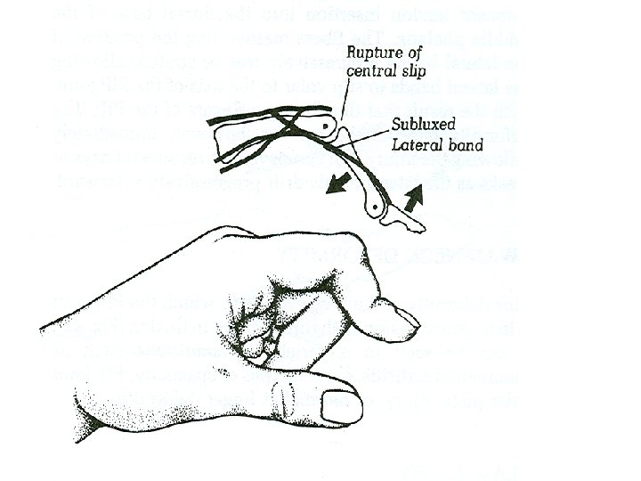
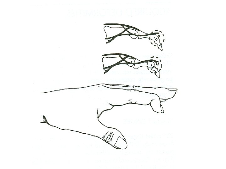
Go to: Immob_Early_Motion_Guides. html to see details of each - Type of Fracture, Immobilization/Early Dislocation, Motion Timelines Ligament Rupture Common Complications (Secondary Problems) Boutonniere PIP flexion contx/extensor lag if not splinted in full extension; Adhesion of intrinsic lateral band resulting in PIP, DIP extension contracture; PIP splinted in extension for up to 4 weeks with DIP free for flexion exercise to prevent adhesion of lateral bands; leave in splint to avoid flexion contx/extensor lag for up to 8 weeks (remove daily for AROM and mild AAROM).
• After any fx, postponing therapy can result in edema and decreased ROM • Always be concerned about joints NOT immobilized (esp. shoulder) and educate in AROM (multiple-daily) and tendon gliding (Figure 42 -3 in Radomski & Trombly Latham 6 th Ed. ) in fxs of MC and phal.
Major Issues re Tendon Injury and Repair Main goal is to place tendon on slack and minimize tension on the repair until it heals as the sutures are NEVER strong enough to sustain the tension developed by active contraction and full passive stretch (as in passive insufficiency).
See details: tendon_injuries. html Zones of the Volar Hand: Zone 1: Insertion of FDS to insertion of FDP Zone 2: "No man's land"; Distal palmar crease to insertion of FDS. Zone 3: Distal edge of volar carpal ligament (roof of carpal tunnel) to distal palmar crease (actually, A 1 pulley). Zone 4: Carpal tunnel area Zone 5: Distal volar wrist
See details: tendon_injuries. html www. uphs. upenn. edu/ortho/oj/1997/oj 10 sp 97 p 5. htmll
See details: tendon_injuries. html Initial Precautions/Contraindications: • No active use of muscle whose tendon was cut (not even a "trace" contraction). • No passive motion which would put tension in the system, in other words, only controlled passive motion Don't forget there may be concomitant injuries (vascular, peripheral nerve, fx, healing wound). • Make sure individual is crystal clear on precautions, for example, leaving the splint on, moving ONLY as instructed, returning for follow-up, non use of extremity even with splint in place, edema precautions, ROM maintenance for other uninvolved (proximal) joints.
See details: tendon_injuries. html General Principles of Treatment: Dorsal splint is applied immediately post op positioning the flexor tendons on the slack: wrist 45 deg. Flex; MCPs 45 deg. (or more) flex; IPs allowed to extend fully. Traction (Kleinert) into flexion applied through rubber bands on the digits with repaired tendons - provides PASSIVE flexion to discourage the use of the flexors. Must allow FULL extension of the IP joints while in the splint.
See details: tendon_injuries. html Example of protocol for zone 2: Zone 2: 1 to 4 1/2 weeks: Splint in dorsal splint as described under "Treatment" above. Perform active extension and passive flexion within the splint 10 times per hour and gentle passive flexion of PIP and DIP 1 O reps, 4 times a day. With any sign of flexion contracture in the PIP or DIP, perform early controlled passive motion = Create slack in the flexor tendons by fully flexing the MCP before gently passively extending the PIP. Similarly, passively flex the MCP and PIP and passively extend the DIP. This is all done in the dorsal splint.
See details: tendon_injuries. html Zone 2 cont: 4 1/2 to 6 weeks: Wrist cuff and rubber band traction as described above. THERE IS STILL NO active flexion or SIMULTANEOUS passive extension of IP, MCP and wrist joints.
See details: tendon_injuries. html Zone 2 cont: 6 to 8 (or 10) weeks: Remove wrist cuff and begin ACTIVE FLEXION FOR THE FIRST TIME. Begin differential tendon gliding exercises (1 O reps per hour) = (1) Flexion and extension of PIP and DIP with MCP held in full extension. (Helps to do this in supination with the back of the hand proximal phalanx in contact with the table to ensure no MCP flexion. ) (2) Flexion of PIP and DIP followed by flexion of MCP to full fist, then full extension of fingers (all joints). (3) Flexion and extension of PIP and MCP WITHOUT flexion of DIP (held in extension). 8 to 10 weeks: Mild resisted flexion exercise for the first time. Some light ADL but be very careful. 10 to 12 weeks: Heavy resistive work.
• Tendons of FDP and FDS glide together through the various pulleys • Tendons also must be able to glide separately to be able to flex the DIP joints – that is, both tendons together can cause MCP and PIP flexion; NEXT, the FDP glides further (additional proximal excursion) to flex the DIP joints (pics from: communityorthopedics. com & wheelessonline. com )
• MC and prox phal fxs immob in clam-digger can lead to intrinsic tightness; need to instruct in intrinsic stretch exercises - see OH = think of what intrinsics do and move fingers in opposite direction • Watch for CRPS
• By the way – a little review: Here is a great picture – what diagnosis does it show? What is the complaint of the person?
You. Tube and Tendon Injuries! Example of flexor tendon synovectomy - good review of FDS versus FDP - watch the DIP and see whether it flexes or not when the various flexor tendons a pulled at the end of the surgery: (If online, click on http: //www. youtube. com/watch? v=mi. Vaev. YB ou. M&feature=related)
Okay - here is a riddle! Watch this guy's DIP on little finger - does he have a flexor tendon problem (like trigger finger? ) or an extensor tendon problem (ruptured ED or maybe trigger finger of extensor tendon? ? ) OR WHAT? ? ? : Click on http: //www. youtube. com/watch? v=C 1 sl. CB 7 X 0 y 0 One more riddle - what is the problem here? (if online, click on http: //www. youtube. com/watch? v=HRYZ 7 kub. Ek. Y&feature=related ) Extensor tendon injury very similar to flexor tendon injury and repair but opposite: Guarded active flexion to splint Passive extension via rubber band traction The following is an extensor digitorum reinforcement - protection from ED tendons "falling off" the metacarpal heads leading to ulnar drift at the MCP joints (as in RA): click on http: //www. youtube. com/watch? v=Lipb. Vg. TPems&feature=related
Major Issues re CRPS Main goal is to avoid any intervention that causes pain while at the same time trying to avoid contractures likely due to lack of motion and edema. An ESSENTIAL reference for treatment: http: //www. rsdfoundation. org/en/en_clinical_practice_ guidelines. html
CRPS also called. . . (1) Shoulder hand syndrome (2) Reflex sympathetic dystrophy (3) Major & minor causalgia (4) While shoulder range can be restricted, and pain complaintes include the shoulder, this syndrome is a pain syndrome loosly linked to anatomical damage in the shoulder – so should be differentiated from other shoulder problems. . .
For example. . . Problems Related to “Painful Arc” 60 - 120 degrees (resisted) abduction implies supraspinatus tear; 120 -180 degrees pain implies AC joint problem. These are primary, anatomicallylinked shoulder problems – and are not to be confused with CRPS
Major Issues re CRPS Major symptoms: (1) Pain (2) decreased ROM (3) changes in appearance: First appearance is pale, feels cool to touch, increased sweating, soft (pitting) edema, lasts 1 -3 months; Second hand is red, dry, warm to touch, brawny edema; Third pale, cool, dry, skin appears glossy, periarticular edema (4) edema
Major Issues re CRPS Pain and osteoporosis increases throughout. A real understanding of RSD exemplifies the need for a broad approach to treatment planning. What would be missed if approached strictly from biomechanics - or even biomechanics and compensatory? ?
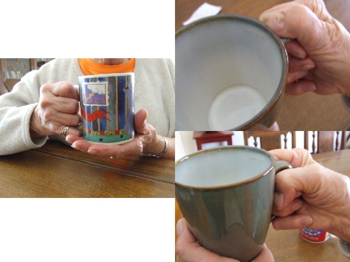
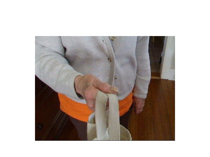
Major Issues re CRPS 1. Physical agent modalities (PAMs); 2. stress loading (1) Scrub portion (compression) using a brush or "Dystrophile" - either involving weight bearing through the arm. (2) Carrying (stretch/tension) - 1 - 5 pounds of weight throughout the day.
Major Issues re CRPS 3. exercise/splinting to increase ROM Generally any movement away from the static resting position could be called exercise; active and active/assisted are best followed by passive and splinting with dynamic components if tolerated. Static resting hand splint would only be indicated for night use if it helps with pain - would want to discourage use in day - WHY DO YOU THINK? ?
Major Issues re CRPS 4. edema control Through elevation when not in use; coban wraps or gloves in conjunction with as much movement as possible; extremity pumps and contrast baths also. 5. functional activities/work Probably the best - with an eye on return to more normal activities and functions (and roles). Can incorporate edema treatment with all functional activities. http: //www. rsdfoundation. org/en/en_clinical_practice_guidelines. html#Physical_ Therapy http: //www. energywellnessproducts. com/rsdmarastory. htm
Assessments: • A/PROM • Isolated muscle strength, pinch and grip strength (unless contraindicated by Dx, timing and stability of fracture) • Edema • COPM • Dash & Quick. Dash
Review
Oh yea – there are extensor tendon zones too. . .
Dupytren’s Contracture
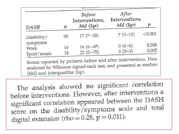
Evaluation of Activity Limitation and Digital i. Extension i n Dupytren’s. . .