Classification of Tissues Histology Study Guide Simple squamous
 Study Guide")


.")


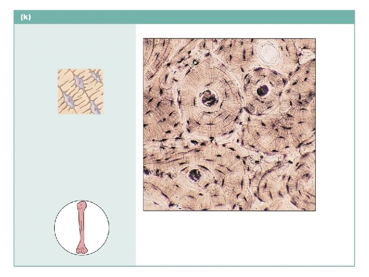
 Description: Hard, calcified matrix containing many collagen fibers; osteocytes lie")
.")


- Slides: 49
Classification of Tissues (Histology) Study Guide
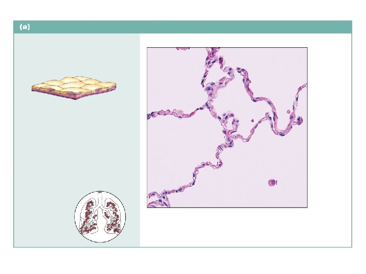
Simple squamous epithelium Description: Single layer of flattened cells with disc-shaped central nuclei and sparse cytoplasm; the simplest of the epithelia. Function: Allows passage of materials by diffusion and filtration in sites where protection is not important; produces lubricating fluid in serosae. Air sacs of lung tissue Nuclei of squamous epithelial cells Location: Kidney glomeruli; air sacs of lungs; lining of heart, blood vessels, and lymphatic vessels; lining of ventral body cavity (serosae). Photomicrograph: Simple squamous epithelium forming part of the alveolar (air sac) walls (140 ).
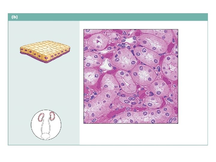
Simple cuboidal epithelium Description: Single layer of cubelike cells with large, spherical central nuclei. Simple cuboidal epithelial cells Function: Secretion and absorption. Basement membrane Location: Kidney tubules; ducts and secretory portions of small glands; ovary surface. Connective tissue Photomicrograph: Simple cuboidal epithelium in kidney tubules (430 ).
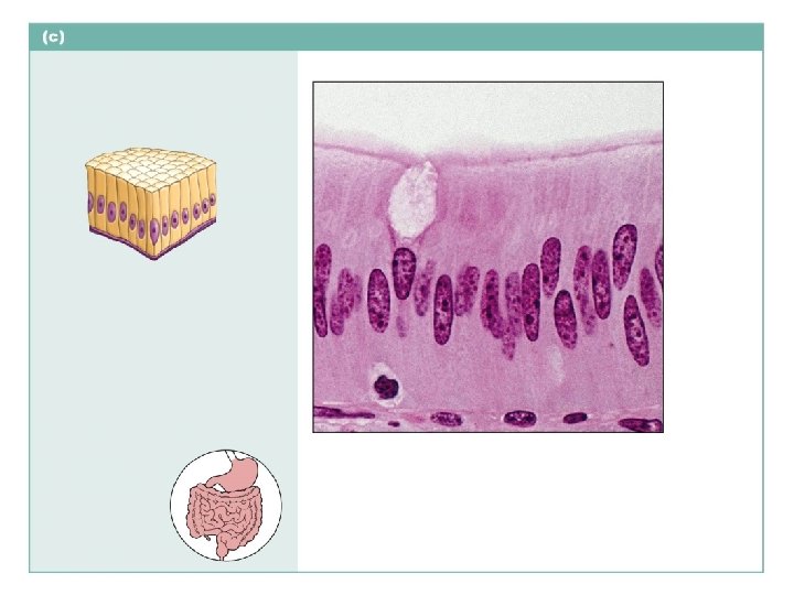
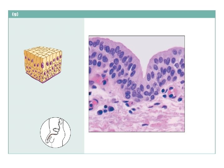
Simple columnar epithelium Description: Single layer of tall cells with round to oval nuclei; some cells bear cilia; layer may contain mucus-secreting unicellular glands (goblet cells). Microvilli Goblet cell Simple columnar epithelial cell Function: Absorption; secretion of mucus, enzymes, and other substances; ciliated type propels mucus (or reproductive cells) by ciliary action. Location: Nonciliated type lines most of the digestive tract (stomach to anal canal), gallbladder, and excretory ducts of some glands; ciliated variety lines small bronchi, uterine tubes, and some regions of the uterus. Basement membrane Photomicrograph: Simple columnar epithelium of the small intestine (650 ).
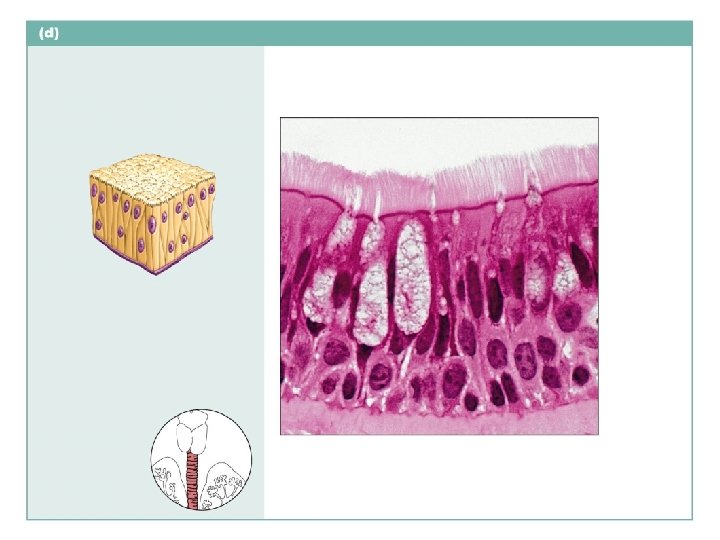
Pseudostratified columnar epithelium Description: Single layer of cells of different heights, some not reaching the free surface; nuclei seen at different levels; may contain mucus-secreting goblet cells and bear cilia. Cilia Goblet cell Pseudostratified epithelial layer Function: Secretion, particularly of mucus; propulsion of mucus by ciliary action. Location: Nonciliated type in male’s sperm-carrying ducts and ducts of large glands; ciliated variety lines the trachea, most of the upper respiratory tract. Trachea Basement membrane Photomicrograph: Pseudostratified ciliated columnar epithelium lining the human trachea (780 ).
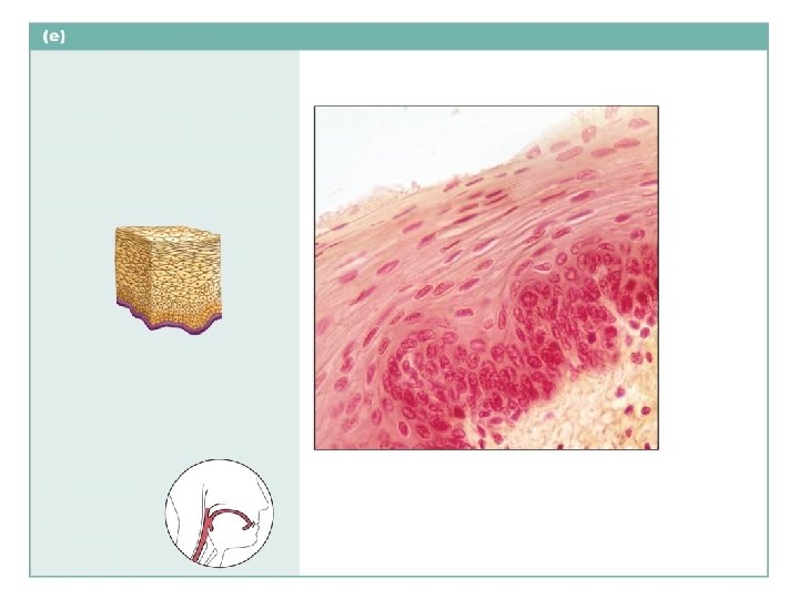
Stratified squamous epithelium Description: Thick membrane composed of several cell layers; basal cells are cuboidal or columnar and metabolically active; surface cells are flattened (squamous); in the keratinized type, the surface cells are full of keratin and dead; basal cells are active in mitosis and produce the cells of the more superficial layers. Stratified squamous epithelium Function: Protects underlying tissues in areas subjected to abrasion. Nuclei Basement membrane Location: Nonkeratinized type forms the moist linings of the esophagus, mouth, and vagina; keratinized variety forms the epidermis of the skin, a dry membrane. Connective tissue Photomicrograph: Stratified squamous epithelium lining the esophagus (280 ).
Stratified cuboidal epithelium Description: Generally two layers of cubelike cells. Basement membrane Function: Protection. Location: Largest ducts of sweat glands, mammary glands, and salivary glands. Cuboidal epithelial cells Duct lumen Photomicrograph: Stratified cuboidal epithelium forming a salivary gland duct (290 ).
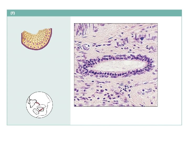
Stratified columnar epithelium Description: Several cell layers; basal cells usually cuboidal; superficial cells elongated and columnar. Stratified columnar epithelium Function: Protection; secretion. Location: Rare in the body; small amounts in male urethra and in large ducts of some glands. Urethra Basement membrane Underlying connective tissue Photomicrograph: Stratified columnar epithelium lining the male urethra (360 ).
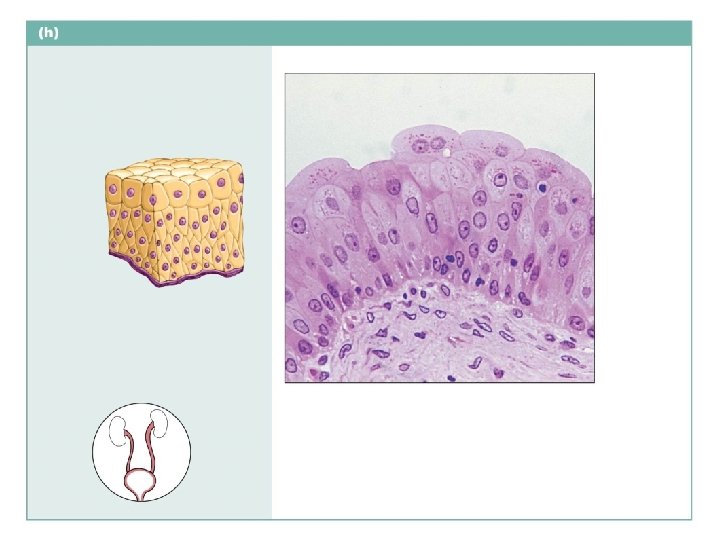
Transitional epithelium Description: Resembles both stratified squamous and stratified cuboidal; basal cells cuboidal or columnar; surface cells dome shaped or squamous-like, depending on degree of organ stretch. Transitional epithelium Function: Stretches readily and permits distension of urinary organ by contained urine. Location: Lines the ureters, bladder, and part of the urethra. Photomicrograph: Transitional epithelium lining the bladder, relaxed state (365 ); note the bulbous, or rounded, appearance of the cells at the surface; these cells flatten and become elongated when the bladder is filled with urine. Basement membrane Connective tissue
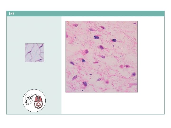
Embryonic connective tissue: mesenchyme Description: Embryonic connective tissue; gel-like ground substance containing fibers; star-shaped mesenchymal cells. Mesenchymal cells Ground substance Function: Gives rise to all other connective tissue types. Fibers Location: Primarily in embryo. Photomicrograph: Mesenchyme, an embryonic connective tissue (385 ). The matrix is composed of the fluid ground substance (clear-appearing background) and fine, sparse fibers.
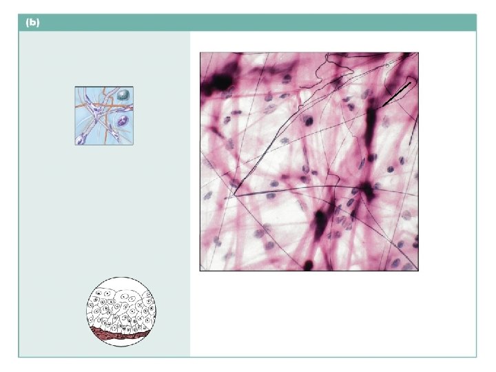
Connective tissue proper: loose connective tissue, areolar Description: Gel-like matrix with all three fiber types; cells: fibroblasts, macrophages, mast cells, and some white blood cells. Elastic fibers Ground substance Function: Wraps and cushions organs; its macrophages phagocytize bacteria; plays important role in inflammation; holds and conveys tissue fluid. Fibroblast nuclei Collagen fibers Location: Widely distributed under epithelia of body, e. g. , forms lamina propria of mucous membranes; packages organs; surrounds capillaries. Photomicrograph: Areolar connective tissue, a soft packaging tissue of the body (340 ). Epithelium Lamina propria
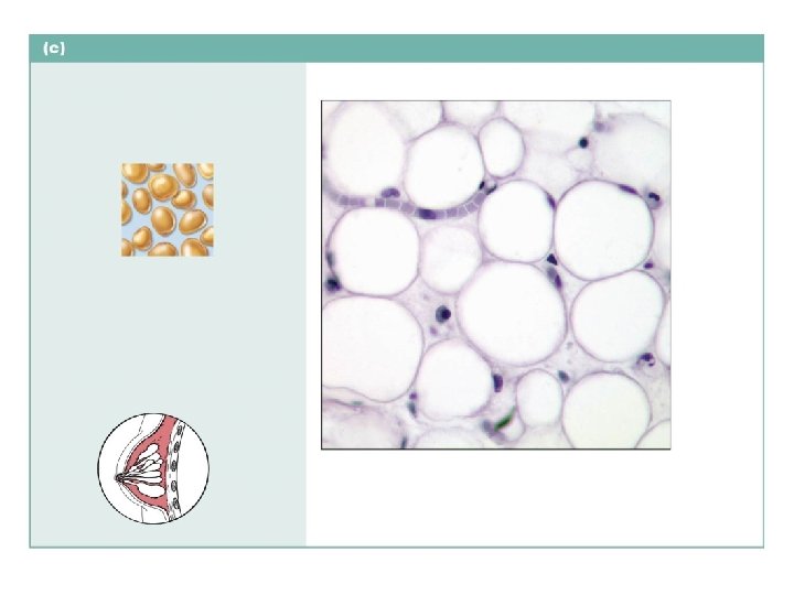
Connective tissue proper: loose connective tissue, adipose Description: Matrix as in areolar connective tissue, but very sparse; closely packed adipocytes, or fat cells, have nucleus pushed to the side by large fat droplet. Nucleus of fat cell Function: Provides reserve food fuel; insulates against heat loss; supports and protects organs. Vacuole containing fat droplet Location: Under skin in the hypodermis; around kidneys and eyeballs; within abdomen; in breasts. Adipose tissue Photomicrograph: Adipose tissue from the subcutaneous layer under the skin (350 ). Mammary glands
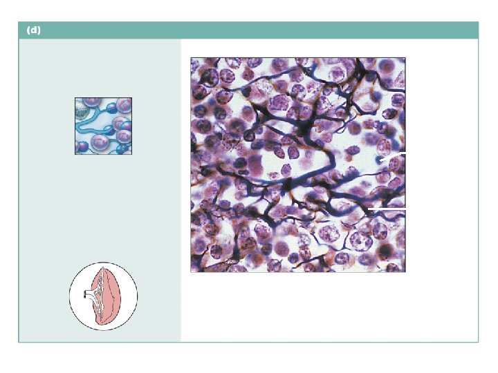
Connective tissue proper: loose connective tissue, reticular Description: Network of reticular fibers in a typical loose ground substance; reticular cells lie on the network. White blood cell (lymphocyte) Function: Fibers form a soft internal skeleton (stroma) that supports other cell types including white blood cells, mast cells, and macrophages. Reticular fibers Location: Lymphoid organs (lymph nodes, bone marrow, and spleen). Spleen Photomicrograph: Dark-staining network of reticular connective tissue fibers forming the internal skeleton of the spleen (350 ).
Connective tissue proper: dense connective tissue, dense irregular Description: Primarily irregularly arranged collagen fibers; some elastic fibers; major cell type is the fibroblast; defense cells and fat cells are also present. Nuclei of fibroblasts Function: Able to withstand tension exerted in many directions; provides structural strength. Collagen fibers Location: Fibrous capsules of organs and of joints; dermis of the skin; submucosa of digestive tract. Fibrous layer of joint capsule Photomicrograph: Dense irregular connective tissue from the dermis of the skin (300 ).
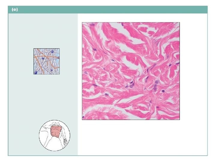
Connective tissue proper: dense connective tissue, dense regular Description: Primarily parallel collagen fibers; a few elastic fibers; major cell type is the fibroblast. Collagen fibers Nuclei of fibroblasts Function: Attaches muscles to bones or to muscles; attaches bones to bones; withstands great tensile stress when pulling force is applied in one direction. Location: Tendons, most ligaments, aponeuroses. Shoulder joint Ligament Tendon Photomicrograph: Dense regular connective tissue from a tendon (425 ).
Connective tissue proper: dense connective tissue, elastic Description: Dense regular connective tissue containing a high proportion of elastic fibers. Elastic fibers Function: Allows recoil of tissue following stretching; maintains pulsatile flow of blood through arteries; aids passive recoil of lungs following inspiration. Location: Walls of large arteries; within certain ligaments associated with the vertebral column; within the walls of the bronchial tubes. Aorta Heart Photomicrograph: Elastic connective tissue in the wall of the aorta (250 ).
Photomicrograph: Elastic connective tissue in the wall of the aorta (250 ).
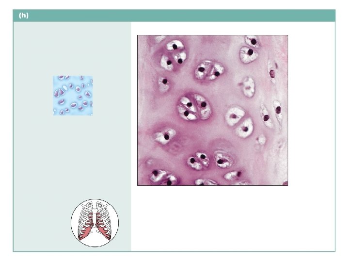
Cartilage: hyaline Description: Amorphous but firm matrix; collagen fibers form an imperceptible network; chondroblasts produce the matrix and, when mature (chondrocytes), lie in lacunae. Chondrocyte in lacuna Matrix Function: Supports and reinforces; serves as resilient cushion; resists compressive stress. Location: Forms most of the embryonic skeleton; covers the ends of long bones in joint cavities; forms costal cartilages of the ribs; cartilages of the nose, trachea, and larynx. Costal cartilages Photomicrograph: Hyaline cartilage from a costal cartilage of a rib (470 ).
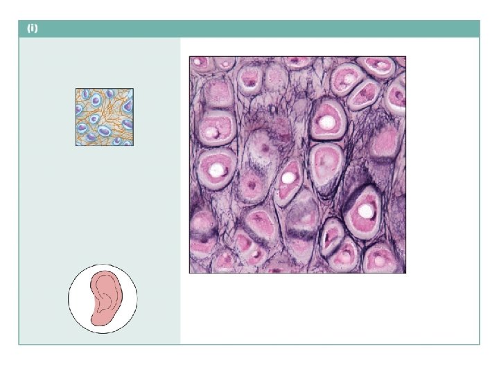
Cartilage: elastic Description: Similar to hyaline cartilage, but more elastic fibers in matrix. Chondrocyte in lacuna Function: Maintains the shape of a structure while allowing great flexibility. Matrix Location: Supports the external ear (pinna); epiglottis. Photomicrograph: Elastic cartilage from the human ear pinna; forms the flexible skeleton of the ear (510 ).
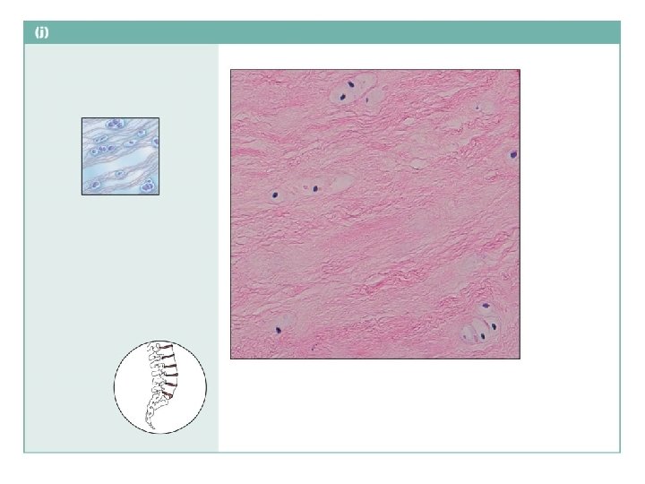
Cartilage: fibrocartilage Description: Matrix similar to but less firm than that in hyaline cartilage; thick collagen fibers predominate. Function: Tensile strength with the ability to absorb compressive shock. Collagen fibers Location: Intervertebral discs; pubic symphysis; discs of knee joint. Chondrocytes in lacunae Intervertebral discs Photomicrograph: Fibrocartilage from an intervertebral disc (175 ).
Others: bone (osseous tissue) Description: Hard, calcified matrix containing many collagen fibers; osteocytes lie in lacunae. Very well vascularized. Central canal Lacunae Function: Supports and protects (by enclosing); provides levers for the muscles to act on; stores calcium and other minerals and fat; marrow inside bones is the site for blood cell formation (hematopoiesis). Lamella Location: Bones. Photomicrograph: Cross-sectional view of bone (175 ).
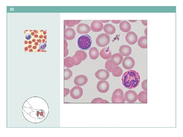
Connective tissue: blood Description: Red and white blood cells in a fluid matrix (plasma). Red blood cells (erythrocytes) White blood cells: • Lymphocyte • Neutrophil Function: Transport respiratory gases, nutrients, wastes, and other substances. Location: Contained within blood vessels. Plasma Photomicrograph: Smear of human blood (1650 ); shows two white blood cells surrounded by red blood cells.
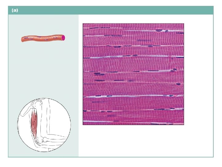
Skeletal muscle Description: Long, cylindrical, multinucleate cells; obvious striations. Striations Nuclei Function: Voluntary movement; locomotion; manipulation of the environment; facial expression. Location: In skeletal muscles attached to bones or occasionally to skin. Part of muscle fiber (cell) Photomicrograph: Skeletal muscle (450 ). Notice the obvious banding pattern and the fact that these large cells are multinucleate.
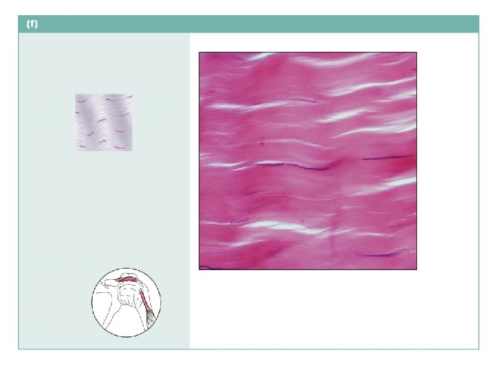
Cardiac muscle Description: Branching, striated, generally uninucleate cells that interdigitate at specialized junctions (intercalated discs). Striations Intercalated discs Function: As it contracts, it propels blood into the circulation; involuntary control. Location: The walls of the heart. Nucleus Photomicrograph: Cardiac muscle (355 ); notice the striations, branching of cells, and the intercalated discs.
Cardiac muscle
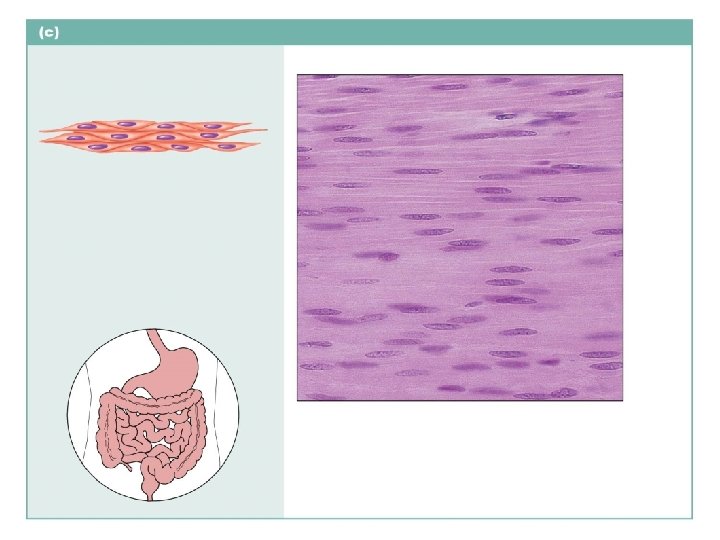
Smooth muscle Description: Spindle-shaped cells with central nuclei; no striations; cells arranged closely to form sheets. Smooth muscle cell Function: Propels substances or objects (foodstuffs, urine, a baby) along internal passageways; involuntary control. Nuclei Location: Mostly in the walls of hollow organs. Photomicrograph: Sheet of smooth muscle from the digestive tract (465 ).
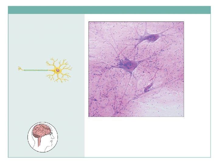
Nervous tissue Description: Neurons are branching cells; cell processes that may be quite long extend from the nucleus-containing cell body; also contributing to nervous tissue are nonconducting supporting cells, neuroglia (not illustrated). Neuron processes Cell body of a neuron Neuron processes Cell body Dendrites Axon Function: Transmit electrical signals from sensory receptors and to effectors (muscles and glands) that control the activity of the effector organs. Location: Brain, spinal cord, and nerves. Nuclei of neuroglia Photomicrograph: Neurons (125 ).