Ultra High Field MRI Charles Dietz MD 2017


")
 MDEFT Barfuss et. al. NMR Biomed: 3(1)1990 (DATA from SIEMENS) SIEMENS,")


 MRI at 7 T with")


 Knee imaging at 7 T")

")
")


 in the Human Brain at 7 Tesla Primary Auditory")
:")


: resonates at 2. 0 40")


 breast MR has focused on")


 400 0 Li, NMR in Biomed: 63 -69;")
 in Neurosciences and Brain disorders Research NIH Brain")


")


: 1565 -1567. 57")
 and Canonical Correlation Analysis (CCA) Mode 58")
 and Canonical Correlation Analysis (CCA) Mode 59")

 • DEVELOPING")


, post-mortem; Phase Shim")
 0 -1")
")

 Small magnet")
- Slides: 75
Ultra High Field MRI Charles Dietz MD 2017
2
Frequency directly related to Magnetic Field 3
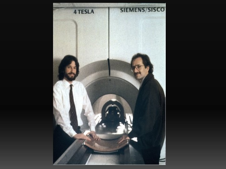
4 Tesla (Installed in CMRR in ~1990)
4 TESLA (1991) MDEFT Barfuss et. al. NMR Biomed: 3(1)1990 (DATA from SIEMENS) SIEMENS, GE, and PHILIPS
BOLD f. MRI a 1991 4 Tesla CMRR R b Full Field Visual Stimulation HEMI Field Visual Stimulation c Ogawa et al. Proc Natl Acad Sci USA (1992) 89, 5951 -
Functional Imaging with Magnetic Resonance Visual Stimulus 4 Tesla 2 1 3 Title Ogawa et al. Proc Natl Acad Sci USA (1992) 89, 5951 -5955
Wei Chen et al. , U of Minnesota, CMRR, 4 T 4% R L R 0. 2% L
Opaque anatomical Image In Grey scale SILENT WORD GENERATION
DEVELOPMENT OF CMRR INSTRUMENTATION for HIGH MAGNETIC FIELD IMAGING and SPECTROSCOPY 4. 7 Tesla/40 cm (~1985) • 4 Tesla Human (1991) 9. 4 Tesla/31 cm (~ 1995) • 7 Tesla Human (~1999) 3 Tesla ~2002 • 9. 4 Tesla Human (~2005) 2 nd 7 T (2010) and 10. 5 T (2015) 16. 4 Tesla /26 cm bore (animal) (2010)
7 T MRI Department of Radiology Charles Dietz
7 Tesla/90 cm bore ~1999
7 Tesla UHF Clinical MRI • Ultra Highfield (UHF) MRI at 7 T with increased S/N ratio • Spectroscopy • Higher Resolution – Improved Diagnostic Accuracy • Shorter Acquisition Time – Higher Clinical Throughput
Patellar Cartilage 7 T 3 T
3 T Knee MRI
7 T Knee MRI
Meniscal Tears 7 T 3 T
3 TESLA SUBTLE FRACTURE
7 TESLA SUBTLE FRACTURE
DETAILED ANATOMY OF TIBIAL NERVE AT 7 T (CMRR) Knee imaging at 7 T a) 16 -element transmit/receive for all knee images. b) Sagittal turbo spin echo (TSE) image (TR/TE = 4210/31 ms, resolution 0. 2 x 3, 39 slices, 1 average, echotrain length 7, 10 min. c) Axial turbo spin echo (TSE) image (TR/TE = 4680/27 ms, resolution 0. 3 x 2, 29 slices, 2 averages. Ellermann et al.
Quantitative cartilage assessment: T 1ρ -maps of articular cartilage Dog model of early osteoathritis at 7 Tesla (CMRR) T 1ρ Surgery preparation module nx 4 hyperbolic secant (HS 1)-pulses pulse duration: 5. 12 ms Control ms control surgery p (two-tail) medial 160± 30 210± 30 0. 01 imaging module in-plane resolution: 620 μm lateral 190± 50 270± 60 0. 7 slice thickness: 1. 5 mm matrix: 128× 128 T 1ρ of the cartilage in the severed joint is longer than in the control # segments: 4 TE/TR = 5. 1 ms/4 s Pepin SR et al: A comparative analysis of 7. 0 -Tesla magnetic resonance imaging and histology measurements of knee articular cartilage in a canine posterolateral knee injury model: a preliminary analysis. Am J Sports Med.
IMAGING AT HIGH AND ULTRAHIGH FIELDS IN THE HUMAN TORSO; THEN (1990; 4 TESLA) 4 Tesla (Data from Siemens) Barfuss et al. NMR Biomed: 3(1)1990 NOW (7 TESLA) 7 Tesla Henry, TR et al. , Radiology 2011; 261(1): 199 -
IMAGING AT HIGH AND ULTRAHIGH FIELDS IN THE HUMAN TORSO; THEN (1990; 4 TESLA) 4 Tesla (Data from Siemens) Barfuss et al. NMR Biomed: 3(1)1990 NOW (2009; 7 TESLA) 7 Tesla (CMRR) Snyder, C. et al MRM 61(3): 517 -524 (2009)
7 TESLA: A 16 -channel combined loop-dipole transceiver array for 7 Tesla body MRI Ertürk et al. MRM (2016). 26
Anatomical contrast @ 7 T T 1 W T 2 W SWI STN SN RN STN: subthalamic nucleus SN: substantia nigra RN: red nucleus SWI: Susceptibility-weighted imaging Abosch, Yacoub, Ugurbil, Harel. 2010
Susceptibility-Weighted Imaging @ 7 T STN SN Magnet: 7 T Resolution: 0. 4 x 0. 8 mm STN = Subthalamic Nucleus SN = Substantia Nigra Dr. Noam Harel, University of Minnesota / CMRR
Susceptibility-Weighted Imaging @ 7 T i P G GP = Globus pallidus e P G Pi e P G SWI @ 7 T (In-vivo) G Schaltenbrand Wahren Atlas (Ex-vivo) Lamina pallidi mediali s Abosch, Yacoub, Ugurbil, Harel. 2010
DBS STN SN Red. N Dr. Noam Harel, University of Minnesota / CMRR
Orientation ODC Phase Left Right 1 mm Yacoub, Shmuel, et al. Yacoub, Harel, Uğurbil Neuroimage (2007) 37(4): 1161 -77 PNAS (2008) 105(30): 10607 -12
Tonotopic Mapping in Human Primary Auditory Cortex 7 T GE f. MRI 1. 2 x 1. 5 x 2. 4 mm 3 CMRR/U Maastrich
Cortex has known neural networks that span cortical layers
High Resolution Frequency maps (tonotopy) in the Human Brain at 7 Tesla Primary Auditory Cortex Laminar Resolution De Martino et al (PNAS 2015) Formisano et al. (Neuron, 2003) Medial Geniculate Body Moerel et al (Scien. Rep. , 5; 17048 (2015)) Inferior Colliculus De Martino et al. (Nature Communications, 2013)
MT Flattened maps of 3 layers surface Zimmermann et al. PLo. S ONE 6(12): e 28716. (2011) deep CMRR & Univ. Maastricht Collaboration 35 PLo. S Zimmermann et al. 0. 8 mm isotropic 6(12): e 28716. (2011)
7 TESLA HUMAN BRAIN 1 H NMR spectrum at NAA STEAM TE = 6 ms TR = 5 s VOI = 8 ml NT = 160 Cr PCr residual water Asp GABA Glc Cr PCr scyllo-Ins Glu Gln Cho GSH Glu Tau Ins Gln NAA Ins 36
lactate: resonates at 1. 3 ppm lipids: resonates at 1. 3 ppm alanine: resonates at 1. 48 ppm N-acetylaspartate (NAA): resonates at 2. 0 glutamine/glutamate: resonates at 2. 2 -2. 4 ppm GABA: resonates at 2. 2 -2. 4 ppm 2 -hydroxyglutarate: resonates at 2. 25 ppm 6 citrate: resonates 2. 6 ppm creatine: resonates at 3. 0 ppm choline: resonates at 3. 2 ppm myo-inositol: resonates at 3. 5 ppm water resonates at 4. 4 ppm 37
7 T, STEAM TR/TE=5000/6 ms VOI=20 x 22 x 20 mm 3 32 scans During visual stimuli, changes of metabolite concentrations were within ± 0. 2 mmol/g 38
Results - 5. 3 min stimulation Group Analysis Error bars = CRLB 39
SPINOCEREBELLAR ATAXIAS N-acetylaspartate (NAA): resonates at 2. 0 40
UHF Prostate GOAL: Develop the imaging hardware and methods to exploit the advantages of UHF imaging in the prostate. • Development of novel RF coils – Surface arrays – Multi-channel endorectal Coils • Improved Imaging and Spectroscopy – Increased resolution anatomic imaging – Increased resolution functional imaging – Improved spectral quantification on spectroscopy • Future Work: evaluate the potential advantages in cancer detection and staging to improve the identification of clinically significant disease.
3 T 7 T Sagittal T 2 Coronal T 2 Axial ADC
7 T Intro Our work on 7 T (UHF) breast MR has focused on RF coil development. We’ve made 4 iterations of RF coils, and while we’ve used them to get some prelim data they are all unsatisfying. The problem is that for contrast-enhanced breast imaging you need relatively uniform flip angles over both breasts and axillae. With the reduced RF wavelength at 7 T (~12 cm) it is very difficult to achive this. This is why we’ve had more success with smaller regions (prostate, cardiac, kidneys) that are more tolerant to flip angle variation.
7 T Breast MR Imaging High SNR allows high-resolution anatomic imaging Axial fat-sat 3 D FLASH 0. 5 x 2 mm Sagittal fat-sat 3 D FLASH 0. 3 x 2. 2 mm
7 T Breast MR Spectroscopy Increased spectral resolution and SNR gives excellent spectra in normals and patients Patient with IDC Normal Volunteer 6 4 ppm 2 0 Haddadin I. et al. , NMR Biomed 2009 6 4 ppm 2 0 Bolan P. J. et al. , ISMRM 2006 choline: resonates at 3. 2 ppm
3 T Advancements • The following slides are 3 T only: they describe the development of highresolution diffusion imaging
High-resolution Breast DWI: RS vs SMS-IPSE T 2 -weighted anatomical Comparing approaches with fixed 5 min acquisition time 0. 8 x 3 mm Standard b = 0 1. 7 x 4 mm RS-EPI b = 0 1. 8 x 2. 4 mm SMS/MB-IPSE b = 0 1. 25 x 2. 5 mm • Std, single-shot EPI • RESOLVE (RS-EPI), following Wisner JMRI 2014 • SMS/MB with in-plane slice encoding SMS/MB-IPSE approaches T 2 resolution & quality
Renal Blood Flow (m. L/100 g/min) 400 0 Li, NMR in Biomed: 63 -69; 2015 Li (2015) ISMRM #3155.
NIH invests substantially (~7 -8 billions/year) in Neurosciences and Brain disorders Research NIH Brain Research through Advancing Innovative Neurotechnologies (BRAIN) Initiative 49
Description of the functional and structural connections among gray matter locations in the human brain • spontaneous fluctuations in an f. MRI time series (i. e. “Resting State” f. MRI ) to deduce ‘functional connectivity’ and/or • Diffusion weighted MRI to infer ‘structural connectivity’ 50
Principal Investigators: David C. Van Essen and Kamil Ugurbil 51
“FUNCTIONAL CONNECTIVITY in the HUMAN BRAIN THROUGH • spontaneous correlated fluctuations in an f. MRI time series (i. e. “Resting State” f. MRI ) to deduce ‘functional connectivity’ Time Spatial patterns of correlated temporal dynamics, resembling 52 activation maps
FUNCTIONAL CONNECTIVITY, DENSE CONNECTOME (from Multiband Resting State f. MRI, 3 T Skyra Connectom) Functional connectivity map (location 1) Functional connectivity map (location 2) Correlation Low High M. Glasser , D. Van Essen et al for the HCP
CMRR 7 T DWI UCLA tractography 54
CMRR 7 T DWI UCLA tractography 55
Wash. U-UMinn-Oxford Consortium. Insan Konnektom Projesi
Nat Neurosci 2015; 18(11): 1565 -1567. 57
Correlation Between each Subject Measures (SM) and Canonical Correlation Analysis (CCA) Mode 58
Correlation Between Subject Measures (SM) and Canonical Correlation Analysis (CCA) Mode 59
60
LIFESPAN - HUMAN CONNECTOME PROJECT • BABY CONNECTOME (birth to 5 years) • DEVELOPING CONNECTOME ( Ages 5 -21) • AGING (Ages 36 - 90+)
SCALES OF BRAINBOW approach to uniquely label individual cells Human Brain Functional Connectivity 62
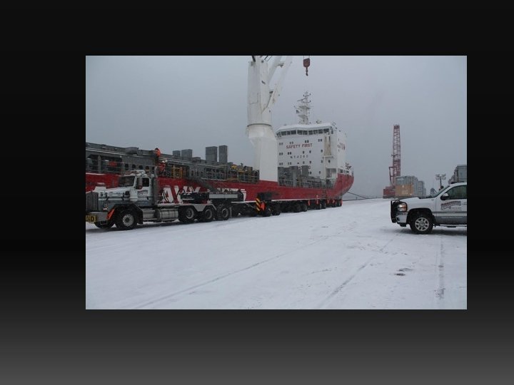
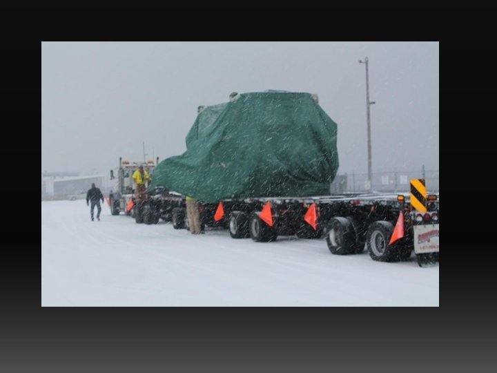
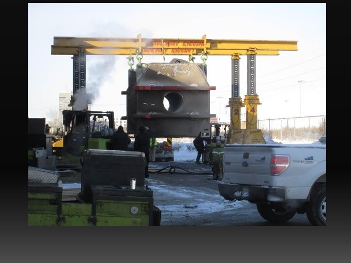
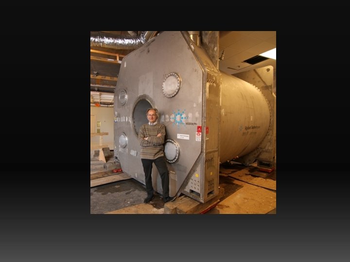
10. 5 T/88 cm; 110 Tons Stored Energy 280 MJ 10. 5 T Conductor length 1160 kms http: //www. healthtalk. umn. edu/2013/12/31/move-110 -ton-imaging-magnet/
68
10. 5 Tesla Images of Porcine head Geometrically Adjustable Dipole Coronal TSEs tse_cor_te=67 ms_bw=500_512_Vref=78 seq = tse 2 d 1_10 SE SKSPOSP Opt: TR/TE = 5000. 00 / 71. 00 ms nominal flipangle = 130. 00 deg Variable: N Excitation Voltage = 48. 05 V Ref. Voltage: 78. 00 V thk = 3. 00 mm etl = 10 ; TA 04: 16 512*512; Fo. V 170*170; Resolution = 0. 33 x 0. 33 mm G. Adriany, P-F. , Van de Moortele et al.
Towards imaging the body at 10. 5 Tesla Swine post-mortem Phase Shim only Swineimaging, (75 kg), post-mortem; A. Ertürk, Y. Eryaman, G. Metzger
Towards imaging the body at 10. 5 Tesla Swine (75 kg), post-mortem; Phase Shim only A. Ertürk, Y. Eryaman, G. Metzger
Log Millimeters 2 1 UHF Functional Imaging with Magnetic Resonance (f. MRI) 0 -1 -2 -3 Calcium or Voltage Imaging with LIGHT
4 Tesla (Installed in CMRR in ~1990)
Center for Magnetic Resonance Research Univ. of MINNESOTA 74
Concept: Future Head MRI PI: M. Garwood, and T. Vaughan (CMRR, Uminn) Small magnet size dictates • The use of high temperature superconductors • Imaging at inhomogeneous magnetic fields