Paediatric Spine MRI Indications for GP referred Medicare


- Slides: 5

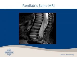
Paediatric Spine MRI

Indications for GP referred Medicare Rebatable MRI spine studies in children under 16 years of age Following radiographic examination for any or the following: • significant trauma • unexplained neck or back pain with associated neurological signs • unexplained back pain where significant pathology is suspected Differs from adult rebate criteria: • No specific spine region. Can investigate lumbar, thoracic or cervical spine (not just cervical). • Can be for back pain without neurological involvement

Traumatic & overuse causes of spinal pain in children • Acute fractures and ligament injury • Pars interarticularis stress fractures (acute spondylolysis) Early detection and management of pars fractures improves chance of union • • Spondylolisthesis Scheuermann’s disease Disc protrusions Facet arthropathy Disc protrusions and facet arthropathy comparatively rarer in adolescence but still seen, particularly those very active in sports • Lumbosacral pseudoarticulation (Bertolotti’s syndrome)

Acute injury: 4 yr boy with a fall on his bottom and ongoing pain L 1 – L 4 UNDISPLACED FRACTURES Bone marrow oedema (bright) within fractured vertebrae Normal adjacent vertebrae NORMAL XRAY This study in a 4 year old was performed without general anaesthetic or sedation. The images are diagnostic.

Non-acute back pain: 15 yr old football player with back pain. Suspected pars fracture. Fracture line appears bright on the T 1 THRIVE sequence (unless inverted as on right). T 1 THRIVE, used as a dedicated MRI sequence for cortical fractures. A sequence is a particular component of an MRI scan, of which each scan has several. Exact same MRI image as left with colours inverted to simulate CT to show the similarity with the cortical bone sequence. Avoids radiation.
